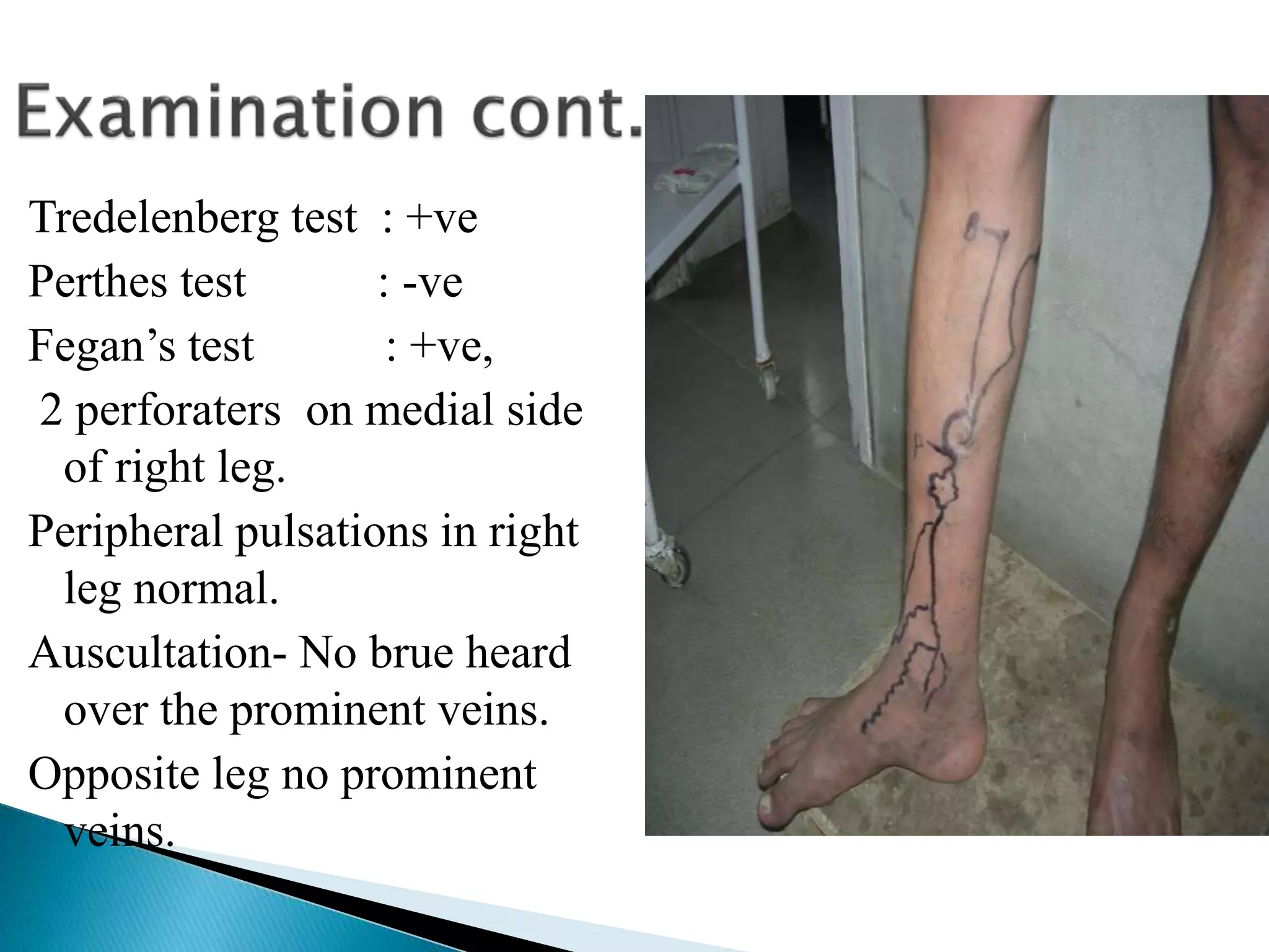
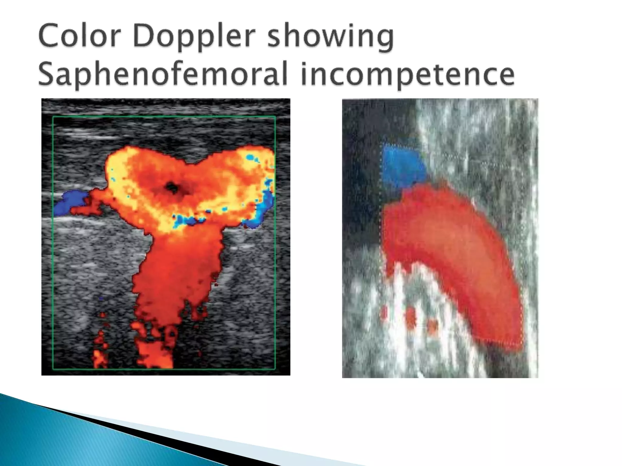
This document describes a 32-year-old male farmer who presented with prominent veins in his right leg for 2 years along with swelling and pain in the right leg for 1 year. On examination, dilated veins were visible on the medial side of the right leg, ankle, and foot extending to the thigh. Tests showed saphenofemoral and perforator incompetence on the right side. The diagnosis was clinical grade 4 varicose veins. Color doppler ultrasound was recommended to further evaluate the perforators and veins, and pre-anaesthetic tests were ordered prior to a planned surgery involving high saphenofemoral junction ligation and stripping of the long saphenous vein along with ligation of perforators and
UG CASE PRESENTATION ON INGUINAL HERNIAAyesha Huma
I have added viva notes after this proforma for quick revision of important stuff asked in exam.
LINK FOR EXAMINATION VIDOES :
1. https://youtu.be/uO-w_9w5okI
2. https://youtu.be/Vc_ZH_-Oad4
Case study on Varicose Veins & Venous UlcersAbhineet Dey
A clinically based study of a case of Varicose Veins & Venous Ulcers.
Moderator:
Dr M. K. Mazumdar
Asst. Professor,
Dept. of Obstetrics and Gynaecology,
Gauhati Medical College & Hospital
Presented by:
29: Abhineet Dey
30: Devasree Kalita
31: Parishmita Sharma
32: Samadrita Borkakoty
33: Ankur Jain
34: Dhurjyoti Nath
35: Mousumi Mehtaz
Students of 8th Semester,
Gauhati Medical College & Hospital, Guwahati, Assam
Examination of Swelling in a patient is always a task for MBBS students. This PPT provides the students, how to elicit a history & also the easy way to examine a swelling.
This is a small booklet in an outline format to assist undergraduate medical students to aid in writing case write ups. This mainly contains how to elicit symptoms and signs.
anatomy of the lower extremity veins, CVI , ambulatory venous hypertension, varicose veins , clinical examination and performance of various tests of the varicose veins
This is a case study done by me as a part of my in-service education progamme in my institution...hope this may help all nurses who wants to do a case study.
It is estimated that 20% of American women and 7% of American men suffer from venous disease. Venous disease results in symptoms such as aching, fatigue, swelling, and pain in the legs which can interfere with daily living.Cosmetic issues may affect quality of life.
At least 20% of patients with venous disease will develop leg ulcers. This presentation outlines the normal anatomy and physiology of venous drainage of the extremities as well as the common venous disorders such as varicose veins and deep vein thrombosis.
to down load this presentation from this link
https://mohmmed-ink.blogspot.com/2020/11/deep-vein-thrombosis-dvt.html
deep vein thrombosis, diagnosis and managment.
Deep Vein Thrombosis is an important and frequently missed out diagnosis that can often lead to sudden death in post operative patients. Did this powerpoint for an O&G seminar. Mainly focusses on DVT in OBG and its management and prevention. Kindly leave a comment and let me know what you think.
Thyroid Swelling: A practical guide on writing and presenting a clinical caseMuskaan Khosla
Every book tells us how to examine and what to examine in a clinical case. But, no book tells us exactly how to write a sheet and how to word the case. Here is a practical example ! Hope it helps!
Comparing Evolved Extractive Text Summary Scores of Bidirectional Encoder Rep...University of Maribor
Slides from:
11th International Conference on Electrical, Electronics and Computer Engineering (IcETRAN), Niš, 3-6 June 2024
Track: Artificial Intelligence
https://www.etran.rs/2024/en/home-english/
Richard's aventures in two entangled wonderlandsRichard Gill
Since the loophole-free Bell experiments of 2020 and the Nobel prizes in physics of 2022, critics of Bell's work have retreated to the fortress of super-determinism. Now, super-determinism is a derogatory word - it just means "determinism". Palmer, Hance and Hossenfelder argue that quantum mechanics and determinism are not incompatible, using a sophisticated mathematical construction based on a subtle thinning of allowed states and measurements in quantum mechanics, such that what is left appears to make Bell's argument fail, without altering the empirical predictions of quantum mechanics. I think however that it is a smoke screen, and the slogan "lost in math" comes to my mind. I will discuss some other recent disproofs of Bell's theorem using the language of causality based on causal graphs. Causal thinking is also central to law and justice. I will mention surprising connections to my work on serial killer nurse cases, in particular the Dutch case of Lucia de Berk and the current UK case of Lucy Letby.
A brief information about the SCOP protein database used in bioinformatics.
The Structural Classification of Proteins (SCOP) database is a comprehensive and authoritative resource for the structural and evolutionary relationships of proteins. It provides a detailed and curated classification of protein structures, grouping them into families, superfamilies, and folds based on their structural and sequence similarities.
Slide 1: Title Slide
Extrachromosomal Inheritance
Slide 2: Introduction to Extrachromosomal Inheritance
Definition: Extrachromosomal inheritance refers to the transmission of genetic material that is not found within the nucleus.
Key Components: Involves genes located in mitochondria, chloroplasts, and plasmids.
Slide 3: Mitochondrial Inheritance
Mitochondria: Organelles responsible for energy production.
Mitochondrial DNA (mtDNA): Circular DNA molecule found in mitochondria.
Inheritance Pattern: Maternally inherited, meaning it is passed from mothers to all their offspring.
Diseases: Examples include Leber’s hereditary optic neuropathy (LHON) and mitochondrial myopathy.
Slide 4: Chloroplast Inheritance
Chloroplasts: Organelles responsible for photosynthesis in plants.
Chloroplast DNA (cpDNA): Circular DNA molecule found in chloroplasts.
Inheritance Pattern: Often maternally inherited in most plants, but can vary in some species.
Examples: Variegation in plants, where leaf color patterns are determined by chloroplast DNA.
Slide 5: Plasmid Inheritance
Plasmids: Small, circular DNA molecules found in bacteria and some eukaryotes.
Features: Can carry antibiotic resistance genes and can be transferred between cells through processes like conjugation.
Significance: Important in biotechnology for gene cloning and genetic engineering.
Slide 6: Mechanisms of Extrachromosomal Inheritance
Non-Mendelian Patterns: Do not follow Mendel’s laws of inheritance.
Cytoplasmic Segregation: During cell division, organelles like mitochondria and chloroplasts are randomly distributed to daughter cells.
Heteroplasmy: Presence of more than one type of organellar genome within a cell, leading to variation in expression.
Slide 7: Examples of Extrachromosomal Inheritance
Four O’clock Plant (Mirabilis jalapa): Shows variegated leaves due to different cpDNA in leaf cells.
Petite Mutants in Yeast: Result from mutations in mitochondrial DNA affecting respiration.
Slide 8: Importance of Extrachromosomal Inheritance
Evolution: Provides insight into the evolution of eukaryotic cells.
Medicine: Understanding mitochondrial inheritance helps in diagnosing and treating mitochondrial diseases.
Agriculture: Chloroplast inheritance can be used in plant breeding and genetic modification.
Slide 9: Recent Research and Advances
Gene Editing: Techniques like CRISPR-Cas9 are being used to edit mitochondrial and chloroplast DNA.
Therapies: Development of mitochondrial replacement therapy (MRT) for preventing mitochondrial diseases.
Slide 10: Conclusion
Summary: Extrachromosomal inheritance involves the transmission of genetic material outside the nucleus and plays a crucial role in genetics, medicine, and biotechnology.
Future Directions: Continued research and technological advancements hold promise for new treatments and applications.
Slide 11: Questions and Discussion
Invite Audience: Open the floor for any questions or further discussion on the topic.
Observation of Io’s Resurfacing via Plume Deposition Using Ground-based Adapt...Sérgio Sacani
Since volcanic activity was first discovered on Io from Voyager images in 1979, changes
on Io’s surface have been monitored from both spacecraft and ground-based telescopes.
Here, we present the highest spatial resolution images of Io ever obtained from a groundbased telescope. These images, acquired by the SHARK-VIS instrument on the Large
Binocular Telescope, show evidence of a major resurfacing event on Io’s trailing hemisphere. When compared to the most recent spacecraft images, the SHARK-VIS images
show that a plume deposit from a powerful eruption at Pillan Patera has covered part
of the long-lived Pele plume deposit. Although this type of resurfacing event may be common on Io, few have been detected due to the rarity of spacecraft visits and the previously low spatial resolution available from Earth-based telescopes. The SHARK-VIS instrument ushers in a new era of high resolution imaging of Io’s surface using adaptive
optics at visible wavelengths.
(May 29th, 2024) Advancements in Intravital Microscopy- Insights for Preclini...Scintica Instrumentation
Intravital microscopy (IVM) is a powerful tool utilized to study cellular behavior over time and space in vivo. Much of our understanding of cell biology has been accomplished using various in vitro and ex vivo methods; however, these studies do not necessarily reflect the natural dynamics of biological processes. Unlike traditional cell culture or fixed tissue imaging, IVM allows for the ultra-fast high-resolution imaging of cellular processes over time and space and were studied in its natural environment. Real-time visualization of biological processes in the context of an intact organism helps maintain physiological relevance and provide insights into the progression of disease, response to treatments or developmental processes.
In this webinar we give an overview of advanced applications of the IVM system in preclinical research. IVIM technology is a provider of all-in-one intravital microscopy systems and solutions optimized for in vivo imaging of live animal models at sub-micron resolution. The system’s unique features and user-friendly software enables researchers to probe fast dynamic biological processes such as immune cell tracking, cell-cell interaction as well as vascularization and tumor metastasis with exceptional detail. This webinar will also give an overview of IVM being utilized in drug development, offering a view into the intricate interaction between drugs/nanoparticles and tissues in vivo and allows for the evaluation of therapeutic intervention in a variety of tissues and organs. This interdisciplinary collaboration continues to drive the advancements of novel therapeutic strategies.
Deep Behavioral Phenotyping in Systems Neuroscience for Functional Atlasing a...Ana Luísa Pinho
Functional Magnetic Resonance Imaging (fMRI) provides means to characterize brain activations in response to behavior. However, cognitive neuroscience has been limited to group-level effects referring to the performance of specific tasks. To obtain the functional profile of elementary cognitive mechanisms, the combination of brain responses to many tasks is required. Yet, to date, both structural atlases and parcellation-based activations do not fully account for cognitive function and still present several limitations. Further, they do not adapt overall to individual characteristics. In this talk, I will give an account of deep-behavioral phenotyping strategies, namely data-driven methods in large task-fMRI datasets, to optimize functional brain-data collection and improve inference of effects-of-interest related to mental processes. Key to this approach is the employment of fast multi-functional paradigms rich on features that can be well parametrized and, consequently, facilitate the creation of psycho-physiological constructs to be modelled with imaging data. Particular emphasis will be given to music stimuli when studying high-order cognitive mechanisms, due to their ecological nature and quality to enable complex behavior compounded by discrete entities. I will also discuss how deep-behavioral phenotyping and individualized models applied to neuroimaging data can better account for the subject-specific organization of domain-general cognitive systems in the human brain. Finally, the accumulation of functional brain signatures brings the possibility to clarify relationships among tasks and create a univocal link between brain systems and mental functions through: (1) the development of ontologies proposing an organization of cognitive processes; and (2) brain-network taxonomies describing functional specialization. To this end, tools to improve commensurability in cognitive science are necessary, such as public repositories, ontology-based platforms and automated meta-analysis tools. I will thus discuss some brain-atlasing resources currently under development, and their applicability in cognitive as well as clinical neuroscience.
2. Introduction of the patient
Raju
32 yrs male Farmer by occupation
Resident of Village- Shikapura Distt. Karnal
Chief Complaints
Patient came with the chief complaints of
Prominent veins in the right leg for past 2 yrs
Swelling and pain right leg for 1 year
3. History of present illness
Patient was asymptomatic 2 yrs back when he noticed
prominent veins in the right leg on the medial side.
Gradually the prominence of veins went on increasing.
The veins become more prominent on prolonged
standing
He started having swelling in the Right leg and ankle
region I yr back. The pain and swelling was more in the
evening hours and on prolonged standing.
H/o itching and pigmentation in right leg and ankle
region for the last 1 year. There is No H/o any
ulceration and No H/o night cramps.
No H/o fever
No H/o urinary or bowel trouble
4. Past History:
No History suggestive of Deep Vein Thrombosis(DVT)
in past
No H/o TB, Diabetes mellitus, asthma, epilepsy, drug
allergy, Hypertension.
Personal History:
Vegetarian
Smoker and non-alcoholic
Family History:
No family history of varicose veins
5. Patient is conscious, cooperative, well oriented to time
place and person,moderately built and nourished.
No Pallor, icterus, cyanosis, clubbing, Lymphadenopathy.
No Pedal edema, JVP not raised.
Vital Signs:
Pulse Rate- 82/min, regular good volume
Blood Pressure- 130/80 mm Hg, right arm lying down
position
Respiratory Rate- 16/min
Temperature- Normal
6. Inspection:
Dilated veins on the medial side of right leg ,ankle and
foot extending upto thigh region ( Long Saphenous
region)
Ankle flare present
pigmentation present in the leg and ankle area and no
skin ulceration seen.
No prominent veins in left leg
Palpation:
Dilated vein palpable on medial side of leg and ankle
extending to mid thigh region.
7. Tredelenberg test : +ve
Perthes test : -ve
Fegan’s test : +ve,
2 perforaters on medial side
of right leg.
Peripheral pulsations in right
leg normal.
Auscultation- No brue heard
over the prominent veins.
Opposite leg no prominent
veins.
8. Inspection: abdomen is normal in shape, umbilicus is
inverted and central in position.
Normal abdominal movements seen with respiration.
Skin over the abdomen is normal. No mass visible
Hernial sites normal.
Palpation: abdomen is soft, non tender
No mass is palpable. Hernial sites normal
9. The diagnosis is Varicose Veins Clinical Grade 4
With saphanofemoral incompetence with perforator
incompetence right side.
We will do colour dopler to find perforators which are
incompetent and to check incompetency of
Saphenofemoral and saphenopopliteal junction.
Deep veins will be looked for any obstruction/DVT
Investigations will be done for pre-anaesthetic check up
and fitness of patient for surgery
10.
11. Surgery :
High Saphenofemoral junction ligation with ligation of
all the tributaries with stripping of thigh portion of long
saphenous vein and subfascial ligation of all the
perforators with multiple superficial phlebectomies
( ligation of superficial prominent veins)
Alternatives:
Endovenous laser treatment of varicose veins
Radiofrequency ablation of varicose veins
Foam Sclerotherapy (Ultrasound guided)