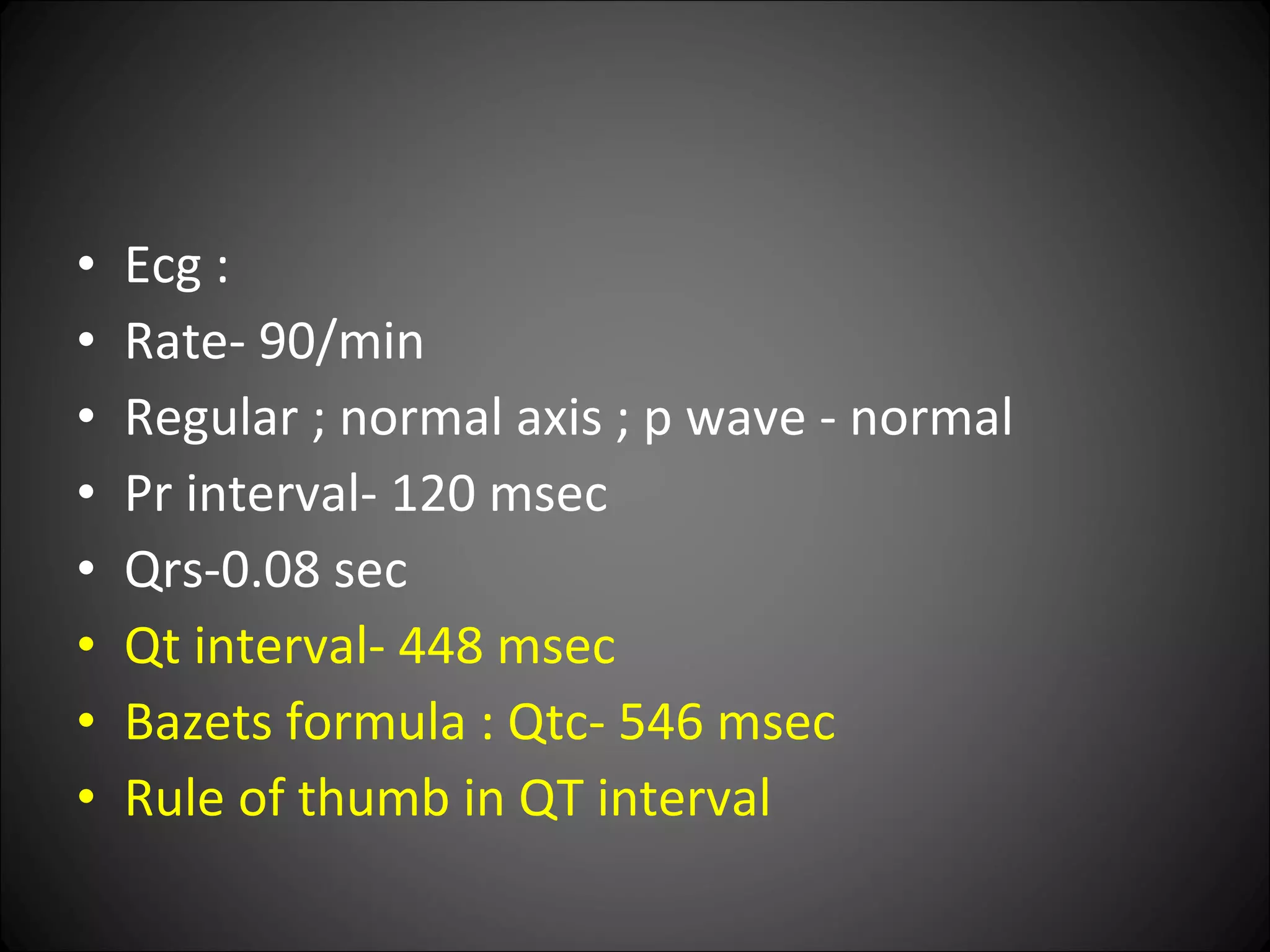
A CASE OF  SEIZURE  in a  Young Girl PROF.DR.MAGESHKUMAR’S UNIT Devendra Patil
HISTORY I7 year old female pt presented with seizures 3 episodes since last 6 months. GTCS; lasted for 5 mins, Associated with frothing from mouth. No h/o tongue bite, bladder and bowel disturbances. With post ictal LOC  No h/o fever, head injury, altered sensorium. No h/o vomiting, head ache, Blurring of vision. No h/o chest pain, palpitation, dyspnea, syncope
PAST HISTORY: Not a known case of DM/SHT/ CHD/BA/ TB No h/o seizure in child hood or perinatal insult. Pt studying in college. Performance is good. Pt not on any chronic medications. No h/o surgery in the past. Personal history: nil significant. Menstrual history: cycles regular, no menorroghoea, dysmenorrhoea,
ON EXAMINATION : Pt conscious, oriented, afebrile. Pulse : 78/ MIN, regular Bp :  110/70 mm hg . RR- 16/ min regular. Not in distress No pallor, icterus, cyanosis, pedal edema, cyanosis, clubbing, lymphadenopathy.
CVS  : S1, S2 heard. No murmurs. RS : NVBS HEARD. No added sounds. P/A : soft , non tender, no organomegaly, no free fluid.
CNS:  Higher functions: normal Cranial nerves: normal  Spinomotor system: Tone – normal No motor deficit. tendon reflexes – normal Plantar- b/l flexor. No cerbellar signs. Fundus- normal
Provisional diagnosis seizures for evaluation
Lab investigations CBC: Tc- 5100/cumm Dc – p -65. l-35 Hb – 11.8 gms Pcv- 35% Platelets- 2 lakhs. RFT: Urea- 34 mg, creat- 0.9 mg Na- 137 meq/l, k- 4, cl- 100, bicarb- 20meq/l Urine routine: Albumin, sugar- nil. Pus cells -1to 3 cells /hpf


Ecg : Rate- 90/min Regular ; normal axis ; p wave - normal Pr interval- 120 msec Qrs-0.08 sec Qt interval- 448 msec Bazets formula : Qtc- 546 msec  Rule of thumb in QT interval
CAUSES OF PROLONGED QTC Hypokalemia Hypocalcemia  CARDIAC -  Acute myocarditis, acute MI, HOCM, torsade de pointes. CONGENITAL-  Jervell-lange-nielsen syndrome Romano-ward synd Cerebral injury. Hypothermia
DRUGS: QUINIDINE, Procainamide,  Tricyclic antidepressants. Antimalarials- halofantrine
Chest radiograph : – NAD Ct brain:- Normal study EEG : Normal Study
Problems : seizures Prolonged QT in ECG CT – Brain normal
Serum calcium:  5.7 mg/dl[normal-8-10]
Investigations needed to work up a case of HYPOCALCEMIA  Sr. Calcium ( total / ionised / unionised ) Sr. Albumin levels Sr. Phosphate levels Sr. Parathormone levels ABG 24 Hrs urinary calcium Sr. Magnesium Sr. 25 (OH)vit D levels Sr. Alkaline Phosphatase levels
Serum calcium:  5.7 mg/dl[normal-8-10] Serum albumin : 3.8 gm /dl Ionised calcium- 0.452 mmol/l[ 1.16-1.32] Unionised calcium- 0.458 mmol/l Serum Phosphorus : 6.2 (2.5 – 4.5 mmol/dl ) Serum PTH :  5.2pg/ml ( 8-51 pg/ml ) 24 Hrs urine Calcium : 7mg/L  (< 300mg/day) Sr. Alkaline Phosphatase : 37 IU/l ( 33-100)
ABG ( room air , at rest ) Po2- 102.4mm hg Pco2 -39.4 mm hg Ph- 7.427 Na -138.6.  K- 3.17, Cl - 92.9 ( all in meq/l) Hco3 – 25.4 mmol/l Osmololity: 287.6 mosm/kg Impression : normal study.
Functional classification of hypocalcemia PTH low/ absent: PTH ineffective Pth overwhelmed: Hereditary  Acquired Hypomagnesemia CRF Active VIT-D lacking: Reduced intake  Low sunlight Anticonvulsant use Dependant rickets- type1 Active vitamin-d ineffective:  Dependant rickets- type 2 Pseudo hypo PTH severe acute  hyperphosphatemia Tumour lysis ARF Rhabdomyolysis Burns Pancreatitis Osteitis fibrosa after para thyroidectomy
Serum magnesium: 2.1 mg/dl[1.7 to 2.55] History didn’t suggest any cause that could lead to acquired hypoparathyroidism No significant history in family members No facial dysmorphism ; short stature. Skin –No hyperpigmentation / vitiligo / muco-cutaneus candidiasis / abscess n scars / eczematous changes. Thyroid profile :Normal
Further plan was: Anti parathyroid antibodies Double antigen PTH immune assay Screening for subclinical endocrine pathologies
Why this case was different Age of onset  Hypocalcemia without any evidence of signs of peripheral neuro-muscular excitibility. CT brain : not showing the evidence of basal ganglia calcifications Difference of treatment in a case of hypo PTH as compared to HypoCa+ without hypoPTH
Age of onset Idiopathic and hereditary hypoparathyroidism usually manifest in the first decade but can present later. Harrison 17/e
Clinical Manifestation of hypocalemia
Neuromuscular Numbness and tingling sensations in the perioral area or in the fingers and toes Muscle cramps, particularly in the back and lower extremities; may progress to carpopedal spasm (ie, tetany) Wheezing; may develop from bronchospasm Dysphagia  Voice changes (due to laryngospasm) Chovstek n Trosseau Sign ( Latent Tetany ) Neurologic Irritability, impaired intellectual capacity, depression, and personality changes Fatigue Seizures (eg, grand mal, petit mal, focal)  Other uncontrolled movements Cardiac Shortness of breath Symptoms of congestive heart failure (possible) Skin Coarse hair Brittle nails Psoriasis Dry skin Eyes -- subcapsular cataract
 
Few articles
Arq. Neuro-Psiquiatr. vol.62 no.1 São Paulo Mar. 2004 doi: 10.1590/S0004-282X2004000100028   Hypocalcemic myopathy without tetany due to idiopathic hypoparathyroidism: case report Myopathy due to idiopathic hypoparathyroidism is very unusual. We report on a  30 years-old man  referred with complaints of sporadic muscle pain and mild global weakness for 10 years. His physical examination showed normal strength in distal muscle and slightly weakness in the pelvic and scapular girdles with no atrophy.  Deep muscle reflexes were slightly hypoactive. Trousseau's and Chvostek's signs were absent .  He had bilateral cataract and complex partial seizures. His laboratory tests showed decreased ionised and total calcium and parathyroid hormone and increased muscle enzymes. EMG and muscle biopsy was compatible with metabolic myopathy. After treatment with calcium and vitamin D supplementation he showed clinical, neurophisiological and laboratorial improvement. In conclusion: patients with muscle symptoms, even when non-specific and with normal neurological examination, should have serum calcium checked, as myopathy due to idiopathic hypoparathyroidism, even being rare, is treatable and easy to diagnose.
J Med Case Reports. 2007 Dec 3;1:167. Seizures as the first manifestation of chromosome 22q11.2 deletion syndrome in a 40-year old man: a case report. Tonelli AR ,  Kosuri K ,  Wei S ,  Chick D . A 40-year-old man without significant past medical history presented with a new-onset generalized tonic-clonic seizure. He had no personal history of hypocalcemia or seizures. Physical examination was remarkable for short stature, hypertelorism, prominent forehead and nasal voice .   Trousseau's and Chvostek's signs were absent .   His initial laboratory examination showed hypocalcemia ( Calcium 5.2 mg/dl  and Calcium ionized 0.69 mmol/l) with hypoparathyroidism (Parathyroid hormone intact < 2.5 pg/ml. NV: 14-72 pg/ml). Urine Calcium was 3 mg/dl on a spot and 88 mg in a 24-hour urine collection (NV: 100-300 mg/24 hs). The electrocardiogram showed  a prolonged corrected QT  interval. Echocardiogram, abdominal ultrasound and electroencephalogram were normal. A computer tomography of the brain showed basal ganglia calcification. The subtle physical findings and the presence of idiopathic hypoparathyroidism motivated the performance of fluorescent in situ hybridization which demonstrated a microdeletion on one of the homologs 22q11.2. The patient was treated with calcium citrate and calcitriol with good response.
[Idiopathic hypoparathyroidism, a syndrome with various clinical expressions: analysis of 10 cases] Rev Med Chil. 1989 Jun;117(6):647-52. Abstract We report 10 patients with primary hypoparathyroidism. Age at onset varied from 7 months to 52 years (mean 28); 7 were female. Diagnosis was established at a mean of 4.1 years after the appearance of clinical manifestations. Unexplained hypocalcemia (mean 5.3 mg/dl) and hyperphosphatemia (mean 6.4 mg/dl) were present in all patients. Prevalent symptoms included tetany (9 patients), seizures (5) and hypocalcemic cataracts (4 ).   Clinical manifestations may be grouped into 5 types 1) tetany; 2) seizures; 3) other neurologic disorders   (basal ganglia calcification, pseudotumor of the brain, ataxia, nystagmus, hypertonus, paresis); 4) disorders of the lens including fully developed cataracts and 5) skin alterations like psoriasis and others. Some of these run on acute course (seizures, tetany), others a subacute one (skin alterations) while others are rather chronic (cataract and other neurologic disorders ).  Seizures and electroencephalographic disorders predominate in younger patients while tetany is more prevalent in older subjects.
The Journal of Clinical Endocrinology & Metabolism Vol. 88, No. 4 1476-1477 , 2003 IMAGES IN CLINICAL ENDOCRINOLOGY Calcification of the Basal Ganglia in Chronic Hypoparathyroidism  An 80-yr-old man with a more than 60-yr history of postsurgical   hypoparathyroidism was evaluated for hypocalcemia detected during   hospitalization after a fall. The patient had long-standing   intermittent paresthesias in his distal extremities, but denied   muscle cramps, tetany, or seizures. His past medical history   was remarkable for an episode of nephrolithiasis in his 20s.   He had been taking up to 4000 mg calcium carbonate (oral, daily)   before admission .  On examination, Chvostek and Trousseau signs,   tremor, and rigidity were absent .  Laboratory studies included:   5.9 mg/dl serum calcium (normal range, 8.4–10.5), 0.78   mmol/liter ionized calcium (normal range, 1.13–1.32),   6.7 mg/dl phosphate (normal range, 2.7–4.5), 3.2 g/dl   albumin (normal range, 3.5–5.3), and 1 pg/ml intact PTH   (normal range, 10–65). In Fig. 1, brain computed tomography   showed diffuse, symmetric parenchymal calcifications involving   the dentate nuclei (A), thalami (B,  curved arrow ), globus palladi   (B,  arrowhead ), caudate heads (B,  straight arrow ), and deep   cerebral white matter (C). The patient was treated with 0.5   µg calcitriol and 1000 mg calcium carbonate (oral, daily).   After 1 week, his chronic paresthesias resolved and serum calcium   rose to 9.2 mg/dl.
Absence of Basal Ganglia calcification Upto 30 % of cases of Hypocalcemia have basal ganglia calcification BASAL GANGLIA CALCIFICATION Birth anoxia  Idiopathic (most common) bilateral and symmetrical  Toxoplasmosis / CMV - usually not limited to basal ganglia  Hypoparathyroidism / pseudohypoparathyroidism  Fahr syndrome Cockayne syndrome
Garg R, Garg N, Tandon N, Khurana ML, Ammini AC. Idiopathic hypoparathyroidism presenting as epilepsy in a 40 years female. Neurol India 1999;47:244 The patient was  a 40 year old female   who complained of generalized tonic clonic seizures for the last 4 years. The seizures used to occur once in every 2-3 months.  positive Chvostek's and Trousseau's signs .  Nervous system examination was normal. Investigations revealed a serum calcium level of 6.4 mg/dl (normal - 8.5-10.5 mg/dl) and serum phosphate level of 6.4 mg/dl (normal - 2.5-4.5 mg/dl). Serum albumin was 4.0 G/dl (normal - 3.8-4.0 G/dl) and renal function tests were normal. Serum alkaline phosphatase level was 8.8 KA units (normal - 3-13 KA units). Serum parathyroid hormone levels were 28 pg/ml (normal - 11-55 pg/ml) which were inappropriately low in the presence of hypocalcaemia. Plasma cortisol, thyroxin and gonadotropins were within normal range. EEG showed generalized spike and wave pattern.  CT scan of the head was normal.
Idiopathic hypoparathyroidism presenting as dementia. D Eraut Br Med J. 1974 March 9; 1(5905): 429–430.  PMCID: PMC1633226 Summary: 80 yr old male ……
Difference of treatment  1. High dose of vitamin D ( 20000 IU /day ) ( 100-120 times the RDA of Vit D ) Why ? 2. High dose of oral Calcium  3. Monitoring of urinary Ca to avoid the risk of nephrolithiasis Prophylactic use of Thiazide
Final Diagnosis : Hypocalcemia induced seizures Hypoparathyroidism Mostly Idiopathic variety
 
THANK  -  YOU
Hereditary hypoparathyroidism May occur without any other endocrinological and dermatological manifestations. Manifest within first decade or later. Syndrome associated with hypopara thyroidism Di George syndrome[ DSG1-chromosome-22. DSG1-Chromosome10]. Kenney- Caffey syndrome: hypo parathyroidism, short stature, osteo sclerosis, thick cortical bones. Sanjad-Sakati synd: growth failure and dysmorphic features. Basal ganglia calcification and extra pyramidal symptoms are more common and earlier in onset.
Poly glandular auto immune type-1 deficiency: Mutation in APECED GENE. childhood onset.  Disease association: muco cuateneous candidiasis, hypo parathyroidism, adrenal insufficiency, hypogonadism, alopecia, hypothyroidism, dental enamel hypoplasia, mal absorption, vitiligo, chronic active hepatitis, pernicious anemia. Kearns - Sayre and MELAS  syndrome
AD- hypocalacemic hypercalciuria : Abnormality in calcium sensing receptor. Receptor senses normal calcium level as excessive and suppresses pth secretion. Increased renal excretion of calcium. External calcium and vit-D supplements exacerbate the symptoms leading to irreversible renal damage due to renal stones or ectopic calcifications.
Acquired hypoparathyroidism Surgery- para thyroidectomy for hyper para thyroidism. Irradiation . Hemochromatosis. hemosiderosis.
 
Calcium Regulation 99% of body calcium in skeleton Miscible Pool: 40% bound to protein, 13% complexed w/ anions, 47% free ionized PTH: Increased Ca, Decreased PO4, Increased Vitamin D Vitamin D: Increased Ca, Increased PO4, Decreased PTH (slow) Kidney, Bones, GI Tract
Acute Hypocalcemia: Parathyroidectomy PTH undetectable at 8 hours Normalizes by 30 hours Calcium nadir at 20 hours, normal Day 2-3 Hungry bone syndrome – Old, Labs Medications: I-131, Cimetidine, ETOH, Cisplatin, Digoxin, Ampho-B Hypomagnesemia Hypocalcemia/Hypoparathyroidism
Hypocalcemia/ Hypoparathyroidism Neuro: Paresthesias, fasciculations, muscle spasm, tetany, irritability, movement disorder, SEIZURE, laryngeal spasm. Visual: Cataracts, optic neuritis, papilledema- in association with increased ict Pulmonary: Bronchospasm CV: Prolonged QT, CHF, Hypotension, digitalis effect may be reduced. GI: Dysphagia, abdominal pain, biliary colic GU: Preterm labor Psychiatric- depression, psychosis. Chvostek’s sign and trousseau’s sgn can be used latent tetany.
Hypoparathyroidism: Treatment Acute severe hypocalcemia Check ionized Ca 100-300 mg (10-30 ml) 10% Ca-gluconate in 150 cc D5W over 10 minutes Continuous infusion at 0.5 mg/kg/hr EKG monitoring Check Magnesium Check Phosphorous
Hypoparathyroidism: Treatment
TREATMENT  Vitamin-D - 40000 to 1,20,000 iu/day[ 1-3 mg /day] combined with elemental calcium is satisfactory. Calcitriol- 0.5 to 1 micro gram/day. They do not reverse the lowered urinary calcium reabsorption. Thiazide diuretics lower urinary calcium in hypoparathyroid pts on vit-D.
HYPOMAGNESEMIA Levels less than 0.4 mmol/l[ 0.8 meq/l] Reduced PTH secretion or reduced response to PTH Severe hypomagnesemia is paradoxically associated with low PTH level. Cause unknown. Serum phosphate levels are usually not elevated. When magnesium is repleted restoration of PTH levels may preceed serum calcium by several days. Treatment- IV magnesium.
PTH overwhelmed Loss of ca from ECF is so severe that PTH cannot compensate. Pancreatitis, Acute hyper phosphatemia- due to cell destruction, tissue damage. Renal failure. PTH raises in response to hypocalcemia but serum calcium do not return to normal.
Differential diagnosis of hypoclcemia Low calcium, high phosphate-  hypoparathroidism, psudo hypoparathyroidism. Low calcium, low phosphate- absent   or ineffective vitamin D. Hypo magnesemia. Nutritional deficiency in alcohol.

An Interesting Case of Seizure

  • 1.
    A CASE OF SEIZURE in a Young Girl PROF.DR.MAGESHKUMAR’S UNIT Devendra Patil
  • 2.
    HISTORY I7 yearold female pt presented with seizures 3 episodes since last 6 months. GTCS; lasted for 5 mins, Associated with frothing from mouth. No h/o tongue bite, bladder and bowel disturbances. With post ictal LOC No h/o fever, head injury, altered sensorium. No h/o vomiting, head ache, Blurring of vision. No h/o chest pain, palpitation, dyspnea, syncope
  • 3.
    PAST HISTORY: Nota known case of DM/SHT/ CHD/BA/ TB No h/o seizure in child hood or perinatal insult. Pt studying in college. Performance is good. Pt not on any chronic medications. No h/o surgery in the past. Personal history: nil significant. Menstrual history: cycles regular, no menorroghoea, dysmenorrhoea,
  • 4.
    ON EXAMINATION :Pt conscious, oriented, afebrile. Pulse : 78/ MIN, regular Bp : 110/70 mm hg . RR- 16/ min regular. Not in distress No pallor, icterus, cyanosis, pedal edema, cyanosis, clubbing, lymphadenopathy.
  • 5.
    CVS :S1, S2 heard. No murmurs. RS : NVBS HEARD. No added sounds. P/A : soft , non tender, no organomegaly, no free fluid.
  • 6.
    CNS: Higherfunctions: normal Cranial nerves: normal Spinomotor system: Tone – normal No motor deficit. tendon reflexes – normal Plantar- b/l flexor. No cerbellar signs. Fundus- normal
  • 7.
  • 8.
    Lab investigations CBC:Tc- 5100/cumm Dc – p -65. l-35 Hb – 11.8 gms Pcv- 35% Platelets- 2 lakhs. RFT: Urea- 34 mg, creat- 0.9 mg Na- 137 meq/l, k- 4, cl- 100, bicarb- 20meq/l Urine routine: Albumin, sugar- nil. Pus cells -1to 3 cells /hpf
  • 9.
  • 10.
  • 11.
  • 12.
  • 13.
    Ecg : Rate-90/min Regular ; normal axis ; p wave - normal Pr interval- 120 msec Qrs-0.08 sec Qt interval- 448 msec Bazets formula : Qtc- 546 msec Rule of thumb in QT interval
  • 14.
    CAUSES OF PROLONGEDQTC Hypokalemia Hypocalcemia CARDIAC - Acute myocarditis, acute MI, HOCM, torsade de pointes. CONGENITAL- Jervell-lange-nielsen syndrome Romano-ward synd Cerebral injury. Hypothermia
  • 15.
    DRUGS: QUINIDINE, Procainamide, Tricyclic antidepressants. Antimalarials- halofantrine
  • 16.
    Chest radiograph :– NAD Ct brain:- Normal study EEG : Normal Study
  • 17.
    Problems : seizuresProlonged QT in ECG CT – Brain normal
  • 18.
    Serum calcium: 5.7 mg/dl[normal-8-10]
  • 19.
    Investigations needed towork up a case of HYPOCALCEMIA Sr. Calcium ( total / ionised / unionised ) Sr. Albumin levels Sr. Phosphate levels Sr. Parathormone levels ABG 24 Hrs urinary calcium Sr. Magnesium Sr. 25 (OH)vit D levels Sr. Alkaline Phosphatase levels
  • 20.
    Serum calcium: 5.7 mg/dl[normal-8-10] Serum albumin : 3.8 gm /dl Ionised calcium- 0.452 mmol/l[ 1.16-1.32] Unionised calcium- 0.458 mmol/l Serum Phosphorus : 6.2 (2.5 – 4.5 mmol/dl ) Serum PTH : 5.2pg/ml ( 8-51 pg/ml ) 24 Hrs urine Calcium : 7mg/L (< 300mg/day) Sr. Alkaline Phosphatase : 37 IU/l ( 33-100)
  • 21.
    ABG ( roomair , at rest ) Po2- 102.4mm hg Pco2 -39.4 mm hg Ph- 7.427 Na -138.6. K- 3.17, Cl - 92.9 ( all in meq/l) Hco3 – 25.4 mmol/l Osmololity: 287.6 mosm/kg Impression : normal study.
  • 22.
    Functional classification ofhypocalcemia PTH low/ absent: PTH ineffective Pth overwhelmed: Hereditary Acquired Hypomagnesemia CRF Active VIT-D lacking: Reduced intake Low sunlight Anticonvulsant use Dependant rickets- type1 Active vitamin-d ineffective: Dependant rickets- type 2 Pseudo hypo PTH severe acute hyperphosphatemia Tumour lysis ARF Rhabdomyolysis Burns Pancreatitis Osteitis fibrosa after para thyroidectomy
  • 23.
    Serum magnesium: 2.1mg/dl[1.7 to 2.55] History didn’t suggest any cause that could lead to acquired hypoparathyroidism No significant history in family members No facial dysmorphism ; short stature. Skin –No hyperpigmentation / vitiligo / muco-cutaneus candidiasis / abscess n scars / eczematous changes. Thyroid profile :Normal
  • 24.
    Further plan was:Anti parathyroid antibodies Double antigen PTH immune assay Screening for subclinical endocrine pathologies
  • 25.
    Why this casewas different Age of onset Hypocalcemia without any evidence of signs of peripheral neuro-muscular excitibility. CT brain : not showing the evidence of basal ganglia calcifications Difference of treatment in a case of hypo PTH as compared to HypoCa+ without hypoPTH
  • 26.
    Age of onsetIdiopathic and hereditary hypoparathyroidism usually manifest in the first decade but can present later. Harrison 17/e
  • 27.
  • 28.
    Neuromuscular Numbness andtingling sensations in the perioral area or in the fingers and toes Muscle cramps, particularly in the back and lower extremities; may progress to carpopedal spasm (ie, tetany) Wheezing; may develop from bronchospasm Dysphagia  Voice changes (due to laryngospasm) Chovstek n Trosseau Sign ( Latent Tetany ) Neurologic Irritability, impaired intellectual capacity, depression, and personality changes Fatigue Seizures (eg, grand mal, petit mal, focal)  Other uncontrolled movements Cardiac Shortness of breath Symptoms of congestive heart failure (possible) Skin Coarse hair Brittle nails Psoriasis Dry skin Eyes -- subcapsular cataract
  • 29.
  • 30.
  • 31.
    Arq. Neuro-Psiquiatr. vol.62 no.1 São Paulo Mar. 2004doi: 10.1590/S0004-282X2004000100028  Hypocalcemic myopathy without tetany due to idiopathic hypoparathyroidism: case report Myopathy due to idiopathic hypoparathyroidism is very unusual. We report on a 30 years-old man referred with complaints of sporadic muscle pain and mild global weakness for 10 years. His physical examination showed normal strength in distal muscle and slightly weakness in the pelvic and scapular girdles with no atrophy. Deep muscle reflexes were slightly hypoactive. Trousseau's and Chvostek's signs were absent . He had bilateral cataract and complex partial seizures. His laboratory tests showed decreased ionised and total calcium and parathyroid hormone and increased muscle enzymes. EMG and muscle biopsy was compatible with metabolic myopathy. After treatment with calcium and vitamin D supplementation he showed clinical, neurophisiological and laboratorial improvement. In conclusion: patients with muscle symptoms, even when non-specific and with normal neurological examination, should have serum calcium checked, as myopathy due to idiopathic hypoparathyroidism, even being rare, is treatable and easy to diagnose.
  • 32.
    J Med CaseReports. 2007 Dec 3;1:167. Seizures as the first manifestation of chromosome 22q11.2 deletion syndrome in a 40-year old man: a case report. Tonelli AR , Kosuri K , Wei S , Chick D . A 40-year-old man without significant past medical history presented with a new-onset generalized tonic-clonic seizure. He had no personal history of hypocalcemia or seizures. Physical examination was remarkable for short stature, hypertelorism, prominent forehead and nasal voice . Trousseau's and Chvostek's signs were absent . His initial laboratory examination showed hypocalcemia ( Calcium 5.2 mg/dl and Calcium ionized 0.69 mmol/l) with hypoparathyroidism (Parathyroid hormone intact < 2.5 pg/ml. NV: 14-72 pg/ml). Urine Calcium was 3 mg/dl on a spot and 88 mg in a 24-hour urine collection (NV: 100-300 mg/24 hs). The electrocardiogram showed a prolonged corrected QT interval. Echocardiogram, abdominal ultrasound and electroencephalogram were normal. A computer tomography of the brain showed basal ganglia calcification. The subtle physical findings and the presence of idiopathic hypoparathyroidism motivated the performance of fluorescent in situ hybridization which demonstrated a microdeletion on one of the homologs 22q11.2. The patient was treated with calcium citrate and calcitriol with good response.
  • 33.
    [Idiopathic hypoparathyroidism, asyndrome with various clinical expressions: analysis of 10 cases] Rev Med Chil. 1989 Jun;117(6):647-52. Abstract We report 10 patients with primary hypoparathyroidism. Age at onset varied from 7 months to 52 years (mean 28); 7 were female. Diagnosis was established at a mean of 4.1 years after the appearance of clinical manifestations. Unexplained hypocalcemia (mean 5.3 mg/dl) and hyperphosphatemia (mean 6.4 mg/dl) were present in all patients. Prevalent symptoms included tetany (9 patients), seizures (5) and hypocalcemic cataracts (4 ). Clinical manifestations may be grouped into 5 types 1) tetany; 2) seizures; 3) other neurologic disorders (basal ganglia calcification, pseudotumor of the brain, ataxia, nystagmus, hypertonus, paresis); 4) disorders of the lens including fully developed cataracts and 5) skin alterations like psoriasis and others. Some of these run on acute course (seizures, tetany), others a subacute one (skin alterations) while others are rather chronic (cataract and other neurologic disorders ). Seizures and electroencephalographic disorders predominate in younger patients while tetany is more prevalent in older subjects.
  • 34.
    The Journal ofClinical Endocrinology & Metabolism Vol. 88, No. 4 1476-1477 , 2003 IMAGES IN CLINICAL ENDOCRINOLOGY Calcification of the Basal Ganglia in Chronic Hypoparathyroidism An 80-yr-old man with a more than 60-yr history of postsurgical hypoparathyroidism was evaluated for hypocalcemia detected during hospitalization after a fall. The patient had long-standing intermittent paresthesias in his distal extremities, but denied muscle cramps, tetany, or seizures. His past medical history was remarkable for an episode of nephrolithiasis in his 20s. He had been taking up to 4000 mg calcium carbonate (oral, daily) before admission . On examination, Chvostek and Trousseau signs, tremor, and rigidity were absent . Laboratory studies included: 5.9 mg/dl serum calcium (normal range, 8.4–10.5), 0.78 mmol/liter ionized calcium (normal range, 1.13–1.32), 6.7 mg/dl phosphate (normal range, 2.7–4.5), 3.2 g/dl albumin (normal range, 3.5–5.3), and 1 pg/ml intact PTH (normal range, 10–65). In Fig. 1, brain computed tomography showed diffuse, symmetric parenchymal calcifications involving the dentate nuclei (A), thalami (B, curved arrow ), globus palladi (B, arrowhead ), caudate heads (B, straight arrow ), and deep cerebral white matter (C). The patient was treated with 0.5 µg calcitriol and 1000 mg calcium carbonate (oral, daily). After 1 week, his chronic paresthesias resolved and serum calcium rose to 9.2 mg/dl.
  • 35.
    Absence of BasalGanglia calcification Upto 30 % of cases of Hypocalcemia have basal ganglia calcification BASAL GANGLIA CALCIFICATION Birth anoxia Idiopathic (most common) bilateral and symmetrical Toxoplasmosis / CMV - usually not limited to basal ganglia Hypoparathyroidism / pseudohypoparathyroidism Fahr syndrome Cockayne syndrome
  • 36.
    Garg R, GargN, Tandon N, Khurana ML, Ammini AC. Idiopathic hypoparathyroidism presenting as epilepsy in a 40 years female. Neurol India 1999;47:244 The patient was a 40 year old female who complained of generalized tonic clonic seizures for the last 4 years. The seizures used to occur once in every 2-3 months. positive Chvostek's and Trousseau's signs . Nervous system examination was normal. Investigations revealed a serum calcium level of 6.4 mg/dl (normal - 8.5-10.5 mg/dl) and serum phosphate level of 6.4 mg/dl (normal - 2.5-4.5 mg/dl). Serum albumin was 4.0 G/dl (normal - 3.8-4.0 G/dl) and renal function tests were normal. Serum alkaline phosphatase level was 8.8 KA units (normal - 3-13 KA units). Serum parathyroid hormone levels were 28 pg/ml (normal - 11-55 pg/ml) which were inappropriately low in the presence of hypocalcaemia. Plasma cortisol, thyroxin and gonadotropins were within normal range. EEG showed generalized spike and wave pattern. CT scan of the head was normal.
  • 37.
    Idiopathic hypoparathyroidism presentingas dementia. D Eraut Br Med J. 1974 March 9; 1(5905): 429–430. PMCID: PMC1633226 Summary: 80 yr old male ……
  • 38.
    Difference of treatment 1. High dose of vitamin D ( 20000 IU /day ) ( 100-120 times the RDA of Vit D ) Why ? 2. High dose of oral Calcium 3. Monitoring of urinary Ca to avoid the risk of nephrolithiasis Prophylactic use of Thiazide
  • 39.
    Final Diagnosis :Hypocalcemia induced seizures Hypoparathyroidism Mostly Idiopathic variety
  • 40.
  • 41.
    THANK - YOU
  • 42.
    Hereditary hypoparathyroidism Mayoccur without any other endocrinological and dermatological manifestations. Manifest within first decade or later. Syndrome associated with hypopara thyroidism Di George syndrome[ DSG1-chromosome-22. DSG1-Chromosome10]. Kenney- Caffey syndrome: hypo parathyroidism, short stature, osteo sclerosis, thick cortical bones. Sanjad-Sakati synd: growth failure and dysmorphic features. Basal ganglia calcification and extra pyramidal symptoms are more common and earlier in onset.
  • 43.
    Poly glandular autoimmune type-1 deficiency: Mutation in APECED GENE. childhood onset. Disease association: muco cuateneous candidiasis, hypo parathyroidism, adrenal insufficiency, hypogonadism, alopecia, hypothyroidism, dental enamel hypoplasia, mal absorption, vitiligo, chronic active hepatitis, pernicious anemia. Kearns - Sayre and MELAS syndrome
  • 44.
    AD- hypocalacemic hypercalciuria: Abnormality in calcium sensing receptor. Receptor senses normal calcium level as excessive and suppresses pth secretion. Increased renal excretion of calcium. External calcium and vit-D supplements exacerbate the symptoms leading to irreversible renal damage due to renal stones or ectopic calcifications.
  • 45.
    Acquired hypoparathyroidism Surgery-para thyroidectomy for hyper para thyroidism. Irradiation . Hemochromatosis. hemosiderosis.
  • 46.
  • 47.
    Calcium Regulation 99%of body calcium in skeleton Miscible Pool: 40% bound to protein, 13% complexed w/ anions, 47% free ionized PTH: Increased Ca, Decreased PO4, Increased Vitamin D Vitamin D: Increased Ca, Increased PO4, Decreased PTH (slow) Kidney, Bones, GI Tract
  • 48.
    Acute Hypocalcemia: ParathyroidectomyPTH undetectable at 8 hours Normalizes by 30 hours Calcium nadir at 20 hours, normal Day 2-3 Hungry bone syndrome – Old, Labs Medications: I-131, Cimetidine, ETOH, Cisplatin, Digoxin, Ampho-B Hypomagnesemia Hypocalcemia/Hypoparathyroidism
  • 49.
    Hypocalcemia/ Hypoparathyroidism Neuro:Paresthesias, fasciculations, muscle spasm, tetany, irritability, movement disorder, SEIZURE, laryngeal spasm. Visual: Cataracts, optic neuritis, papilledema- in association with increased ict Pulmonary: Bronchospasm CV: Prolonged QT, CHF, Hypotension, digitalis effect may be reduced. GI: Dysphagia, abdominal pain, biliary colic GU: Preterm labor Psychiatric- depression, psychosis. Chvostek’s sign and trousseau’s sgn can be used latent tetany.
  • 50.
    Hypoparathyroidism: Treatment Acutesevere hypocalcemia Check ionized Ca 100-300 mg (10-30 ml) 10% Ca-gluconate in 150 cc D5W over 10 minutes Continuous infusion at 0.5 mg/kg/hr EKG monitoring Check Magnesium Check Phosphorous
  • 51.
  • 52.
    TREATMENT Vitamin-D- 40000 to 1,20,000 iu/day[ 1-3 mg /day] combined with elemental calcium is satisfactory. Calcitriol- 0.5 to 1 micro gram/day. They do not reverse the lowered urinary calcium reabsorption. Thiazide diuretics lower urinary calcium in hypoparathyroid pts on vit-D.
  • 53.
    HYPOMAGNESEMIA Levels lessthan 0.4 mmol/l[ 0.8 meq/l] Reduced PTH secretion or reduced response to PTH Severe hypomagnesemia is paradoxically associated with low PTH level. Cause unknown. Serum phosphate levels are usually not elevated. When magnesium is repleted restoration of PTH levels may preceed serum calcium by several days. Treatment- IV magnesium.
  • 54.
    PTH overwhelmed Lossof ca from ECF is so severe that PTH cannot compensate. Pancreatitis, Acute hyper phosphatemia- due to cell destruction, tissue damage. Renal failure. PTH raises in response to hypocalcemia but serum calcium do not return to normal.
  • 55.
    Differential diagnosis ofhypoclcemia Low calcium, high phosphate- hypoparathroidism, psudo hypoparathyroidism. Low calcium, low phosphate- absent or ineffective vitamin D. Hypo magnesemia. Nutritional deficiency in alcohol.