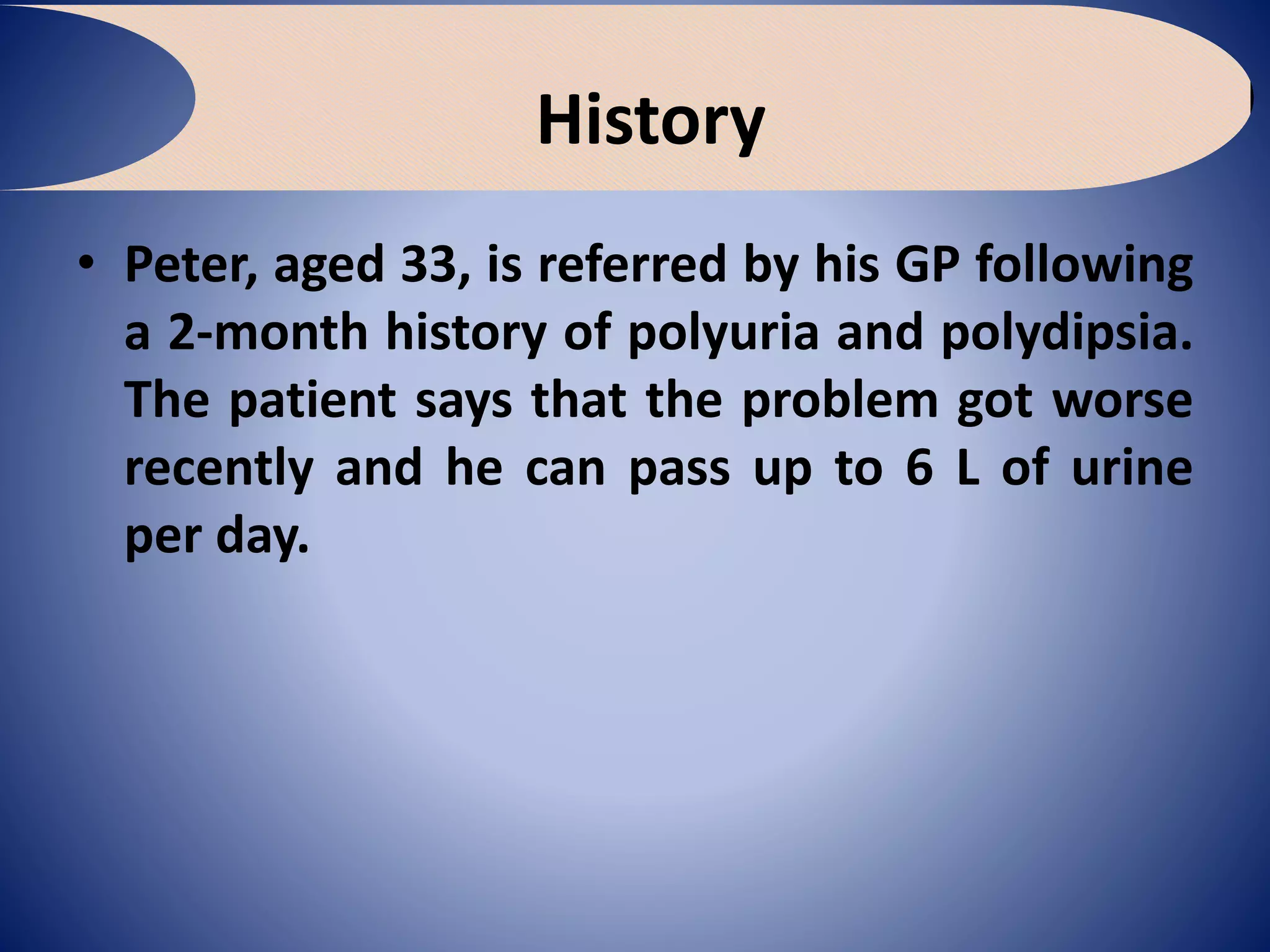
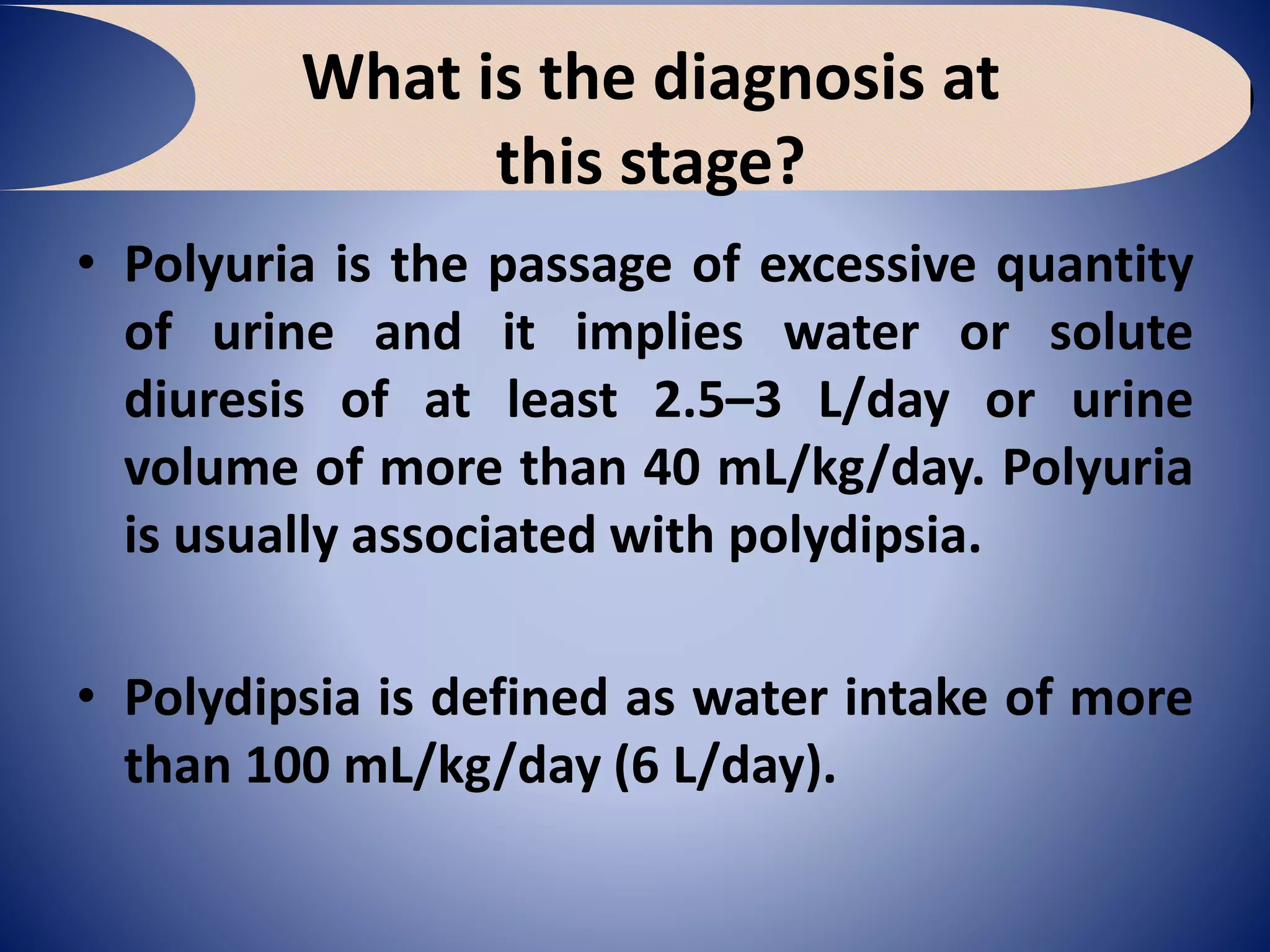
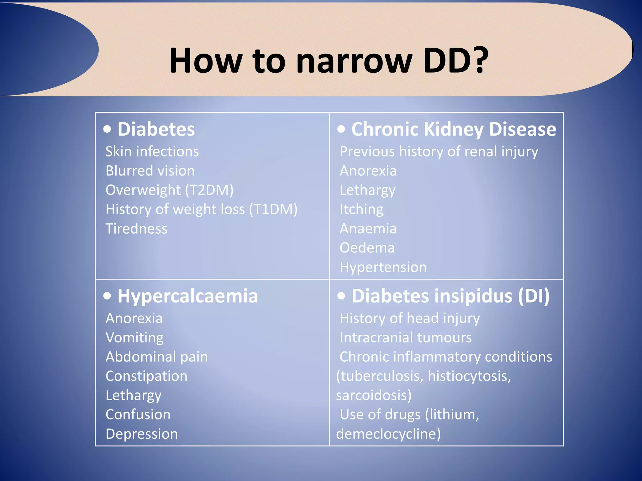
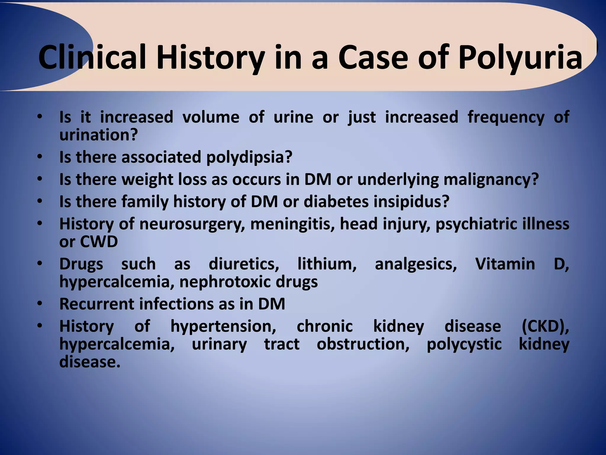
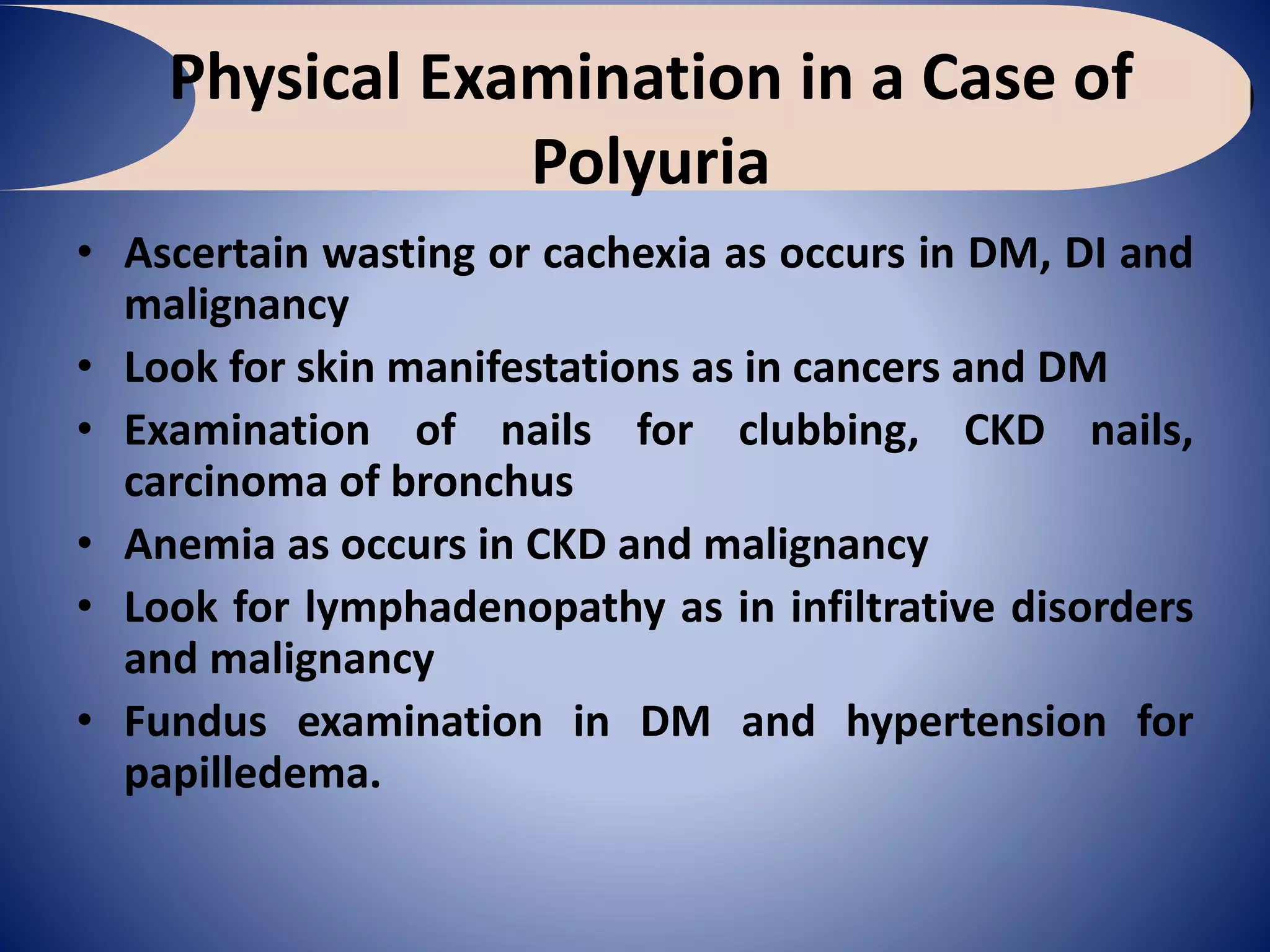
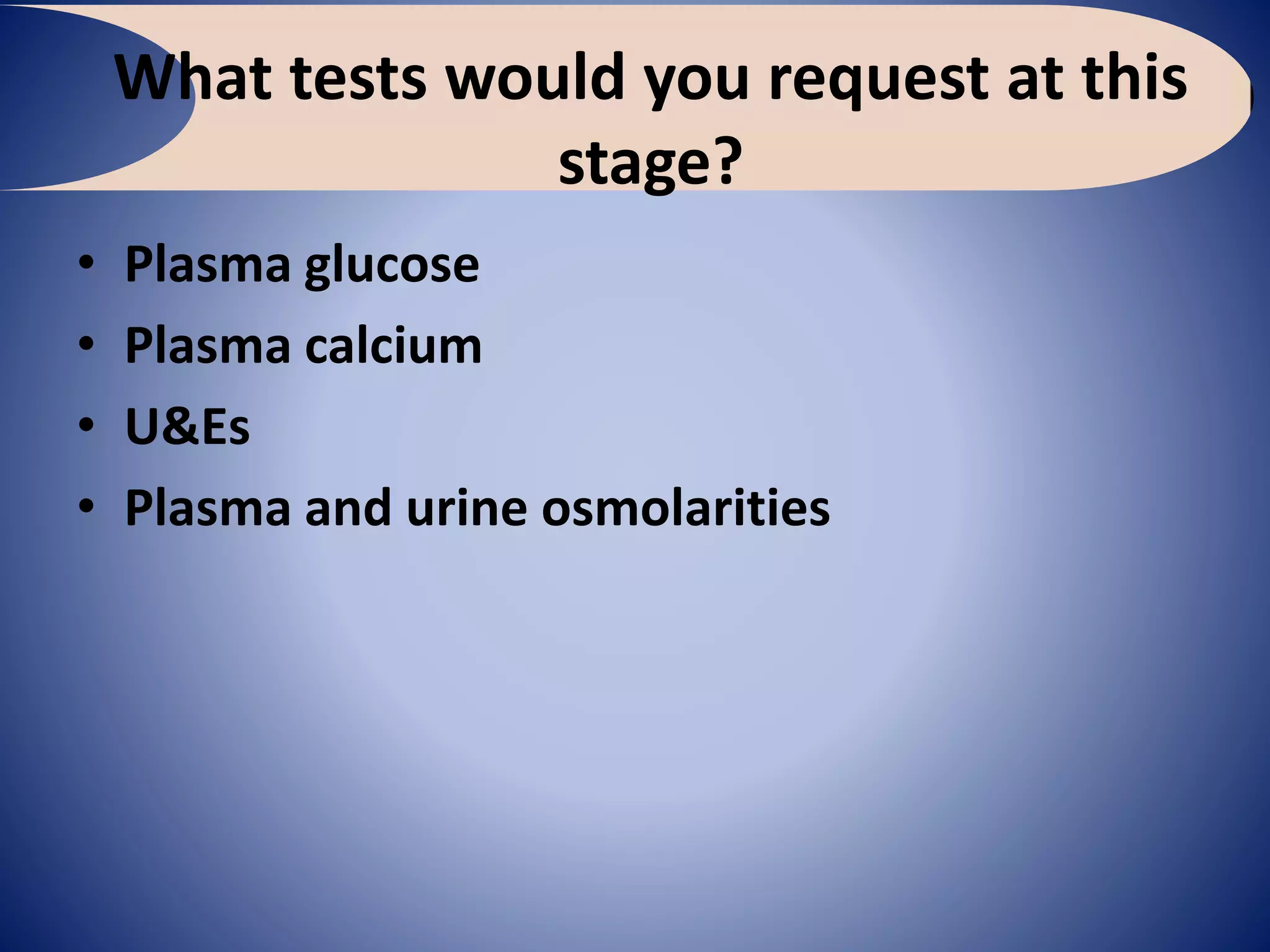
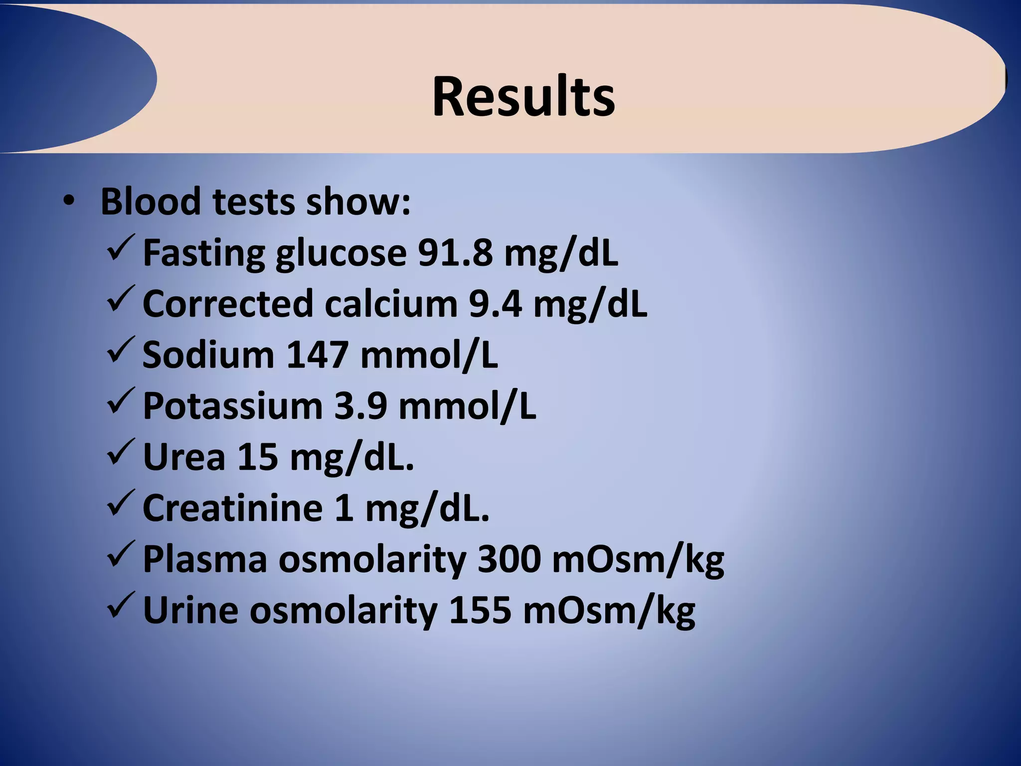
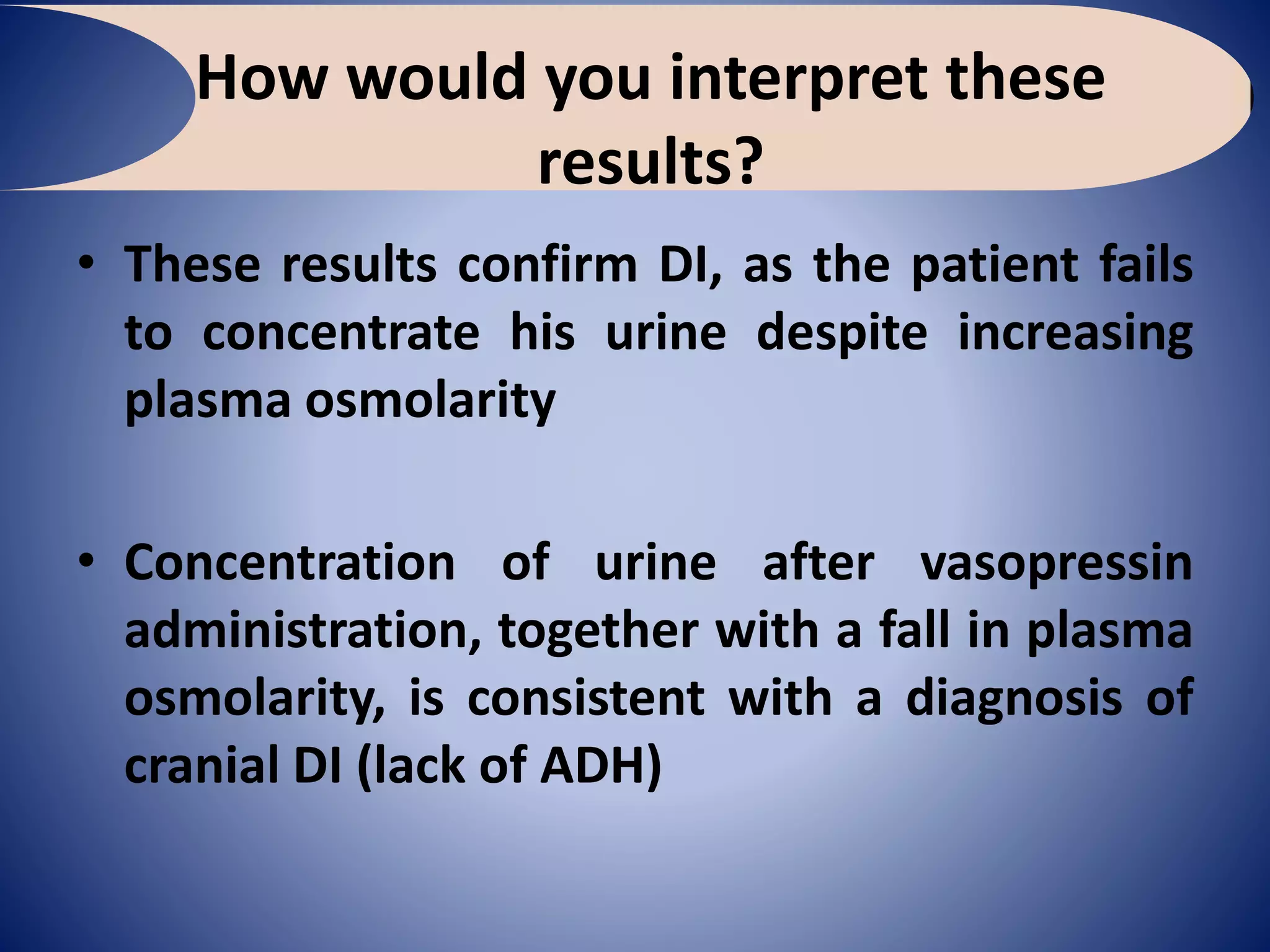
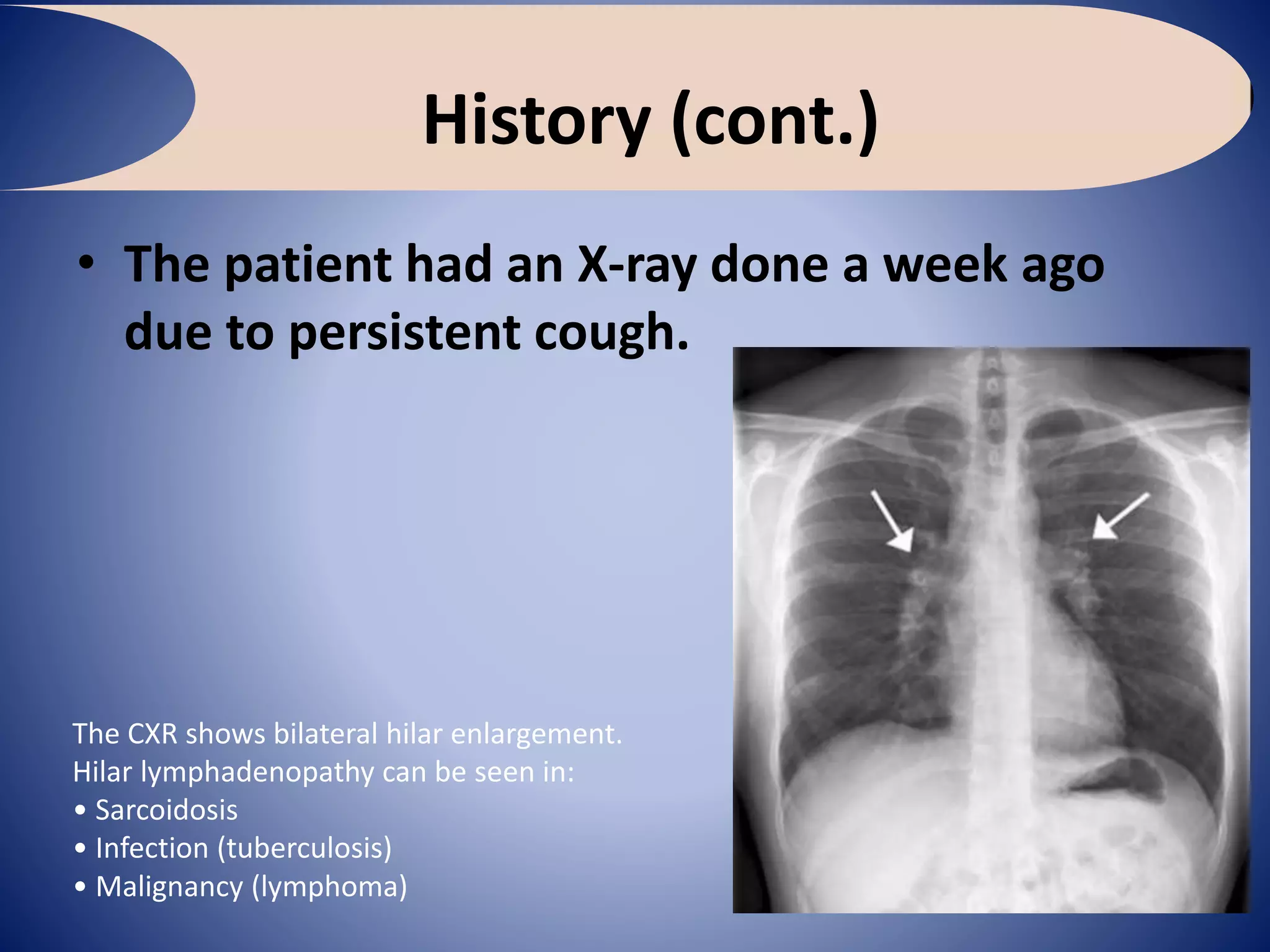
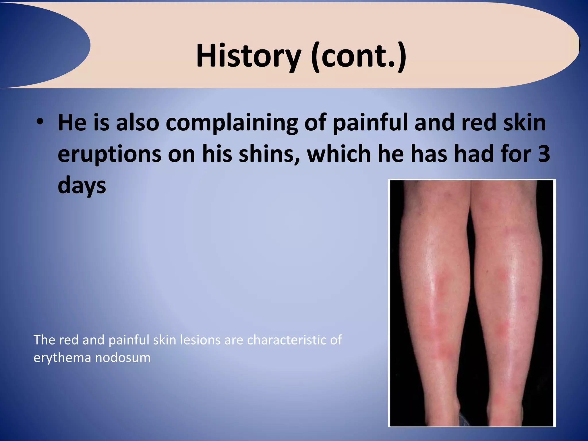
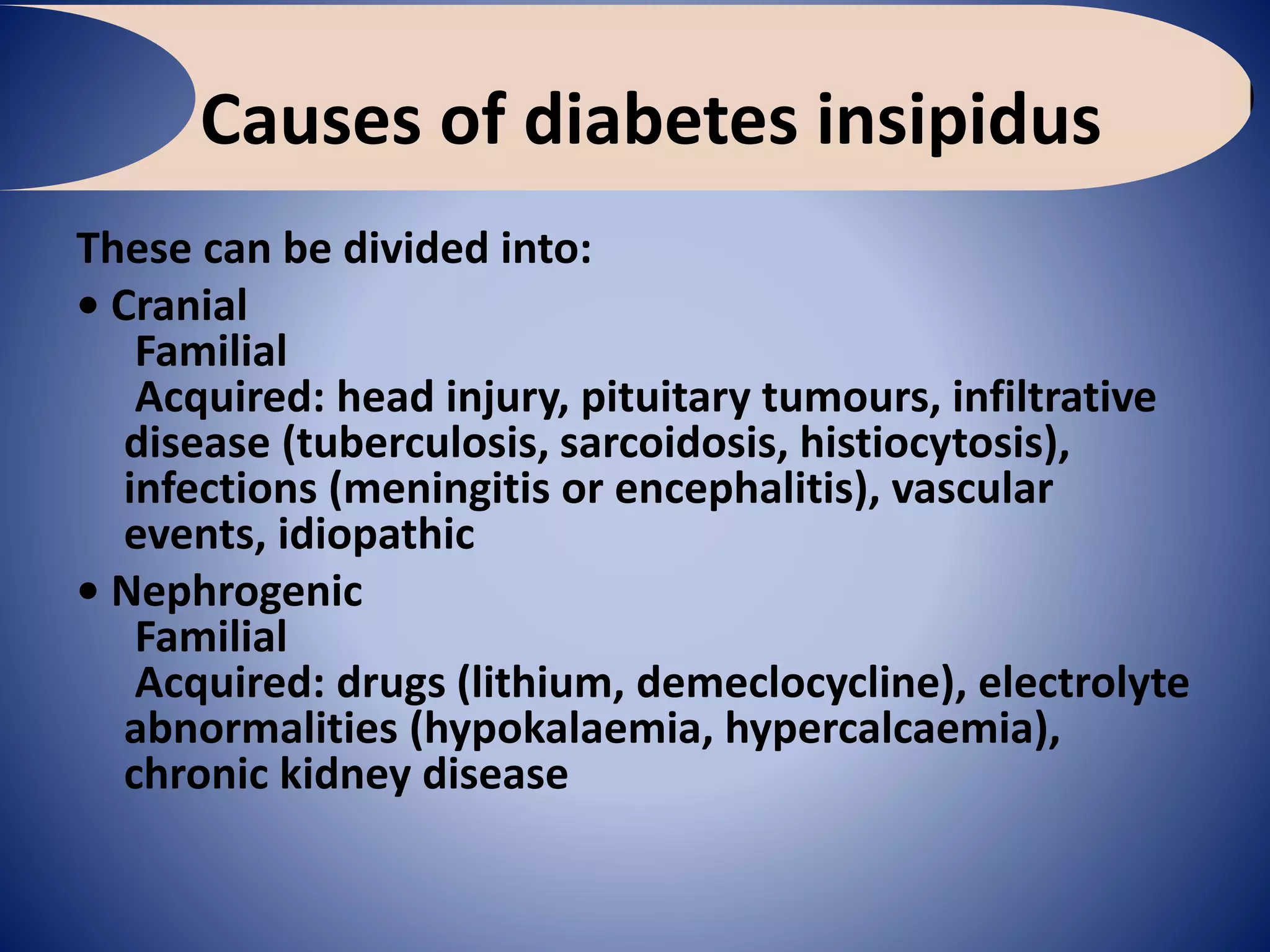
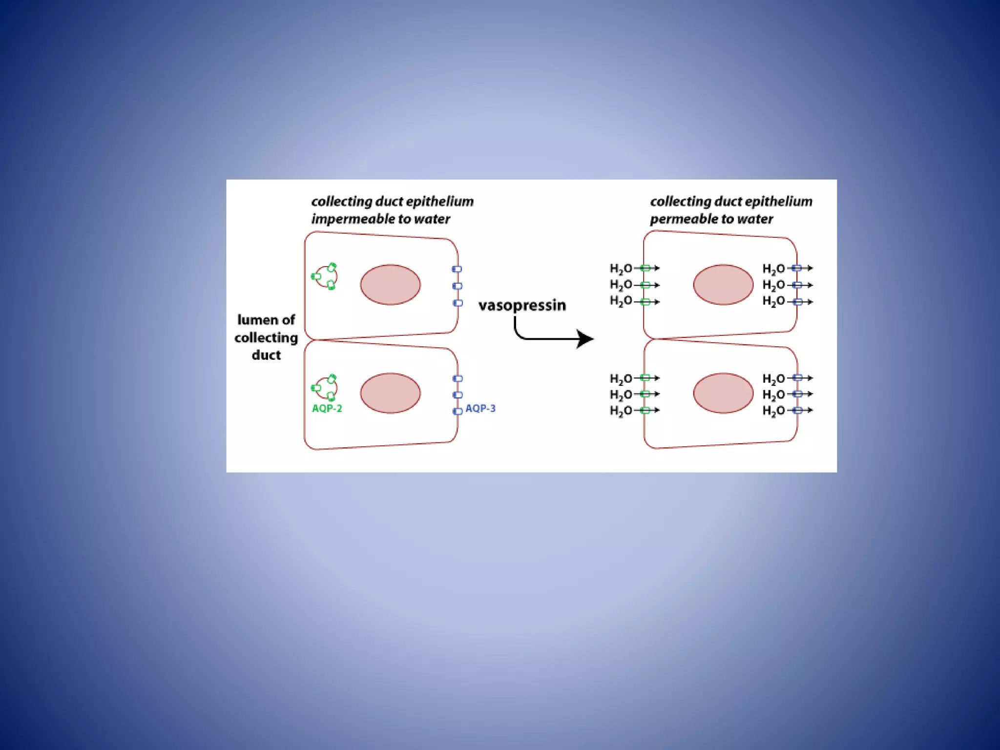
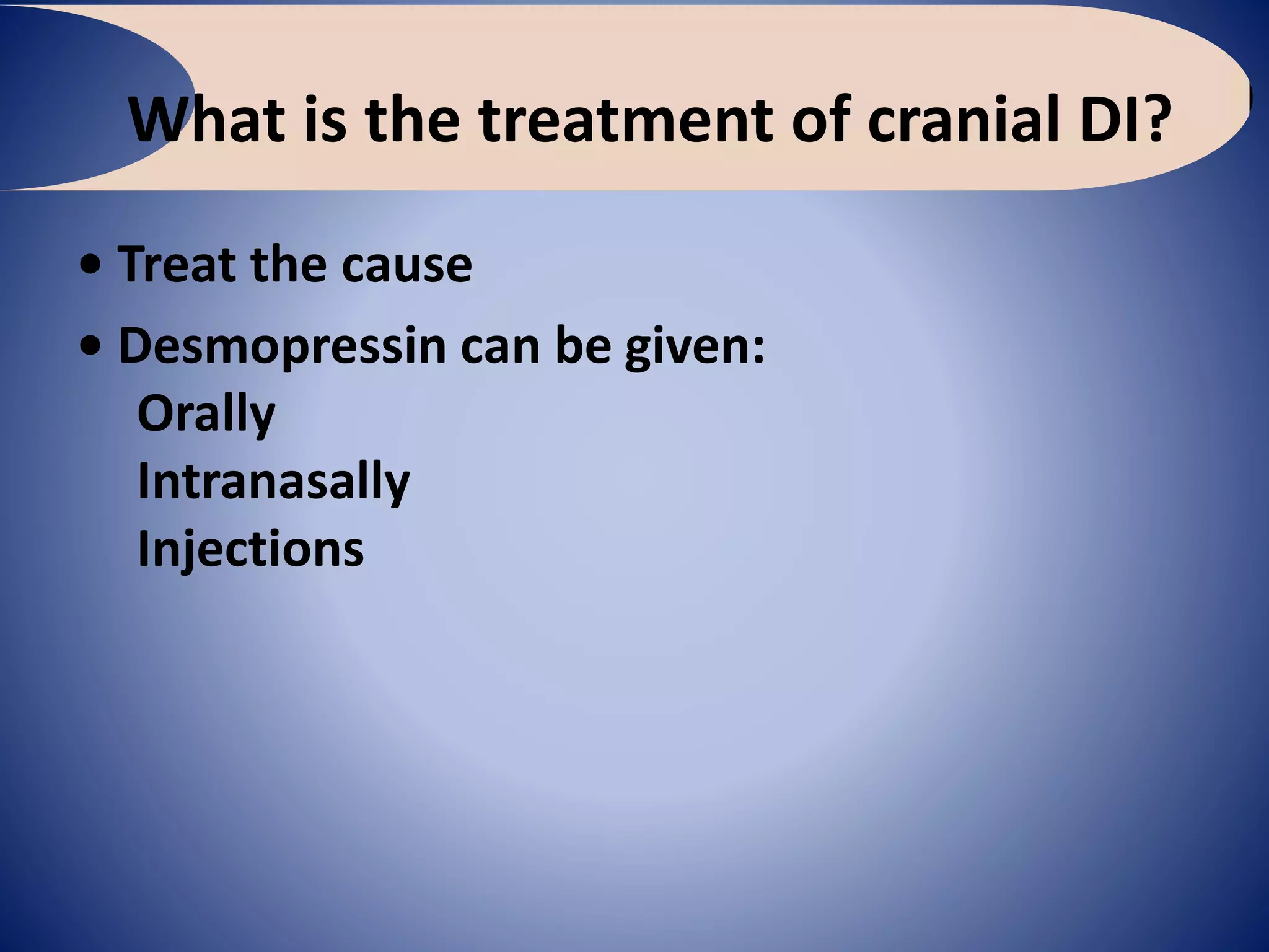
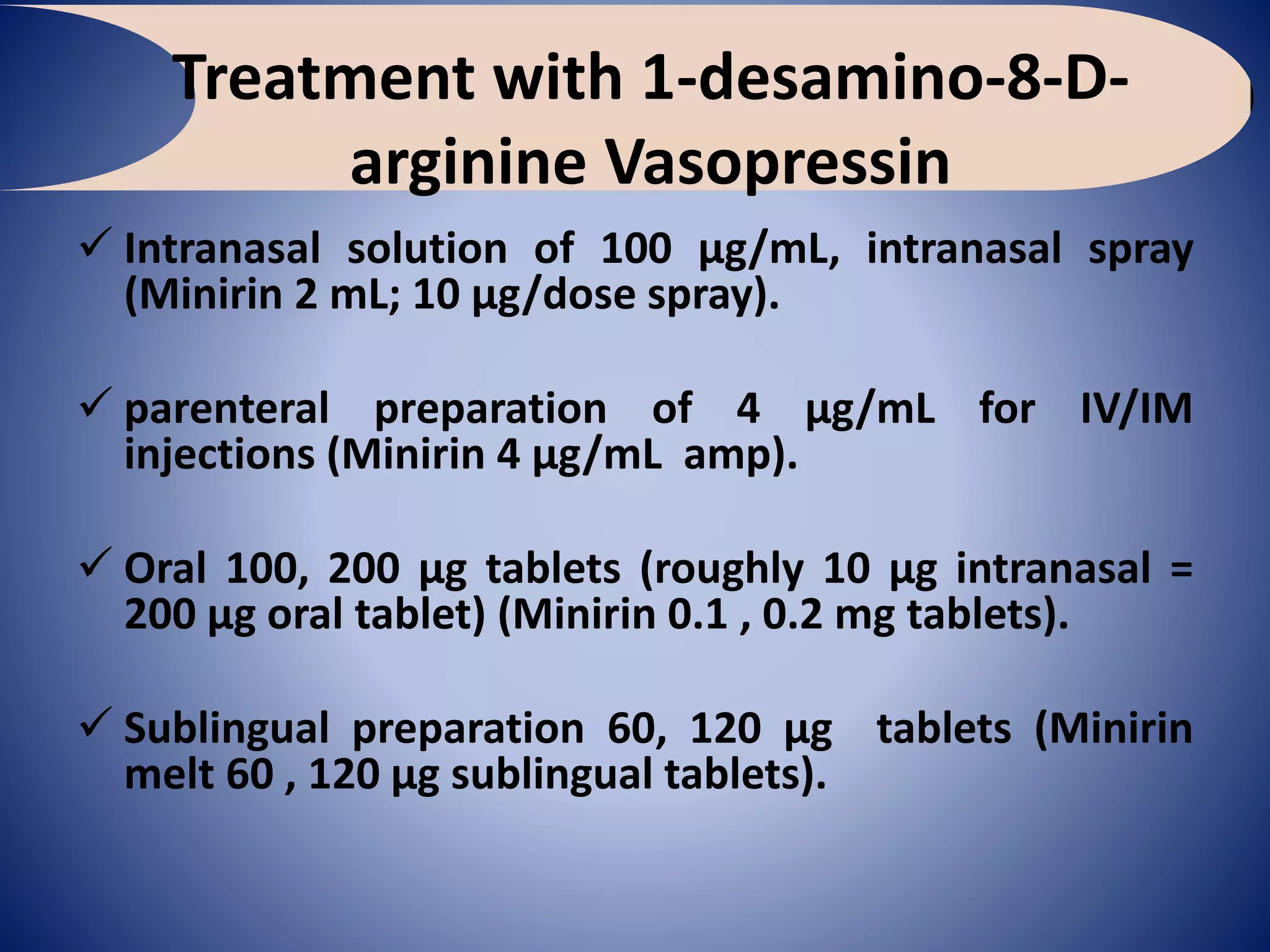
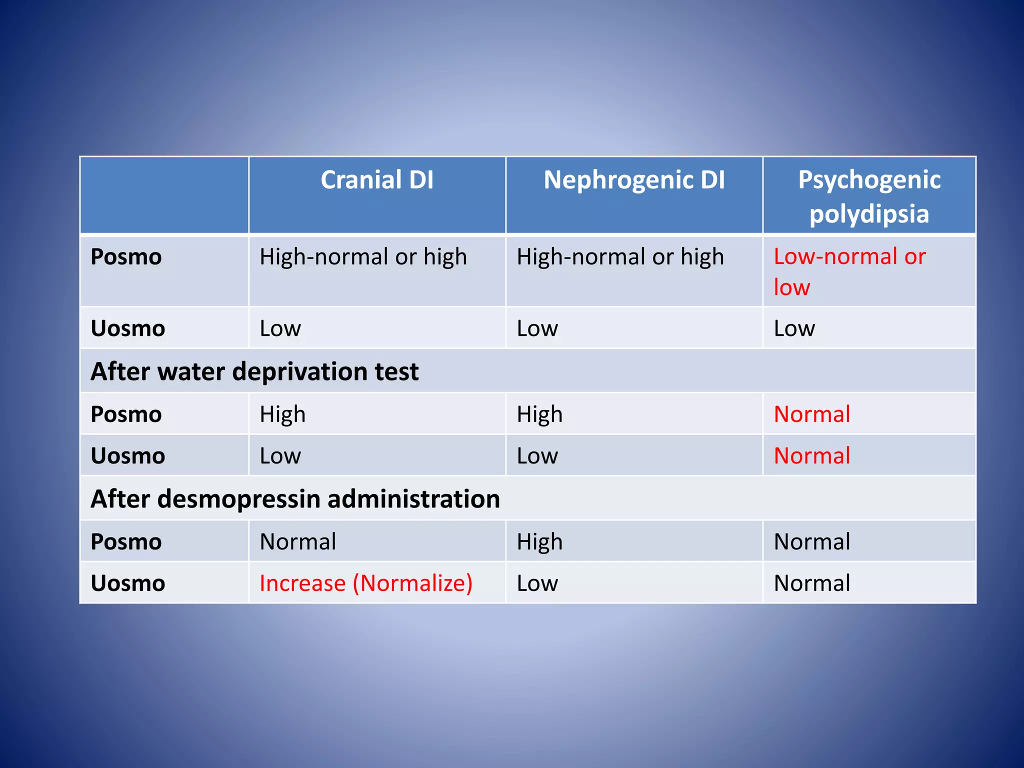
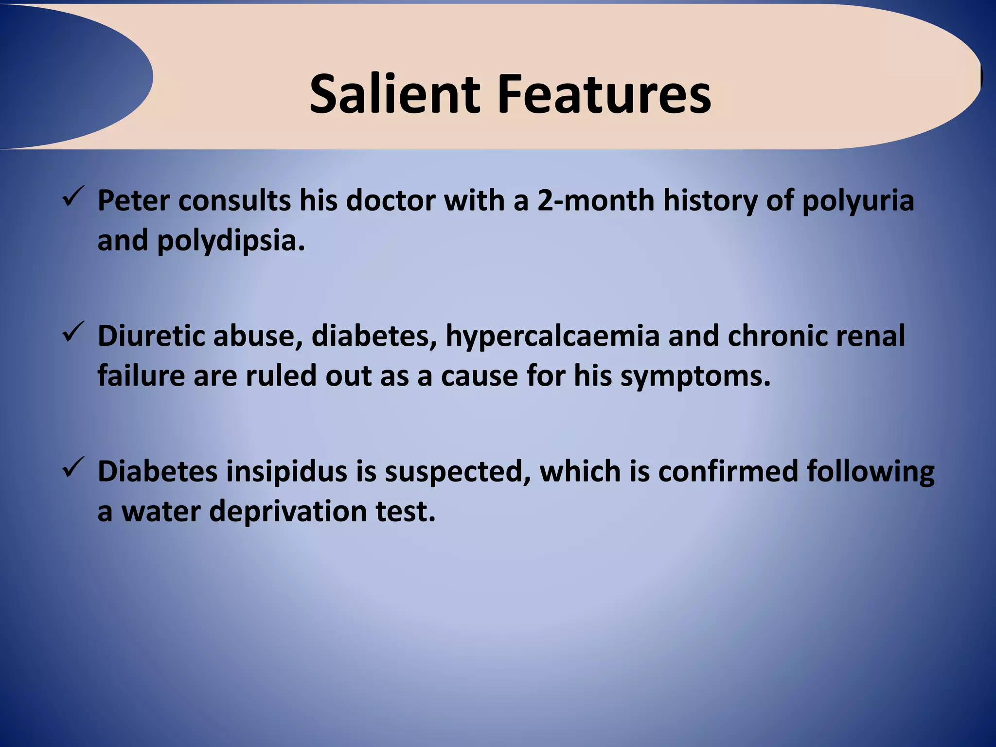
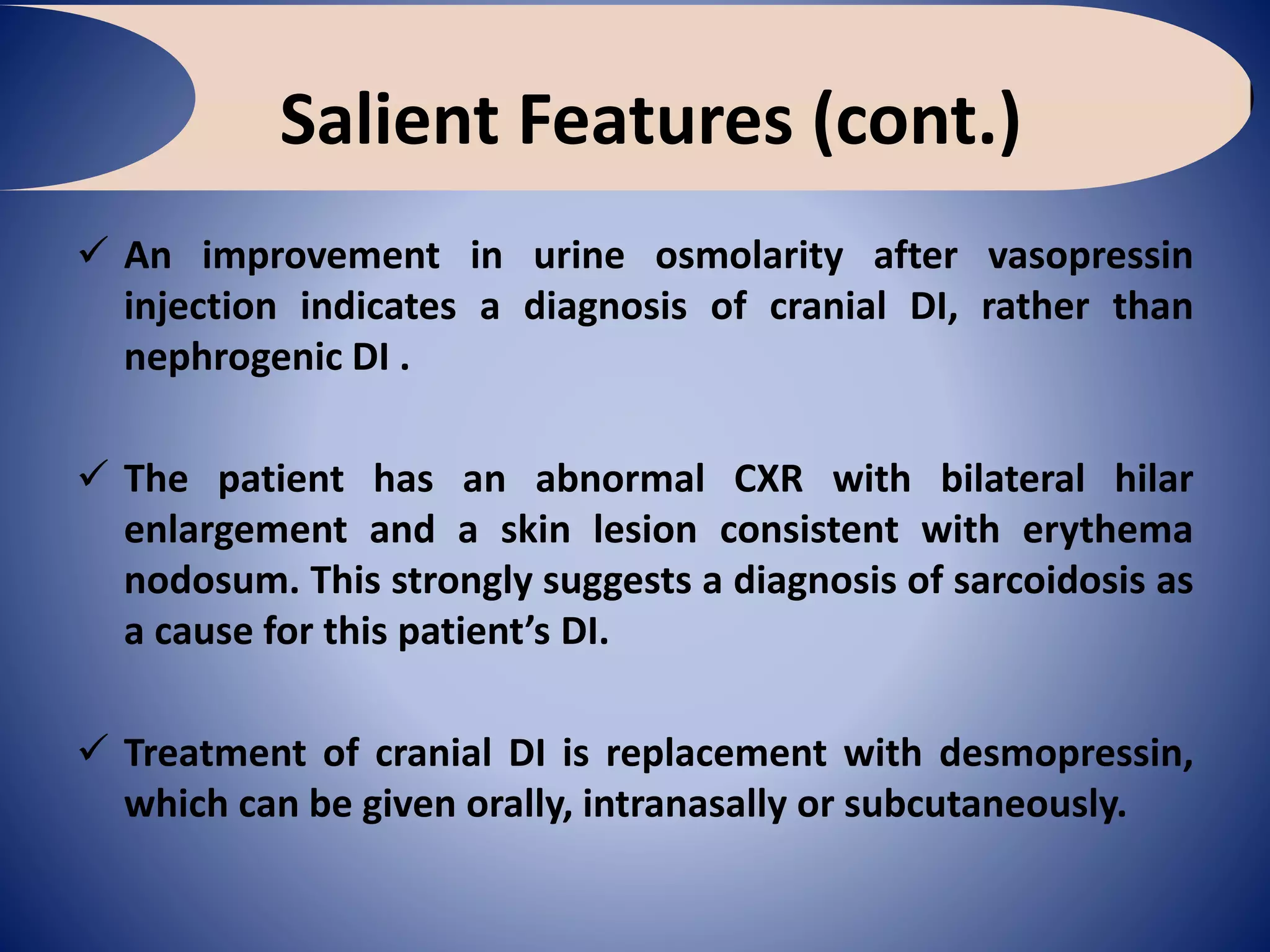
A 33-year-old man with polyuria
and polydipsia
Clinical Case uncovered (CCU) series
Endocrinology & Diabetes
Case 11
Usama Ragab Youssif (Msc.)
Assistant Lecturer of Medicine
(27, March, 2017)
History
• Peter, aged 33, is referred by his GP following
a 2-month history of polyuria and polydipsia.
The patient says that the problem got worse
recently and he can pass up to 6 L of urine
per day.
What is the diagnosis at
this stage?
• Polyuria is the passage of excessive quantity
of urine and it implies water or solute
diuresis of at least 2.5–3 L/day or urine
volume of more than 40 mL/kg/day. Polyuria
is usually associated with polydipsia.
• Polydipsia is defined as water intake of more
than 100 mL/kg/day (6 L/day).
What is the differential diagnosis at
this stage?
• Diabetes (type 1 or type 2)
• Hypercalcaemia
• Chronic kidney disease
• Diabetes insipidus
• Diuretic abuse
• Psychogenic; Compulsive water drinking.
It is important to note that some patients complain of
polyuria without an actual increase in their urine output
and urine volumes should be measured over 24 h to
confirm the diagnosis of polyuria in uncertain cases.
Mechanisms explain polyuria
1. Increased intake of fluids as in psychogenic causes,
stress and anxiety
2. Increased GFR as in hyperthyroidism, fever,
hypermetabolic states
3. Increased output of solutes as occurs in DM,
hyperthyroidism, hyperparathyroidism, use of
diuretics (which present more solute at the DCT)
4. Inability of the kidney to reabsorb water in DCT as in
CDI, NDI, drugs and chronic renal failure (CRF).
5. Other causes; SVT*
How to narrow DD?
• Chronic Kidney Disease
Previous history of renal injury
Anorexia
Lethargy
Itching
Anaemia
Oedema
Hypertension
• Diabetes
Skin infections
Blurred vision
Overweight (T2DM)
History of weight loss (T1DM)
Tiredness
• Diabetes insipidus (DI)
History of head injury
Intracranial tumours
Chronic inflammatory conditions
(tuberculosis, histiocytosis,
sarcoidosis)
Use of drugs (lithium,
demeclocycline)
• Hypercalcaemia
Anorexia
Vomiting
Abdominal pain
Constipation
Lethargy
Confusion
Depression
Clinical History in a Case of Polyuria
• Is it increased volume of urine or just increased frequency of
urination?
• Is there associated polydipsia?
• Is there weight loss as occurs in DM or underlying malignancy?
• Is there family history of DM or diabetes insipidus?
• History of neurosurgery, meningitis, head injury, psychiatric illness
or CWD
• Drugs such as diuretics, lithium, analgesics, Vitamin D,
hypercalcemia, nephrotoxic drugs
• Recurrent infections as in DM
• History of hypertension, chronic kidney disease (CKD),
hypercalcemia, urinary tract obstruction, polycystic kidney
disease.
Physical Examination in a Case of
Polyuria
• Ascertain wasting or cachexia as occurs in DM, DI and
malignancy
• Look for skin manifestations as in cancers and DM
• Examination of nails for clubbing, CKD nails,
carcinoma of bronchus
• Anemia as occurs in CKD and malignancy
• Look for lymphadenopathy as in infiltrative disorders
and malignancy
• Fundus examination in DM and hypertension for
papilledema.
What tests would you request at this
stage?
• Plasma glucose
• Plasma calcium
• U&Es
• Plasma and urine osmolarities
Results
• Blood tests show:
Fasting glucose 91.8 mg/dL
Corrected calcium 9.4 mg/dL
Sodium 147 mmol/L
Potassium 3.9 mmol/L
Urea 15 mg/dL.
Creatinine 1 mg/dL.
Plasma osmolarity 300 mOsm/kg
Urine osmolarity 155 mOsm/kg
What do these results indicate?
• The above results rule out the following as the
cause of this patient’s symptoms:
Diabetes
Hypercalcaemia
Chronic kidney disease
The high plasma sodium and increased plasma
osmolarity with inappropriately low urine
osmolarity suggest a diagnosis of diabetes
insipidus.
What test would you request to
confirm the diagnosis?
• A water deprivation test (Hare-Hickey Test
) with desmopressin administration.
Urine OsmolalityPlasma OsmolalityTime
1583000 h
1543064 h
1553085 h (i.m.
Vasopressin given)
8352958 h
Water Deprivation Test
How would you interpret these
results?
• These results confirm DI, as the patient fails
to concentrate his urine despite increasing
plasma osmolarity
• Concentration of urine after vasopressin
administration, together with a fall in plasma
osmolarity, is consistent with a diagnosis of
cranial DI (lack of ADH)
History (cont.)
• The patient had an X-ray done a week ago
due to persistent cough.
The CXR shows bilateral hilar enlargement.
Hilar lymphadenopathy can be seen in:
• Sarcoidosis
• Infection (tuberculosis)
• Malignancy (lymphoma)
History (cont.)
• He is also complaining of painful and red skin
eruptions on his shins, which he has had for 3
days
The red and painful skin lesions are characteristic of
erythema nodosum
Erythema nodosum
• Infections
Bacterial (Streptococcus, tuberculosis)
Viral
Fungal
• Inflammatory bowel disease
Malignancy
Sarcoidosis
Drugs (oral contraceptives, sulphonamides, penicillin)
Taken together, the most likely unifying diagnosis is sarcoidosis causing
DI, secondary to the inflammatory infiltrate in the posterior
pituitary; also termed neurosarcoidosis.
Another remote possibility is tuberculosis.
What imaging would you request in
this patient?
MRI of the pituitary.
Causes of diabetes insipidus
These can be divided into:
• Cranial
Familial
Acquired: head injury, pituitary tumours, infiltrative
disease (tuberculosis, sarcoidosis, histiocytosis),
infections (meningitis or encephalitis), vascular
events, idiopathic
• Nephrogenic
Familial
Acquired: drugs (lithium, demeclocycline), electrolyte
abnormalities (hypokalaemia, hypercalcaemia),
chronic kidney disease
What is the treatment of cranial DI?
• Treat the cause
• Desmopressin can be given:
Orally
Intranasally
Injections
Treatment with 1-desamino-8-D-
arginine Vasopressin
 Intranasal solution of 100 µg/mL, intranasal spray
(Minirin 2 mL; 10 µg/dose spray).
 parenteral preparation of 4 µg/mL for IV/IM
injections (Minirin 4 µg/mL amp).
 Oral 100, 200 µg tablets (roughly 10 µg intranasal =
200 µg oral tablet) (Minirin 0.1 , 0.2 mg tablets).
 Sublingual preparation 60, 120 µg tablets (Minirin
melt 60 , 120 µg sublingual tablets).
What is the treatment of nephrogenic
DI?
• Treat the cause and maintain adequate fluid
intake
• Drug treatment (only partially effective)
includes:
Thiazide diuretics
Prostaglandin synthase inhibitors such as
indomethacin
What is the main difference in serum
and urine osmolarities comparing DI
with psychogenic polydipsia?
• In both DI and psychogenic polydipsia the
urine osmolarity is low
• Plasma osmolarity is high in DI but low or
low-normal in psychogenic polydipsia
• In difficult cases, a water deprivation test
should be performed to differentiate
between these two conditions
Psychogenic
polydipsia
Nephrogenic DICranial DI
Low-normal or
low
High-normal or highHigh-normal or highPosmo
LowLowLowUosmo
After water deprivation test
NormalHighHighPosmo
NormalLowLowUosmo
After desmopressin administration
NormalHighNormalPosmo
NormalLowIncrease (Normalize)Uosmo
Salient Features
 Peter consults his doctor with a 2-month history of polyuria
and polydipsia.
 Diuretic abuse, diabetes, hypercalcaemia and chronic renal
failure are ruled out as a cause for his symptoms.
 Diabetes insipidus is suspected, which is confirmed following
a water deprivation test.
Salient Features (cont.)
 An improvement in urine osmolarity after vasopressin
injection indicates a diagnosis of cranial DI, rather than
nephrogenic DI .
 The patient has an abnormal CXR with bilateral hilar
enlargement and a skin lesion consistent with erythema
nodosum. This strongly suggests a diagnosis of sarcoidosis as
a cause for this patient’s DI.
 Treatment of cranial DI is replacement with desmopressin,
which can be given orally, intranasally or subcutaneously.
A 33-year old man with polyuria and polydipsia

A 33-year old man with polyuria and polydipsia

  • 1.
    A 33-year-old manwith polyuria and polydipsia Clinical Case uncovered (CCU) series Endocrinology & Diabetes Case 11 Usama Ragab Youssif (Msc.) Assistant Lecturer of Medicine (27, March, 2017)
  • 3.
    History • Peter, aged33, is referred by his GP following a 2-month history of polyuria and polydipsia. The patient says that the problem got worse recently and he can pass up to 6 L of urine per day.
  • 4.
    What is thediagnosis at this stage? • Polyuria is the passage of excessive quantity of urine and it implies water or solute diuresis of at least 2.5–3 L/day or urine volume of more than 40 mL/kg/day. Polyuria is usually associated with polydipsia. • Polydipsia is defined as water intake of more than 100 mL/kg/day (6 L/day).
  • 5.
    What is thedifferential diagnosis at this stage? • Diabetes (type 1 or type 2) • Hypercalcaemia • Chronic kidney disease • Diabetes insipidus • Diuretic abuse • Psychogenic; Compulsive water drinking. It is important to note that some patients complain of polyuria without an actual increase in their urine output and urine volumes should be measured over 24 h to confirm the diagnosis of polyuria in uncertain cases.
  • 6.
    Mechanisms explain polyuria 1.Increased intake of fluids as in psychogenic causes, stress and anxiety 2. Increased GFR as in hyperthyroidism, fever, hypermetabolic states 3. Increased output of solutes as occurs in DM, hyperthyroidism, hyperparathyroidism, use of diuretics (which present more solute at the DCT) 4. Inability of the kidney to reabsorb water in DCT as in CDI, NDI, drugs and chronic renal failure (CRF). 5. Other causes; SVT*
  • 8.
    How to narrowDD? • Chronic Kidney Disease Previous history of renal injury Anorexia Lethargy Itching Anaemia Oedema Hypertension • Diabetes Skin infections Blurred vision Overweight (T2DM) History of weight loss (T1DM) Tiredness • Diabetes insipidus (DI) History of head injury Intracranial tumours Chronic inflammatory conditions (tuberculosis, histiocytosis, sarcoidosis) Use of drugs (lithium, demeclocycline) • Hypercalcaemia Anorexia Vomiting Abdominal pain Constipation Lethargy Confusion Depression
  • 9.
    Clinical History ina Case of Polyuria • Is it increased volume of urine or just increased frequency of urination? • Is there associated polydipsia? • Is there weight loss as occurs in DM or underlying malignancy? • Is there family history of DM or diabetes insipidus? • History of neurosurgery, meningitis, head injury, psychiatric illness or CWD • Drugs such as diuretics, lithium, analgesics, Vitamin D, hypercalcemia, nephrotoxic drugs • Recurrent infections as in DM • History of hypertension, chronic kidney disease (CKD), hypercalcemia, urinary tract obstruction, polycystic kidney disease.
  • 10.
    Physical Examination ina Case of Polyuria • Ascertain wasting or cachexia as occurs in DM, DI and malignancy • Look for skin manifestations as in cancers and DM • Examination of nails for clubbing, CKD nails, carcinoma of bronchus • Anemia as occurs in CKD and malignancy • Look for lymphadenopathy as in infiltrative disorders and malignancy • Fundus examination in DM and hypertension for papilledema.
  • 11.
    What tests wouldyou request at this stage? • Plasma glucose • Plasma calcium • U&Es • Plasma and urine osmolarities
  • 12.
    Results • Blood testsshow: Fasting glucose 91.8 mg/dL Corrected calcium 9.4 mg/dL Sodium 147 mmol/L Potassium 3.9 mmol/L Urea 15 mg/dL. Creatinine 1 mg/dL. Plasma osmolarity 300 mOsm/kg Urine osmolarity 155 mOsm/kg
  • 13.
    What do theseresults indicate? • The above results rule out the following as the cause of this patient’s symptoms: Diabetes Hypercalcaemia Chronic kidney disease The high plasma sodium and increased plasma osmolarity with inappropriately low urine osmolarity suggest a diagnosis of diabetes insipidus.
  • 14.
    What test wouldyou request to confirm the diagnosis? • A water deprivation test (Hare-Hickey Test ) with desmopressin administration. Urine OsmolalityPlasma OsmolalityTime 1583000 h 1543064 h 1553085 h (i.m. Vasopressin given) 8352958 h
  • 15.
  • 16.
    How would youinterpret these results? • These results confirm DI, as the patient fails to concentrate his urine despite increasing plasma osmolarity • Concentration of urine after vasopressin administration, together with a fall in plasma osmolarity, is consistent with a diagnosis of cranial DI (lack of ADH)
  • 17.
    History (cont.) • Thepatient had an X-ray done a week ago due to persistent cough. The CXR shows bilateral hilar enlargement. Hilar lymphadenopathy can be seen in: • Sarcoidosis • Infection (tuberculosis) • Malignancy (lymphoma)
  • 18.
    History (cont.) • Heis also complaining of painful and red skin eruptions on his shins, which he has had for 3 days The red and painful skin lesions are characteristic of erythema nodosum
  • 19.
    Erythema nodosum • Infections Bacterial(Streptococcus, tuberculosis) Viral Fungal • Inflammatory bowel disease Malignancy Sarcoidosis Drugs (oral contraceptives, sulphonamides, penicillin) Taken together, the most likely unifying diagnosis is sarcoidosis causing DI, secondary to the inflammatory infiltrate in the posterior pituitary; also termed neurosarcoidosis. Another remote possibility is tuberculosis.
  • 20.
    What imaging wouldyou request in this patient? MRI of the pituitary.
  • 21.
    Causes of diabetesinsipidus These can be divided into: • Cranial Familial Acquired: head injury, pituitary tumours, infiltrative disease (tuberculosis, sarcoidosis, histiocytosis), infections (meningitis or encephalitis), vascular events, idiopathic • Nephrogenic Familial Acquired: drugs (lithium, demeclocycline), electrolyte abnormalities (hypokalaemia, hypercalcaemia), chronic kidney disease
  • 25.
    What is thetreatment of cranial DI? • Treat the cause • Desmopressin can be given: Orally Intranasally Injections
  • 26.
    Treatment with 1-desamino-8-D- arginineVasopressin  Intranasal solution of 100 µg/mL, intranasal spray (Minirin 2 mL; 10 µg/dose spray).  parenteral preparation of 4 µg/mL for IV/IM injections (Minirin 4 µg/mL amp).  Oral 100, 200 µg tablets (roughly 10 µg intranasal = 200 µg oral tablet) (Minirin 0.1 , 0.2 mg tablets).  Sublingual preparation 60, 120 µg tablets (Minirin melt 60 , 120 µg sublingual tablets).
  • 27.
    What is thetreatment of nephrogenic DI? • Treat the cause and maintain adequate fluid intake • Drug treatment (only partially effective) includes: Thiazide diuretics Prostaglandin synthase inhibitors such as indomethacin
  • 28.
    What is themain difference in serum and urine osmolarities comparing DI with psychogenic polydipsia? • In both DI and psychogenic polydipsia the urine osmolarity is low • Plasma osmolarity is high in DI but low or low-normal in psychogenic polydipsia • In difficult cases, a water deprivation test should be performed to differentiate between these two conditions
  • 29.
    Psychogenic polydipsia Nephrogenic DICranial DI Low-normalor low High-normal or highHigh-normal or highPosmo LowLowLowUosmo After water deprivation test NormalHighHighPosmo NormalLowLowUosmo After desmopressin administration NormalHighNormalPosmo NormalLowIncrease (Normalize)Uosmo
  • 30.
    Salient Features  Peterconsults his doctor with a 2-month history of polyuria and polydipsia.  Diuretic abuse, diabetes, hypercalcaemia and chronic renal failure are ruled out as a cause for his symptoms.  Diabetes insipidus is suspected, which is confirmed following a water deprivation test.
  • 31.
    Salient Features (cont.) An improvement in urine osmolarity after vasopressin injection indicates a diagnosis of cranial DI, rather than nephrogenic DI .  The patient has an abnormal CXR with bilateral hilar enlargement and a skin lesion consistent with erythema nodosum. This strongly suggests a diagnosis of sarcoidosis as a cause for this patient’s DI.  Treatment of cranial DI is replacement with desmopressin, which can be given orally, intranasally or subcutaneously.

Editor's Notes

  • #7 * polyuria during SVT is attributed mainly to the inhibition of ADH release and that the natriuresis after SVT is due not only to the increased ANP but also to the increased renal prostaglandin E2 probably stimulated by ADH.
  • #8 Water reabsorption is a passive process: water is reabsorbed by osmosis. In most of the nephron, water reabsorption is unregulated and coupled to solute reabsorption. The ability to concentrate urine depends upon the generation of a hyperosmolar environment in the medulla. In the thick ascending limb, there is active reabsorption of ions, but this segment is relatively impermeable to water. As a result, these cells can make the interstitial fluid hyperosmotic relative to the fluid inside the tubule (the filtrate). In the descending limb, the opposite occurs. These cells don’t allow ions to leave the filtrate, but water can freely leave, and it does, being drawn out by the high osmolarity created by the nearby thick ascending limb. This causes the filtrate in the descending limb to become concentrated.
  • #22 Central diabetes insipidus: may be caused by a genetic mutation where vasopressin is missing or defective. Head trauma and injury to the posterior pituitary may also cause central diabetes insipidus. Central diabetes insipidus is treated with vasopressin replacement therapy. Nephrogenic diabetes insipidus: May be caused by a defect in the vasopressin receptor. Another type of mutation that causes the disorder involves a defect in the gene for AQP2. This defect prevents the proper localization of AQP2 proteins on the apical membrane of collecting duct cells. ----------------------------------------------------------------- Dipsogenic Dipsogenic DI or primary polydipsia results from excessive intake of fluids as opposed to deficiency of arginine vasopressin. It may be due to a defect or damage to the thirst mechanism, located in the hypothalamus; or due to mental illness. Treatment with DDAVP may lead to water intoxication. Gestational Gestational DI occurs only during pregnancy and the postpartum period. During pregnancy, women produce vasopressinase in the placenta, which breaks down ADH. Gestational DI is thought to occur with excessive production and/or impaired clearance of vasopressinase. Most cases of gestational DI can be treated with desmopressin (ddAVP), but not vasopressin. In rare cases, however, an abnormality in the thirst mechanism causes gestational DI, and desmopressin should not be used. Diabetes insipidus is also associated with some serious diseases of pregnancy, including pre-eclampsia, HELLP syndrome and acute fatty liver of pregnancy. These cause DI by impairing hepatic clearance of circulating vasopressinase. It is important to consider these diseases if a woman presents with diabetes insipidus in pregnancy, because their treatments require delivery of the baby before the disease will improve. Failure to treat these diseases promptly can lead to maternal or perinatal mortality.
  • #24 The permeability of cell membranes to water depends upon the presence of water channels known as aquaporins. There is a family of aquaporin proteins, with different types being expressed in different tissues. AQP3 (blue in figure) is constitutively expressed on the basolateral surface of cells in the collecting duct. AQP2 is found on the apical surface of these cells, but the number of AQP2 channels on the membrane is regulated by the hormone vasopressin (also known as antidiuretic hormone). When vasopressin binds to its receptor on the collecting duct cells, it stimulates the translocation of AQP2 to the membrane by causing vesicles containing the protein to fuse with the plasma membrane.
  • #25 The answer happens at the thick ascending loop of Henle. One of the (passive) pumps there takes 1 Na+, 1 K+ and 2 Cl- ions from the tubular lumen into the cell. This is the major way that Na+ is absorbed in this area (but not the major way overall - most sodium is reabsorbed much earlier). However, there is a problem: there are much less K+ than Na+ ions, and if K+ runs short, the pump doesn't work. The body gets around this by allowing some K+ to leak back from the cell (where it's just been transported) into the tubular lumen again. Thus, the pump can continue to operate. Already, the mechanism of polyuria in hypokalaemia is obvious. If there is low potassium in the blood, there will be low potassium in the tubules (since tubular fluid is an ultrafiltrate of blood). And if there is low potassium in the tubular fluid, there won't be enough to allow the 1Na+/2Cl-/1K+ pump to work (even if some does leak back into the lumen). With less Na+ absorbed due to the above pump's failure, there is more Na+ in the tubular lumen. And since Na+ is so osmotically active, water is retained alongside it: polyuria. A similar thing happens in the collecting tubule later one (this time with a Na+/K+ pump). In hypercalcemia: The high tubular concentration of calcium makes the net charge of the tubular fluid more positive (Ca2+). This tends to inhibit the back-leak of K+ ions, since they are also positively charged, and 'like' charges repel. Again, this inhibits the above pump, resulting in more Na+, and thus more water, in the lumen. Nephrogenic diabetes insipidus
  • #28 Desmopressin will be ineffective in nephrogenic DI and is treated by reversing the underlying cause (if possible) and replacing the free water deficit. The diuretic hydrochlorothiazide (a thiazide diuretic) or indomethacin can be used to create mild hypovolemia which encourages salt and water uptake in proximal tubule and thus improve nephrogenic diabetes insipidus. Amiloride has additional benefit of blocking Na uptake. Thiazide diuretics are sometimes combined with amiloride to prevent hypokalemia. It seems paradoxical to treat an extreme diuresis with a diuretic, and the exact mechanism of action is unknown but the thiazide diuretics will decrease distal convoluted tubule reabsorption of sodium and water, thereby causing diuresis. This decreases plasma volume, thus lowering the glomerular filtration rate and enhancing the absorption of sodium and water in the proximal nephron. Less fluid reaches the distal nephron, so overall fluid conservation is obtained. Lithium-induced nephrogenic DI may be effectively managed with the administration of amiloride, a potassium-sparing diuretic often used in conjunction with thiazide or loop diuretics. Clinicians have been aware of lithium toxicity for many years, and traditionally have administered thiazide diuretics for lithium-induced polyuria and nephrogenic diabetes insipidus. However, amiloride has recently been shown to be a successful treatment for this condition.