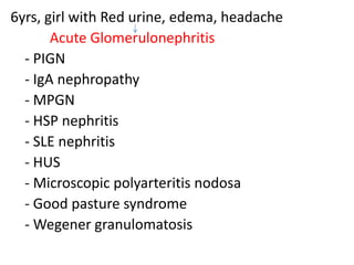
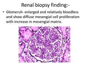
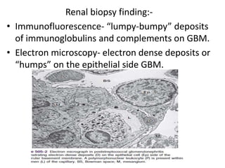
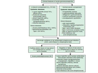
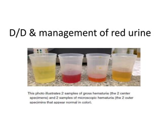
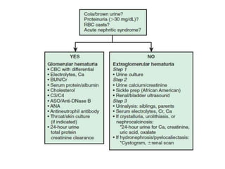
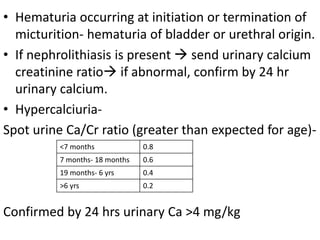
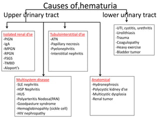
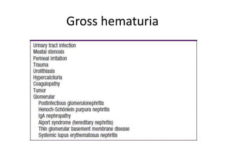
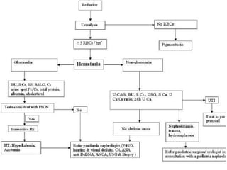
Approach to Acute Glomerulonephritis
Dr. Dilip Choudhary
Department of Pediatrics,
Mata Chanan Devi Hospital, New Delhi
Approach to a case of 6 years old
girl with h/o red coloured urine ,
edema, & headache . She had a
h/o sore throat 10 days back.
Discuss D/D & Mx of red coloured
urine
Objectives
• Differential diagnosis and approach
to a case of Acute glomerulonephritis (AGN)
• Management of AGN
• Causes of red urine
• Approach to hematuria
6yrs, girl with Red urine, edema, headache
Acute Glomerulonephritis
- PIGN
- IgA nephropathy
- MPGN
- HSP nephritis
- SLE nephritis
- HUS
- Microscopic polyarteritis nodosa
- Good pasture syndrome
- Wegener granulomatosis
If H/O infection is present-
• PIGN- URI / skin infection
• HSP/ IgA / Alport’s syndrome- URI infetion
• HUS- GI infection
Is it Henoch- Scholein Purpura Nephritis?-
Answer- NO
Because-
No rashes (palpable purpura)
No GI manifestation (Abdo pain, diarrhea, vomiting)
No musculoskeletal symptoms( arthritis, arthralgia)
Renal involvement- wks to months after initial c/f
Is it IgA nephropathy?
No
Because-
• Common at 10-35 yr of age.
• Hematuria occurs within 1-2 days of onset of URI.
• Very rarely leads to acute renal failure.
PIGN ( Postinfectious GN)
PIGN/ PSGN
Epidemiology:-
• Group A β- hemolytic streptococcal infection
• Follows infection of throat or skin by
“nephritogenic” strains.
• Streptococcal pharyngitis (M1, M4, M25 and
some strain of M12) in winter season.
• Skin infection (M49 serotype) in summer.
Pathogenesis of APSGN:-
• Immune complex mediated
• Molecular mimicry- antibodies elicited by
streptococcal antigens react with normal
glomerular antigens, immune complex formation
and glomerular deposition.
• Complement activation by directly deposited
streptococcal antigens ( serum C3 ↓ed)
Clinical manifestation of APSGN:-
• Most common in 5-12 yr age group, uncommon
before the age of 3 yrs.
• Acute nephritic syndrome-
1-2 wk after streptococcal pharyngitis
3-6 wk after streptococcal pyoderma
• Severity of kidney involvement- from
asymptomatic microscopic hematuria with
normal renal function to gross hematuria with
ARF.
Renal involvement
Oliguria
Hypertension (HTn) Hypervolemia
HTn encephalopathy Edema Heart failure
Blurred vision, headache, Pulm edema
altered mental status, RD, cough,
new seizure orthopnea, rales
• as per severity of renal involvement- patient can
develop various degree of edema, HTn and
oliguria.
• Risk for developing encephalopathy and / or
heart failure secondary to HTn or hypervolemia.
• Peripheral edema- results from Salt and water
retention.
Atypical presentation-
- Those with subclinical disease
- Severe symptoms but initial urinary abnormalities
- Who present with purpuric rash (difficult to
distinguish APSGN from HSP nephritis without renal
biopsy).
Duration of disease-
acute phase 6-8 wks
proteinuria and hypertension 4-6 wks
persistent microscopic
hematuria
1-2 yr
Diagnosis of APSGN
Sr. No Name of investigation finding
1. Urine routine & microscpoy RBC, RBC cast, PMN leucocytes, proteinuria
2. CBC Anemia (d/t hemodilution and low grade
hemolysis)
3. Serum C3, CH50 Low
4. Throat culture If positive, support diagnosis or might
represent carrier state
5. Antibody titre to streptococcal antigen
(i) Antistreptolysisn O (ASO) titre
(ii) Anti DNAase B
(iii) Streptozyme screen
- Increased after pharyngitis but rarely
increase after skin infection
- Best for cutaneous streptococcal
infection
- Measures multiple antibodies to
different streptococcal Ag
6. Chest X ray For pulmonary edema
7. MRI (if neurological symptoms) PRES in parieto-occipital area
Renal biopsy indication:
- Presence of ARF
- Presence of nephrotic syndrome
- Absence of evidence of streptococcal infection
- Normal complement level
- Hematuria, proteinuria, impaired renal function
and / or low C3 level persist >2 months after onset
Renal biopsy finding:-
• Glomeruli- enlarged and relatively bloodless
and show diffuse mesengial cell proliferation
with increase in mesengial matrix.
Renal biopsy finding:-
• Immunofluorescence- “lumpy-bumpy” deposits
of immunoglobulins and complements on GBM.
• Electron microscopy- electron dense deposits or
“humps” on the epithelial side GBM.
Treatment of PSGN
• Antibiotic- doesn’t alter the natural history but
10 days of Penicillin recommended to limit the
spread of nephritogenic organism.
• Acute renal insufficiency-
Hypervolemia- restricted fluid, diuretic therapy
Electrolyte imbalance- hyponatremia, hyperkalemia
Metabolic acidosis
Hypocalcemia- by lowering Sr. phosphate,
I.V. calcium only in case of tetany.
• Hypertension-
-Salt and water restriction
-Diuretics
-Long acting agents like Calcium Channel blocker
(amlodipine) or β-blockers (Propranolol, labetolol).
• Neurological symptoms- headache, seizures,
lethargy, confusion (encephalopathy). Caused by
HTn encephalopathy, hyponatremia, cerebral H’ge,
uremia.
-Benzodizepines for acutly controlling seizures
-Correct the precipitating cause
• Anemia-
- Packed RBCs if Hb falls below 7 gm/dl slowly over
4-6 hrs, @10 ml/kg.
- Use fresh, washed RBCs to minimize the risk of
hyperkalemia.
- In the presence of severe hypervolemia or
hyperkalemia, BT during dialysis.
• Nutrition-
- Restricted Na, K, Phosphorus
- Moderately restricted protein while maximizing
calorie intake (to minimize nitrogenous waste
accumulation).
- In critically ill- parenteral hyperalimentation.
• Dialysis indication:-
1. Anuria/ oliguria
2. Volume overload with evidence of HTn and / or
pulmonary edema refectory to diuretic therapy.
3. Persistent hyperkalemia
4. Severe metabolic acidosis unresponsive to
medical management.
5. Uremia (encephalopathy, pericarditis, neuropathy
6. BUN >100-150 mg/dl (or lower if rising rapidly)
D/D & management of red urine
Hematuria
• Microscopic hematuria- at least 5 RBCs/μl of urine.
• Macroscopic hematuria- visible to the naked eyes.
• Significant hematuria- >50 RBCs/μl of urine.
• Bedside investigation- reagent strip test (Dipstick-
sensitivity 100% and specificity 99% in detecting
1-5 RBCs/ hpf).
• False negative dipstick - formalin, high amount of
nitrides, ascorbic acid and high specific gravity.
• False positive dipstick - alkaline urine (pH>8),
contamination with hydrogen peroxide.
• Hydrogen peroxide + chromogen tetramethyl
benzidine
Pseudoperoxidase activity
of Hb/ myoglobin
Oxidized chromoge(green blue color)
• So if dipstick test is +ve, send urine microscopic
examination.
• Confirmation of hematuria is must -Microscopic
analysis of 10-15 ml of freshly voided and
centrifuged urine, suggested by >10 RBCs/μl, or
1+ positive urinary dipstick.
• If dipstick is +ve but no RBC and RBC cast it’s
pigmenturia (hemoglobinuria/ myoglobinuria).
Red urine (send Urine- R/M)
RBC absent RBC +nt
Hematuria
categorise
Heme positive
Hemoglobin
(acute or chronic hemolysis)
Myoglobin
(Rhabdomyolysis)
Heme negative
1. Drugs – Rifampin,
Ibuprofen,
Nitrofuratoin
Metronidazole
Deferoxamine
Iron sorbitol
Phenothiazines
2. Dyes- Beets,
Blackbarries,
Food and candy coloring
3. Metabolites –Homogentisic acid,
Melanin, Methemoglobin,
Porphyrin,
Urates
Features Glomerular Disease Non-glomerular disease
(UTI or Nephrolithiasis)
History
Dysuria
Systemic complaints
Family history
Absent (painless hematuria)
Edema, fever, pharyngitis,
rash, arthralgia
Deafness, renal
failure(alport’s syndrome)
Present (painful hematuria)
Fever with UTI, Pain with calculi
Renal stone, sickle cell disease
Physical
examination
HTn, edema
Rash, arthritis
Abdominal mass
Renal angle
tenderness
Present
+nt (HSP, SLE nephritis)
Absent
Absent
Less common
Absent
+nt in Wilm’s tumor
Present
Urinanalysis
Color
Proteinuria
Dysmorphic RBCs
RBC cast
Brown, tea, cola
2+ or more
>20%
Common
Bright red, clots may present
<2+
Eumorphic
Absent
• Hematuria due to glomerular causes is painless.
• The presence of red cell casts and dysmorphic
cells (>20%) are also consistent with glomerular
bleeding.
• Proteinuria may be present regardless of the
cause of bleeding, but usually does not exceed 2+
(100 mg/dL) on dipstick , if the only source of
protein is from blood in the urine
(extraglomerular hematuria).
• The combination of hematuria and proteinuria
(>100 mg/dL) indicates a significant renal disease
of glomerular origin.
• Hematuria occurring at initiation or termination of
micturition- hematuria of bladder or urethral origin.
• If nephrolithiasis is present  send urinary calcium
creatinine ratio if abnormal, confirm by 24 hr
urinary calcium.
• Hypercalciuria-
Spot urine Ca/Cr ratio (greater than expected for age)-
Confirmed by 24 hrs urinary Ca >4 mg/kg
<7 months 0.8
7 months- 18 months 0.6
19 months- 6 yrs 0.4
>6 yrs 0.2
Causes of hematuria
Upper urinary tract lower urinary tract
-UTI, cystitis, urethritis
-Urolithiasis
-Trauma
-Coagulopathy
-Heavy exercise
-Bladder tumor
Isolated renal d’se
-PIGN
-IgA
-MPGN
-RPGN
-FSGS
-TMBD
-Aloport’s
Multisystem disease
-SLE nephritis
-HSP Nephritis
-HUS
-Polyarteritis Nodosa(PAN)
-Goodpasture syndrome
-Hemoglobinopahty (sickle cell)
-HIV nephropathy
Tubulointerstitial d’se
-ATN
-Papillary necrosis
-Pyelonephritis
-Interstitial nephritis
Anatomical
-Hydronephrosis
-Polycystic kidney d’se
-Multicystic dysplasia
-Renal tumor
Gross hematuria
Recurrent hematuria
Two patterns of recurrent hematuria-
(a) Complete clearing hematuria
(b)Persistent microscopic hematuria between the
episodes of gross hematuria:
-IgA nephropathy (recurrent gross hematuria,
especially when asso. with infection).
-Alport’s syndrome
Familial or hereditary hematuria
• Benign, nonprogressive- Thin basement
membrane disease, sickle cell disease.
• Progressive- Alport’s syndrome
Interesting facts
Q.1 What is cyclical hematuria?
Ans. (a) In females with congenital adrenal
hyperplasia and virilization, who have been
raised as males. In adolescent age, when
menarche starts, cyclical hematuria episodes.
And (b) in females with Vesico-uterine
fistula(VUF) following LSCS(Youssef’s syndrome).
Q.2 What is Urethrorrhagia?
Ans. In prepubertal boys, urethral bleeding in
the absence of urine, is associated with dysuria
and blood spots on underwear after voiding. At
intervals several months, Benign self limited,
<10% have evidence of Glomerulonephritis
Q.3 Normally how much of RBCs are excreted in
urine per day?
Ans- 10 lakhs (1 million)
Q4. What to do for asymptomatic isolated
microscopic hematuria?
Ans- No need of extensive diagnostic evaluation,
often transient and benign. If it persist on at least 3
urinanalysis over a minmum of a 2 wks period
send urine culture. For culture negative, send urine
spot Ca:Cr ratio for hypercalciuria.
Reference
• Nelson Textbook of Pediatrics, 20th edition
Thank you

Acute Glomerulonephritis (AGN) by Dr. Dilip

  • 1.
    Approach to AcuteGlomerulonephritis Dr. Dilip Choudhary Department of Pediatrics, Mata Chanan Devi Hospital, New Delhi
  • 2.
    Approach to acase of 6 years old girl with h/o red coloured urine , edema, & headache . She had a h/o sore throat 10 days back. Discuss D/D & Mx of red coloured urine
  • 3.
    Objectives • Differential diagnosisand approach to a case of Acute glomerulonephritis (AGN) • Management of AGN • Causes of red urine • Approach to hematuria
  • 4.
    6yrs, girl withRed urine, edema, headache Acute Glomerulonephritis - PIGN - IgA nephropathy - MPGN - HSP nephritis - SLE nephritis - HUS - Microscopic polyarteritis nodosa - Good pasture syndrome - Wegener granulomatosis
  • 5.
    If H/O infectionis present- • PIGN- URI / skin infection • HSP/ IgA / Alport’s syndrome- URI infetion • HUS- GI infection
  • 6.
    Is it Henoch-Scholein Purpura Nephritis?- Answer- NO Because- No rashes (palpable purpura) No GI manifestation (Abdo pain, diarrhea, vomiting) No musculoskeletal symptoms( arthritis, arthralgia) Renal involvement- wks to months after initial c/f
  • 7.
    Is it IgAnephropathy? No Because- • Common at 10-35 yr of age. • Hematuria occurs within 1-2 days of onset of URI. • Very rarely leads to acute renal failure.
  • 8.
  • 9.
    PIGN/ PSGN Epidemiology:- • GroupA β- hemolytic streptococcal infection • Follows infection of throat or skin by “nephritogenic” strains. • Streptococcal pharyngitis (M1, M4, M25 and some strain of M12) in winter season. • Skin infection (M49 serotype) in summer.
  • 10.
    Pathogenesis of APSGN:- •Immune complex mediated • Molecular mimicry- antibodies elicited by streptococcal antigens react with normal glomerular antigens, immune complex formation and glomerular deposition. • Complement activation by directly deposited streptococcal antigens ( serum C3 ↓ed)
  • 11.
    Clinical manifestation ofAPSGN:- • Most common in 5-12 yr age group, uncommon before the age of 3 yrs. • Acute nephritic syndrome- 1-2 wk after streptococcal pharyngitis 3-6 wk after streptococcal pyoderma • Severity of kidney involvement- from asymptomatic microscopic hematuria with normal renal function to gross hematuria with ARF.
  • 12.
    Renal involvement Oliguria Hypertension (HTn)Hypervolemia HTn encephalopathy Edema Heart failure Blurred vision, headache, Pulm edema altered mental status, RD, cough, new seizure orthopnea, rales
  • 13.
    • as perseverity of renal involvement- patient can develop various degree of edema, HTn and oliguria. • Risk for developing encephalopathy and / or heart failure secondary to HTn or hypervolemia. • Peripheral edema- results from Salt and water retention.
  • 14.
    Atypical presentation- - Thosewith subclinical disease - Severe symptoms but initial urinary abnormalities - Who present with purpuric rash (difficult to distinguish APSGN from HSP nephritis without renal biopsy). Duration of disease- acute phase 6-8 wks proteinuria and hypertension 4-6 wks persistent microscopic hematuria 1-2 yr
  • 15.
    Diagnosis of APSGN Sr.No Name of investigation finding 1. Urine routine & microscpoy RBC, RBC cast, PMN leucocytes, proteinuria 2. CBC Anemia (d/t hemodilution and low grade hemolysis) 3. Serum C3, CH50 Low 4. Throat culture If positive, support diagnosis or might represent carrier state 5. Antibody titre to streptococcal antigen (i) Antistreptolysisn O (ASO) titre (ii) Anti DNAase B (iii) Streptozyme screen - Increased after pharyngitis but rarely increase after skin infection - Best for cutaneous streptococcal infection - Measures multiple antibodies to different streptococcal Ag 6. Chest X ray For pulmonary edema 7. MRI (if neurological symptoms) PRES in parieto-occipital area
  • 16.
    Renal biopsy indication: -Presence of ARF - Presence of nephrotic syndrome - Absence of evidence of streptococcal infection - Normal complement level - Hematuria, proteinuria, impaired renal function and / or low C3 level persist >2 months after onset
  • 17.
    Renal biopsy finding:- •Glomeruli- enlarged and relatively bloodless and show diffuse mesengial cell proliferation with increase in mesengial matrix.
  • 18.
    Renal biopsy finding:- •Immunofluorescence- “lumpy-bumpy” deposits of immunoglobulins and complements on GBM. • Electron microscopy- electron dense deposits or “humps” on the epithelial side GBM.
  • 19.
    Treatment of PSGN •Antibiotic- doesn’t alter the natural history but 10 days of Penicillin recommended to limit the spread of nephritogenic organism. • Acute renal insufficiency- Hypervolemia- restricted fluid, diuretic therapy Electrolyte imbalance- hyponatremia, hyperkalemia Metabolic acidosis Hypocalcemia- by lowering Sr. phosphate, I.V. calcium only in case of tetany.
  • 20.
    • Hypertension- -Salt andwater restriction -Diuretics -Long acting agents like Calcium Channel blocker (amlodipine) or β-blockers (Propranolol, labetolol). • Neurological symptoms- headache, seizures, lethargy, confusion (encephalopathy). Caused by HTn encephalopathy, hyponatremia, cerebral H’ge, uremia. -Benzodizepines for acutly controlling seizures -Correct the precipitating cause
  • 21.
    • Anemia- - PackedRBCs if Hb falls below 7 gm/dl slowly over 4-6 hrs, @10 ml/kg. - Use fresh, washed RBCs to minimize the risk of hyperkalemia. - In the presence of severe hypervolemia or hyperkalemia, BT during dialysis. • Nutrition- - Restricted Na, K, Phosphorus - Moderately restricted protein while maximizing calorie intake (to minimize nitrogenous waste accumulation). - In critically ill- parenteral hyperalimentation.
  • 22.
    • Dialysis indication:- 1.Anuria/ oliguria 2. Volume overload with evidence of HTn and / or pulmonary edema refectory to diuretic therapy. 3. Persistent hyperkalemia 4. Severe metabolic acidosis unresponsive to medical management. 5. Uremia (encephalopathy, pericarditis, neuropathy 6. BUN >100-150 mg/dl (or lower if rising rapidly)
  • 24.
    D/D & managementof red urine
  • 25.
    Hematuria • Microscopic hematuria-at least 5 RBCs/μl of urine. • Macroscopic hematuria- visible to the naked eyes. • Significant hematuria- >50 RBCs/μl of urine. • Bedside investigation- reagent strip test (Dipstick- sensitivity 100% and specificity 99% in detecting 1-5 RBCs/ hpf).
  • 26.
    • False negativedipstick - formalin, high amount of nitrides, ascorbic acid and high specific gravity. • False positive dipstick - alkaline urine (pH>8), contamination with hydrogen peroxide. • Hydrogen peroxide + chromogen tetramethyl benzidine Pseudoperoxidase activity of Hb/ myoglobin Oxidized chromoge(green blue color)
  • 27.
    • So ifdipstick test is +ve, send urine microscopic examination. • Confirmation of hematuria is must -Microscopic analysis of 10-15 ml of freshly voided and centrifuged urine, suggested by >10 RBCs/μl, or 1+ positive urinary dipstick. • If dipstick is +ve but no RBC and RBC cast it’s pigmenturia (hemoglobinuria/ myoglobinuria).
  • 28.
    Red urine (sendUrine- R/M) RBC absent RBC +nt Hematuria categorise Heme positive Hemoglobin (acute or chronic hemolysis) Myoglobin (Rhabdomyolysis) Heme negative 1. Drugs – Rifampin, Ibuprofen, Nitrofuratoin Metronidazole Deferoxamine Iron sorbitol Phenothiazines 2. Dyes- Beets, Blackbarries, Food and candy coloring 3. Metabolites –Homogentisic acid, Melanin, Methemoglobin, Porphyrin, Urates
  • 29.
    Features Glomerular DiseaseNon-glomerular disease (UTI or Nephrolithiasis) History Dysuria Systemic complaints Family history Absent (painless hematuria) Edema, fever, pharyngitis, rash, arthralgia Deafness, renal failure(alport’s syndrome) Present (painful hematuria) Fever with UTI, Pain with calculi Renal stone, sickle cell disease Physical examination HTn, edema Rash, arthritis Abdominal mass Renal angle tenderness Present +nt (HSP, SLE nephritis) Absent Absent Less common Absent +nt in Wilm’s tumor Present Urinanalysis Color Proteinuria Dysmorphic RBCs RBC cast Brown, tea, cola 2+ or more >20% Common Bright red, clots may present <2+ Eumorphic Absent
  • 30.
    • Hematuria dueto glomerular causes is painless. • The presence of red cell casts and dysmorphic cells (>20%) are also consistent with glomerular bleeding. • Proteinuria may be present regardless of the cause of bleeding, but usually does not exceed 2+ (100 mg/dL) on dipstick , if the only source of protein is from blood in the urine (extraglomerular hematuria). • The combination of hematuria and proteinuria (>100 mg/dL) indicates a significant renal disease of glomerular origin.
  • 32.
    • Hematuria occurringat initiation or termination of micturition- hematuria of bladder or urethral origin. • If nephrolithiasis is present  send urinary calcium creatinine ratio if abnormal, confirm by 24 hr urinary calcium. • Hypercalciuria- Spot urine Ca/Cr ratio (greater than expected for age)- Confirmed by 24 hrs urinary Ca >4 mg/kg <7 months 0.8 7 months- 18 months 0.6 19 months- 6 yrs 0.4 >6 yrs 0.2
  • 33.
    Causes of hematuria Upperurinary tract lower urinary tract -UTI, cystitis, urethritis -Urolithiasis -Trauma -Coagulopathy -Heavy exercise -Bladder tumor Isolated renal d’se -PIGN -IgA -MPGN -RPGN -FSGS -TMBD -Aloport’s Multisystem disease -SLE nephritis -HSP Nephritis -HUS -Polyarteritis Nodosa(PAN) -Goodpasture syndrome -Hemoglobinopahty (sickle cell) -HIV nephropathy Tubulointerstitial d’se -ATN -Papillary necrosis -Pyelonephritis -Interstitial nephritis Anatomical -Hydronephrosis -Polycystic kidney d’se -Multicystic dysplasia -Renal tumor
  • 34.
  • 35.
    Recurrent hematuria Two patternsof recurrent hematuria- (a) Complete clearing hematuria (b)Persistent microscopic hematuria between the episodes of gross hematuria: -IgA nephropathy (recurrent gross hematuria, especially when asso. with infection). -Alport’s syndrome
  • 36.
    Familial or hereditaryhematuria • Benign, nonprogressive- Thin basement membrane disease, sickle cell disease. • Progressive- Alport’s syndrome
  • 37.
    Interesting facts Q.1 Whatis cyclical hematuria? Ans. (a) In females with congenital adrenal hyperplasia and virilization, who have been raised as males. In adolescent age, when menarche starts, cyclical hematuria episodes. And (b) in females with Vesico-uterine fistula(VUF) following LSCS(Youssef’s syndrome).
  • 38.
    Q.2 What isUrethrorrhagia? Ans. In prepubertal boys, urethral bleeding in the absence of urine, is associated with dysuria and blood spots on underwear after voiding. At intervals several months, Benign self limited, <10% have evidence of Glomerulonephritis Q.3 Normally how much of RBCs are excreted in urine per day? Ans- 10 lakhs (1 million)
  • 39.
    Q4. What todo for asymptomatic isolated microscopic hematuria? Ans- No need of extensive diagnostic evaluation, often transient and benign. If it persist on at least 3 urinanalysis over a minmum of a 2 wks period send urine culture. For culture negative, send urine spot Ca:Cr ratio for hypercalciuria.
  • 41.
    Reference • Nelson Textbookof Pediatrics, 20th edition
  • 42.