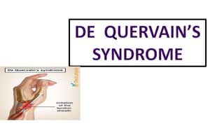
De quervain syndrome
•Download as PPTX, PDF•
8 likes•1,236 views

DE QUERVAIN'S SYNDROME …ANATOMY , ETIOLOGY , CLINICAL FEATURES ,DIAGNOSIS , EXAMINATION AND MANAGEMENT

Recommended
More Related Content
What's hot
What's hot (20)
Similar to De quervain syndrome
Similar to De quervain syndrome (20)
More from ANNIE BLESSIE
More from ANNIE BLESSIE (10)
Recently uploaded
Recently uploaded (20)
De quervain syndrome
- 2. CONTENTS : Definition Anatomy Aetiology Incidence Clinical features Diagnosis Examination Management
- 3. Definition : De Quervain disease is a chronic constrictive tenosynovitis affecting the Abductor Pollicis Longus and Extensor Pollicis Brevis tendon of the thumb at the wrist. These muscles are located on the dorsal side of forearm and go to the lateral side of the thumb through a fibrous osseous tunnel .
- 4. ANATOMY : • The dorsal aspect of wrist contains six compartment that transmit the tendon to the hand. • The first dorsal compartment is located over the radial styloid proximal to the radio- carpal joint . Muscles are : 1) Extensor Pollicis Brevis (EPB) 2) Abductor Pollicis Longus (APB)
- 5. Extensor Pollicis Brevis ( EPB) • Origin - ½ dorsal side of radius • Insertion - base of proximal phalanx of thumb • Action – radial abduction, thumb extension • Nerve supply – radial nerve Abductor Pollicis Longus (APL) • Origin – dorsal side of radius and ulna • Insertion – base of metacarpal • Action – extension of thumb • Nerve supply – radial nerve
- 6. Aetiology : • De Quervain is named after the Swiss surgeon FRITZ DE QUERVAIN Who first described it . • It describes the inflammation of the sheath or tunnel that surround two tendons that control the movement of the thumb . • Main cause is repetitive use of the thumb in combination with radial deviation of the wrist. • Characterized by degeneration and fibrosis of the tendon sheath .
- 7. Tendon of abductor pollicis longus and extensor pollicis brevis are tightly secured against the radial styloid by the overlying retinaculum Acute or repetitive trauma restrains gliding of the tendon result in inflammation of synovial sheath Increases friction Reactive fibrosis and thickening of the sheath Degeneration
- 8. INCIDENCE : • Occurs most often in individuals age between 30 and 50 yrs • It affects women up to six times more often than men • It commonly associated with dominant hand
- 9. Clinical features : - Complain with pain on radial side of wrist that is worsened by moving the wrist or thumb. - Tendon sheath may feel thick and hard - Swelling in anatomical snuff box - Acute Tenderness at tip of radial styloid - Pain aggrevates on grasping object - Wet leather sign - Finkelstein test is positive FINKELSTEIN TEST :- it is provocative test used in diagnosis for de quervain synovitis - Make a fist with the thumb inside - Now ask patient to bend the wrist toward the little finger
- 10. Diagnosis : 1. CMC arthritis of thumb : pain and crepitus present with the thumb “crank and grind test” 2. Chauffeurs fracture 3. Intercarpal instabilities 4. Scaphoid fracture 5. Wartenberg’s syndrome : nerve become compressed btw tendon of Brachioradialis and extensor carpi Radialis Brevis . 6. C6 Cervical Radiopathy 7. Osteoarthritis of 1st CMC 8. Intersection syndrome
- 11. EXAMINATION : 1.] ON OBSERVATION : - Resting posture of hand /thumb - Inflammation around dorsal part of base of thumb 2.] ON PALPATION : - Tenderness over the base of thumb and 1st dorsal compartment extensor tendon - Thickening of synovial sheath 3.] RANGE OF MOTION : -Cervical ROM - Shoulder ,elbow ,forearm ,wrist ROM
- 12. 4.] FIST GRIP STRENGTH 5.] PINCH STRENGTH 6.] NEUROLOGICAL TEST : - Superficial radial nerve - Tinnels sign 7.] NEUROLOGICAL INVOLVEMENT : - dermatome ( C4 - T1 ) - myotome (C4 – T1 ) - reflexes ( C5 C6 C7 )
- 13. MANAGEMENT : GOALS OF TREATMENT :- - Restoration of Normal, painless use of involved hand - Resolution of inflammatory process - Prevention of recurrence - Restoration of pain free movement and strength
- 14. Medical management : 1.) Corticosteroid injection : -can be given to patient with moderate to marked pain with symptoms lasting for more than 3 weeks 2.) NSAIDS : - It is prescribed initially for 6- 8 weeks to reduce pain and inflammation
- 15. Physiotherapy management : 1. Immobilization :- -thumb splint is used to restrict thumb movement so that first dorsal compartment tendon are at rest 2. Cold compression :- -10-12 min over inflamed area 3. Ultrasonic therapy :- - Pulsed mode ,3MHZ ,time 5 min 4. Phonopheresis : -with 10% hydrocortisone 5. Gentle active and passive motion of thumb and wrist 6. Strengthening and stretching exercise 7. Rehabilitation exercise : - Wrist stretch - Wrist flexion extension - Grip strengthening - Finger spring
- 16. Surgery management : Decompression surgery :- after 0-2 days of surgery Immobilization with cast After 48 hours dressing are removed 2-14 days Presurgical splint is worn for comfort and active exercise 2-6 weeks Grip and pinch strengthening exercise may begin at approx. 3 weeks and can be progressed ,by the end of 6 week the patient usually able to resume full activities .