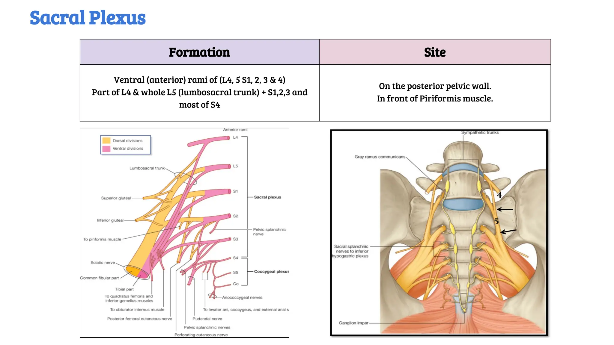
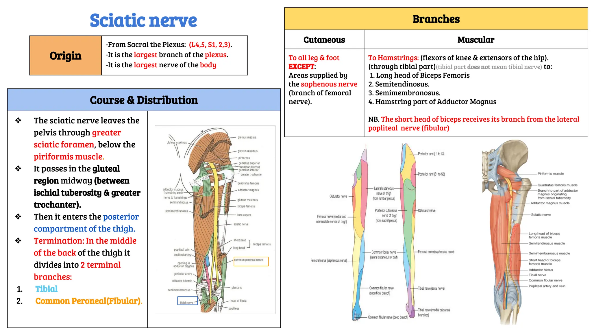
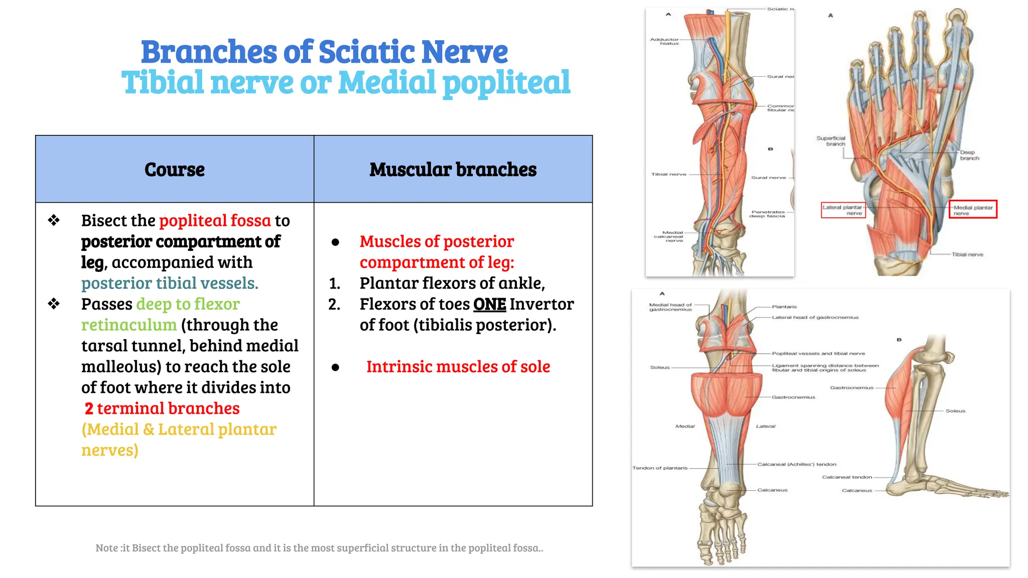
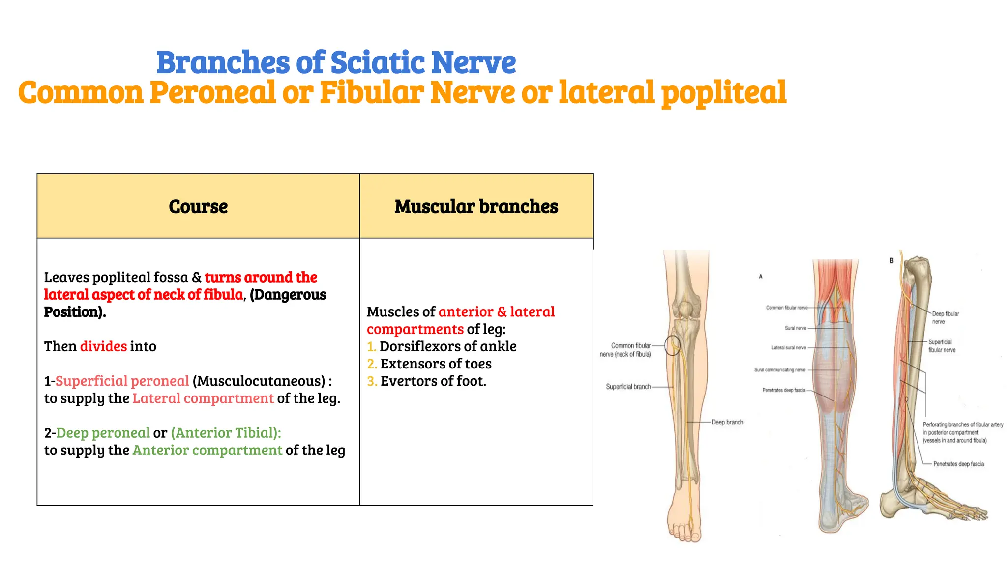
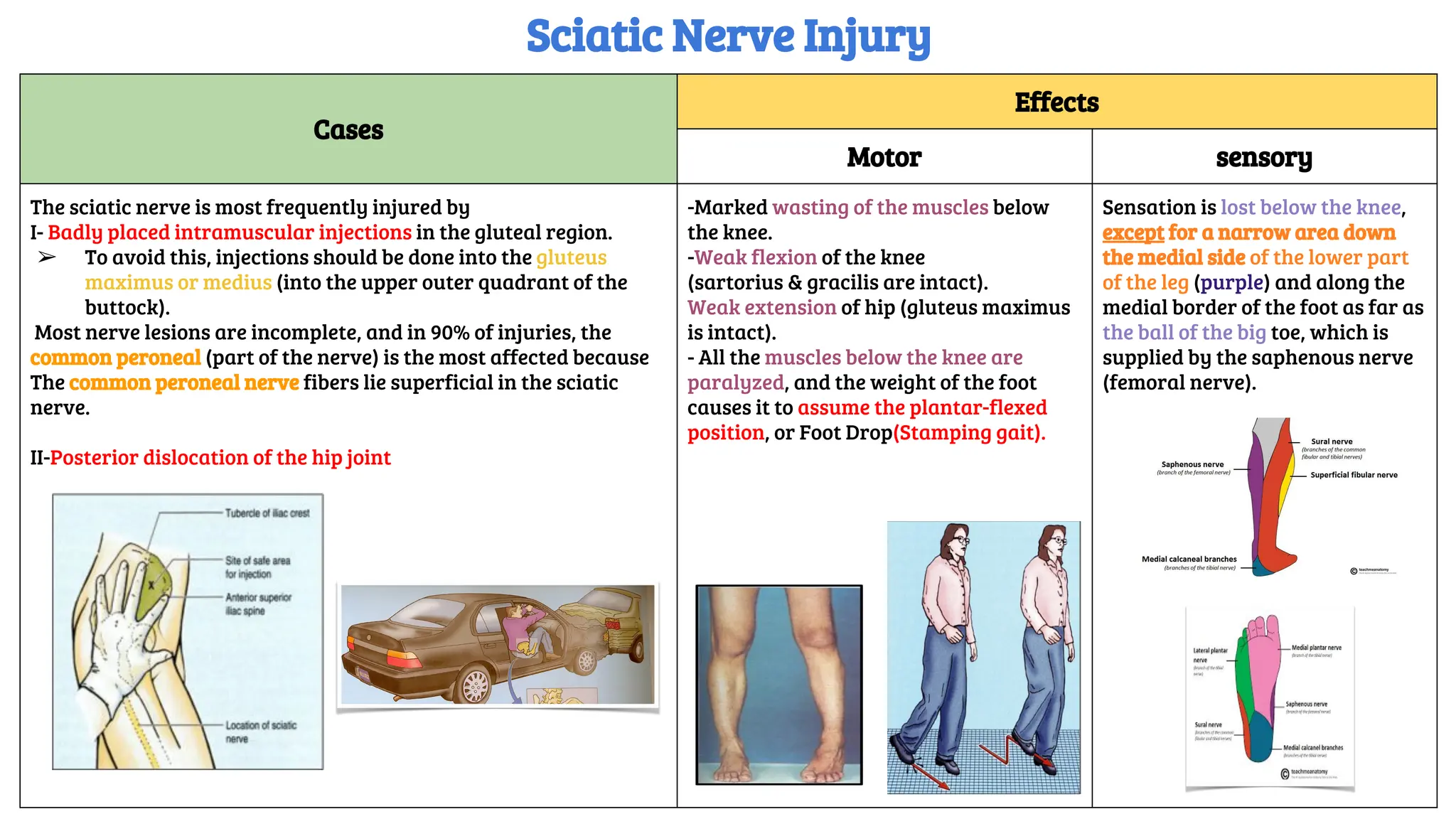
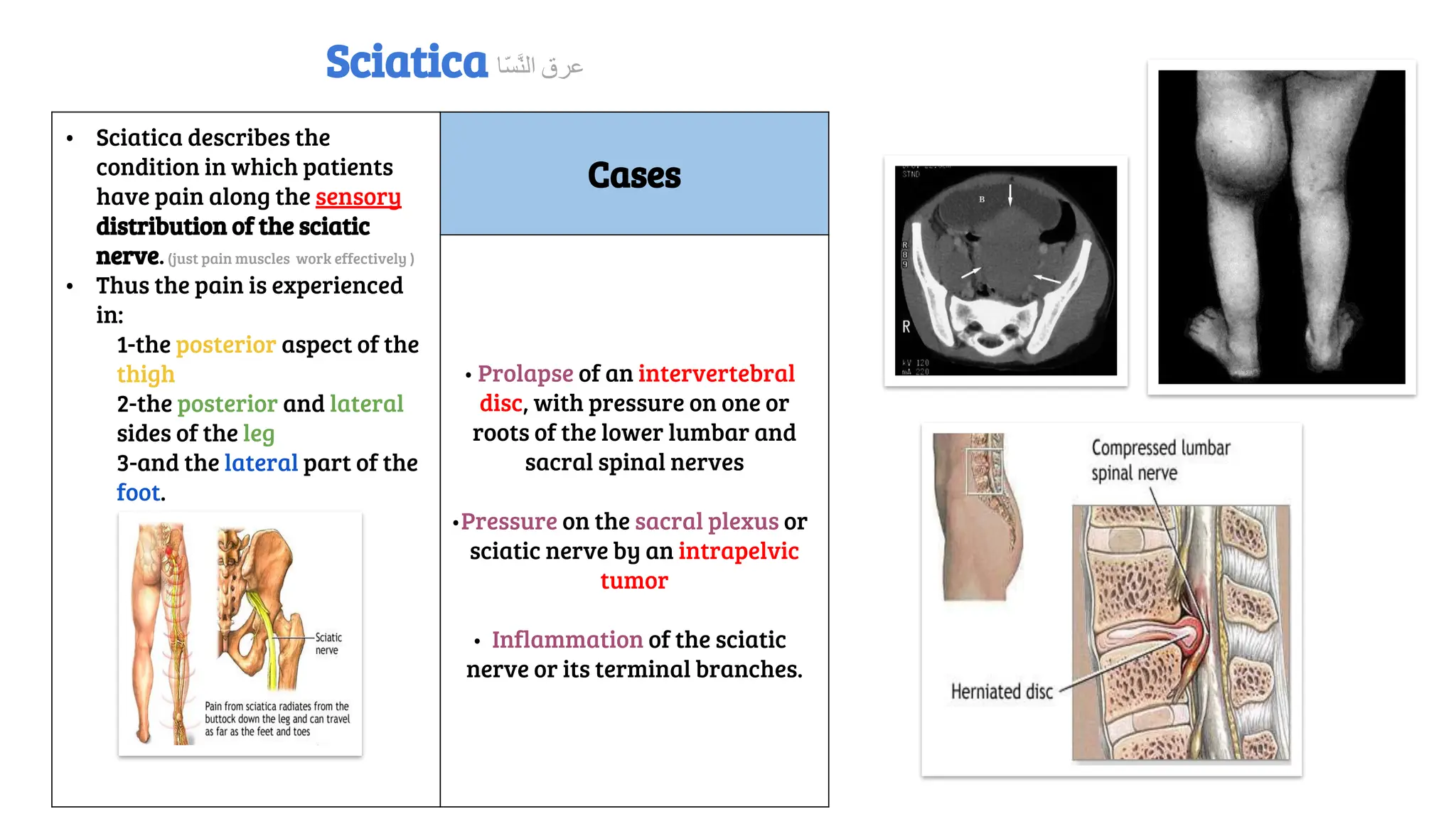
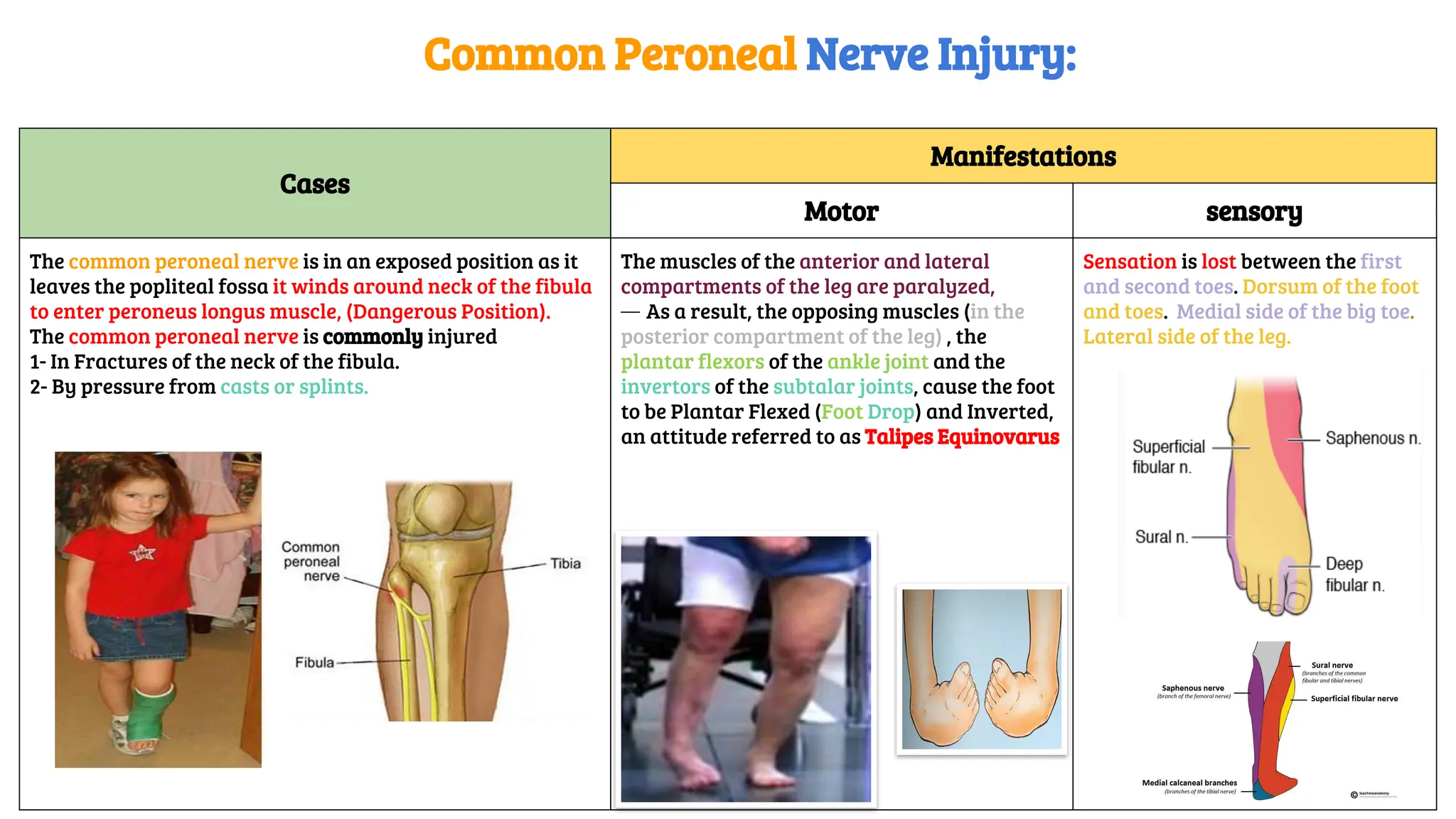
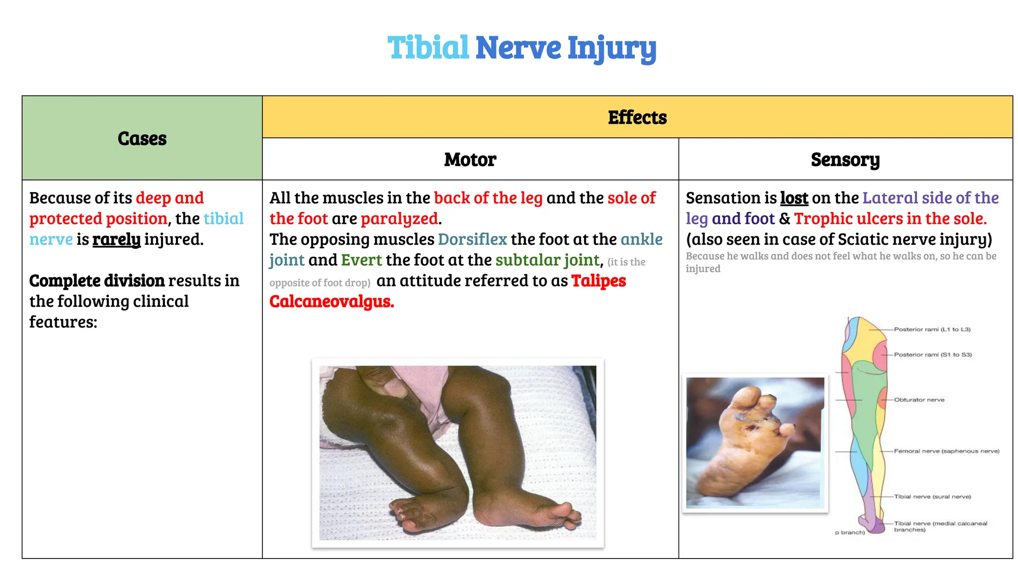
Musculoskeletal block- Anatomy-lecture 13
Editing file
Sciatic Nerve
Objectives
✓Describe the anatomy (origin, course and distribution) of
the sciatic nerve.
✓List the branches of the sciatic nerve.
✓Describe briefly the main motor and sensory
manifestations in case of injury of the sciatic nerve or its
main branches.
Color guide :
Only in boys slides in Blue
Only in girls slides in Purple
important in Red
Doctor note in Green
Extra information in Grey
Formation Site
Ventral (anterior) rami of (L4, 5 S1, 2, 3 & 4)
Part of L4 & whole L5 (lumbosacral trunk) + S1,2,3 and
most of S4
On the posterior pelvic wall.
In front of Piriformis muscle.
Sacral Plexus
Course & Distribution
âť– The sciatic nerve leaves the
pelvis through greater
sciatic foramen, below the
piriformis muscle.
âť– It passes in the gluteal
region midway (between
ischial tuberosity & greater
trochanter).
âť– Then it enters the posterior
compartment of the thigh.
âť– Termination: In the middle
of the back of the thigh it
divides into 2 terminal
branches:
1. Tibial
2. Common Peroneal(Fibular).
Branches
Cutaneous Muscular
To all leg & foot
EXCEPT:
Areas supplied by
the saphenous nerve
(branch of femoral
nerve).
To Hamstrings: (flexors of knee & extensors of the hip).
(through tibial part)(tibial part does not mean tibial nerve) to:
1. Long head of Biceps Femoris
2. Semitendinosus.
3. Semimembranosus.
4. Hamstring part of Adductor Magnus
NB. The short head of biceps receives its branch from the lateral
popliteal nerve (fibular)
Origin
-From Sacral the Plexus: (L4,5, S1, 2,3).
-It is the largest branch of the plexus.
-It is the largest nerve of the body
Sciatic nerve
Course Muscular branches
âť– Bisect the popliteal fossa to
posterior compartment of
leg, accompanied with
posterior tibial vessels.
âť– Passes deep to flexor
retinaculum (through the
tarsal tunnel, behind medial
malleolus) to reach the sole
of foot where it divides into
2 terminal branches
(Medial & Lateral plantar
nerves)
â—Ź Muscles of posterior
compartment of leg:
1. Plantar flexors of ankle,
2. Flexors of toes ONE Invertor
of foot (tibialis posterior).
â—Ź Intrinsic muscles of sole
Branches of Sciatic Nerve
Tibial nerve or Medial popliteal
Note :it Bisect the popliteal fossa and it is the most superficial structure in the popliteal fossa..
Course Muscular branches
Leaves popliteal fossa & turns around the
lateral aspect of neck of fibula, (Dangerous
Position).
Then divides into
1-Superficial peroneal (Musculocutaneous) :
to supply the Lateral compartment of the leg.
2-Deep peroneal or (Anterior Tibial):
to supply the Anterior compartment of the leg
Muscles of anterior & lateral
compartments of leg:
1. Dorsiflexors of ankle
2. Extensors of toes
3. Evertors of foot.
Branches of Sciatic Nerve
Common Peroneal or Fibular Nerve or lateral popliteal
Cases
Effects
Motor sensory
The sciatic nerve is most frequently injured by
I- Badly placed intramuscular injections in the gluteal region.
➢ To avoid this, injections should be done into the gluteus
maximus or medius (into the upper outer quadrant of the
buttock).
Most nerve lesions are incomplete, and in 90% of injuries, the
common peroneal (part of the nerve) is the most affected because
The common peroneal nerve fibers lie superficial in the sciatic
nerve.
II-Posterior dislocation of the hip joint
-Marked wasting of the muscles below
the knee.
-Weak flexion of the knee
(sartorius & gracilis are intact).
Weak extension of hip (gluteus maximus
is intact).
- All the muscles below the knee are
paralyzed, and the weight of the foot
causes it to assume the plantar-flexed
position, or Foot Drop(Stamping gait).
Sensation is lost below the knee,
except for a narrow area down
the medial side of the lower part
of the leg (purple) and along the
medial border of the foot as far as
the ball of the big toe, which is
supplied by the saphenous nerve
(femoral nerve).
Sciatic Nerve Injury
• Sciatica describes the
condition in which patients
have pain along the sensory
distribution of the sciatic
nerve.(just pain muscles work effectively )
• Thus the pain is experienced
in:
1-the posterior aspect of the
thigh
2-the posterior and lateral
sides of the leg
3-and the lateral part of the
foot.
Cases
• Prolapse of an intervertebral
disc, with pressure on one or
roots of the lower lumbar and
sacral spinal nerves
•Pressure on the sacral plexus or
sciatic nerve by an intrapelvic
tumor
• Inflammation of the sciatic
nerve or its terminal branches.
Sciatica ‫ّﺎ‬‫ﺴ‬‫ﱠ‬‫ﻨ‬‫اﻟ‬ ‫ﻋﺮق‬
Cases
Manifestations
Motor sensory
The common peroneal nerve is in an exposed position as it
leaves the popliteal fossa it winds around neck of the fibula
to enter peroneus longus muscle, (Dangerous Position).
The common peroneal nerve is commonly injured
1- In Fractures of the neck of the fibula.
2- By pressure from casts or splints.
The muscles of the anterior and lateral
compartments of the leg are paralyzed,
— As a result, the opposing muscles (in the
posterior compartment of the leg) , the
plantar flexors of the ankle joint and the
invertors of the subtalar joints, cause the foot
to be Plantar Flexed (Foot Drop) and Inverted,
an attitude referred to as Talipes Equinovarus
Sensation is lost between the first
and second toes. Dorsum of the foot
and toes. Medial side of the big toe.
Lateral side of the leg.
Common Peroneal Nerve Injury:
Cases
Effects
Motor Sensory
Because of its deep and
protected position, the tibial
nerve is rarely injured.
Complete division results in
the following clinical
features:
All the muscles in the back of the leg and the sole of
the foot are paralyzed.
The opposing muscles Dorsiflex the foot at the ankle
joint and Evert the foot at the subtalar joint, (it is the
opposite of foot drop) an attitude referred to as Talipes
Calcaneovalgus.
Sensation is lost on the Lateral side of the
leg and foot & Trophic ulcers in the sole.
(also seen in case of Sciatic nerve injury)
Because he walks and does not feel what he walks on, so he can be
injured
Tibial Nerve Injury
MCQs
Question 1: Which of the following nerve is the largest nerve of the body?
A. Sciatic nerve.
B. Radial nerve.
C. Ulnar nerve.
D. Peroneal nerve.
Question 2: The site of sacral plexus:
A.On the anterior wall of the pelvis, in front of piriformis muscle.
B.On the posterior wall of the pelvis, in the back of piriformis muscle.
C.On the posterior wall of the pelvis, in front of piriformis muscle.
D.On the anterior wall of the pelvis, in the back of piriformis muscle.
Question 3: Which muscle of these is not one of the anterior and lateral
compartments of leg?
A.Dorsiflexors of ankle.
B. Evertors of foot.
C. Extensors of toes.
D.Plantar flexors of ankle.
Question 4: Which one of theses muscles is supplied by common peroneal nerve?
A.Long head of biceps.
B. Short head of biceps.
C.Hamstring.
D.Semitendinosus.
Question 5: The most frequent injuries of the sciatic nerve is:
A.Badly placed intramuscular injections in the gluteal region.
B.Posterior dislocation of hip joint.
C.Both a and b.
D.None of the above
Question 6: When all muscles below the knee are paralyzed, the weight of the foot
causes it to assume the:
A.Plantar position.
B.Foot drop.
C.Stamping gait.
D.All of the above.
Question 7: In the tibial nerve course, it descends through popliteal fossa to the:
A.Anterior compartment of the leg.
B.Posterior compartment of the leg.
C.Posterior compartment of thigh.
D.Anterior compartment of the thigh.
Answers: Q1.A Q2.C -Q3.D -Q4.B- Q5.C- Q6.D-Q7.B
Give us your feedback:
Boys team:
â—Ź Khalid AL-Dossari
â—Ź Naif Al-Dossari
â—Ź Faisal Alqifari
â—Ź Salman Alagla
â—Ź Ziyad Al-jofan
â—Ź Suhail Basuhail
â—Ź Ali Aldawood
â—Ź Khalid Nagshabandi
â—Ź Mohammed Al-huqbani
â—Ź Jehad Alorainy
â—Ź Khalid AlKhani
â—Ź Omar Alammari
Girls team :
â—Ź Ajeed Al Rashoud
â—Ź Taif Alotaibi
â—Ź Noura Al Turki
â—Ź Amirah Al-Zahrani
â—Ź Alhanouf Al-haluli
â—Ź Sara Al-Abdulkarem
â—Ź Rawan Al Zayed
â—Ź Reema Al Masoud
â—Ź Renad Al Haqbani
â—Ź Nouf Al Humaidhi
â—Ź Fay Al Buqami
â—Ź Jude Al Khalifah
â—Ź Nouf Al Hussaini
â—Ź Alwateen Al Balawi
â—Ź Rahaf Al Shabri
â—Ź Danah Al Halees
â—Ź Haifa Al Waily
â—Ź Rema Al Mutawa
â—Ź Amirah Al Dakhilallah
â—Ź Maha Al Nahdi
â—Ź Renad Al Mutawa
â—Ź Ghaida Al Braithen
â—Ź Reham Yousef
Team members
Team leaders
â—Ź Abdulrahman Shadid
â—Ź Ateen Almutairi
Good luck
Special thank for
Anatomy team 436
=This lecture done by

Sciatic Nerve and clinical correlation.pdf

  • 1.
    Musculoskeletal block- Anatomy-lecture13 Editing file Sciatic Nerve
  • 2.
    Objectives ✓Describe the anatomy(origin, course and distribution) of the sciatic nerve. ✓List the branches of the sciatic nerve. ✓Describe briefly the main motor and sensory manifestations in case of injury of the sciatic nerve or its main branches. Color guide : Only in boys slides in Blue Only in girls slides in Purple important in Red Doctor note in Green Extra information in Grey
  • 3.
    Formation Site Ventral (anterior)rami of (L4, 5 S1, 2, 3 & 4) Part of L4 & whole L5 (lumbosacral trunk) + S1,2,3 and most of S4 On the posterior pelvic wall. In front of Piriformis muscle. Sacral Plexus
  • 4.
    Course & Distribution âť–The sciatic nerve leaves the pelvis through greater sciatic foramen, below the piriformis muscle. âť– It passes in the gluteal region midway (between ischial tuberosity & greater trochanter). âť– Then it enters the posterior compartment of the thigh. âť– Termination: In the middle of the back of the thigh it divides into 2 terminal branches: 1. Tibial 2. Common Peroneal(Fibular). Branches Cutaneous Muscular To all leg & foot EXCEPT: Areas supplied by the saphenous nerve (branch of femoral nerve). To Hamstrings: (flexors of knee & extensors of the hip). (through tibial part)(tibial part does not mean tibial nerve) to: 1. Long head of Biceps Femoris 2. Semitendinosus. 3. Semimembranosus. 4. Hamstring part of Adductor Magnus NB. The short head of biceps receives its branch from the lateral popliteal nerve (fibular) Origin -From Sacral the Plexus: (L4,5, S1, 2,3). -It is the largest branch of the plexus. -It is the largest nerve of the body Sciatic nerve
  • 5.
    Course Muscular branches âť–Bisect the popliteal fossa to posterior compartment of leg, accompanied with posterior tibial vessels. âť– Passes deep to flexor retinaculum (through the tarsal tunnel, behind medial malleolus) to reach the sole of foot where it divides into 2 terminal branches (Medial & Lateral plantar nerves) â—Ź Muscles of posterior compartment of leg: 1. Plantar flexors of ankle, 2. Flexors of toes ONE Invertor of foot (tibialis posterior). â—Ź Intrinsic muscles of sole Branches of Sciatic Nerve Tibial nerve or Medial popliteal Note :it Bisect the popliteal fossa and it is the most superficial structure in the popliteal fossa..
  • 6.
    Course Muscular branches Leavespopliteal fossa & turns around the lateral aspect of neck of fibula, (Dangerous Position). Then divides into 1-Superficial peroneal (Musculocutaneous) : to supply the Lateral compartment of the leg. 2-Deep peroneal or (Anterior Tibial): to supply the Anterior compartment of the leg Muscles of anterior & lateral compartments of leg: 1. Dorsiflexors of ankle 2. Extensors of toes 3. Evertors of foot. Branches of Sciatic Nerve Common Peroneal or Fibular Nerve or lateral popliteal
  • 7.
    Cases Effects Motor sensory The sciaticnerve is most frequently injured by I- Badly placed intramuscular injections in the gluteal region. ➢ To avoid this, injections should be done into the gluteus maximus or medius (into the upper outer quadrant of the buttock). Most nerve lesions are incomplete, and in 90% of injuries, the common peroneal (part of the nerve) is the most affected because The common peroneal nerve fibers lie superficial in the sciatic nerve. II-Posterior dislocation of the hip joint -Marked wasting of the muscles below the knee. -Weak flexion of the knee (sartorius & gracilis are intact). Weak extension of hip (gluteus maximus is intact). - All the muscles below the knee are paralyzed, and the weight of the foot causes it to assume the plantar-flexed position, or Foot Drop(Stamping gait). Sensation is lost below the knee, except for a narrow area down the medial side of the lower part of the leg (purple) and along the medial border of the foot as far as the ball of the big toe, which is supplied by the saphenous nerve (femoral nerve). Sciatic Nerve Injury
  • 8.
    • Sciatica describesthe condition in which patients have pain along the sensory distribution of the sciatic nerve.(just pain muscles work effectively ) • Thus the pain is experienced in: 1-the posterior aspect of the thigh 2-the posterior and lateral sides of the leg 3-and the lateral part of the foot. Cases • Prolapse of an intervertebral disc, with pressure on one or roots of the lower lumbar and sacral spinal nerves •Pressure on the sacral plexus or sciatic nerve by an intrapelvic tumor • Inflammation of the sciatic nerve or its terminal branches. Sciatica ‫ّﺎ‬‫ﺴ‬‫ﱠ‬‫ﻨ‬‫اﻟ‬ ‫ﻋﺮق‬
  • 9.
    Cases Manifestations Motor sensory The commonperoneal nerve is in an exposed position as it leaves the popliteal fossa it winds around neck of the fibula to enter peroneus longus muscle, (Dangerous Position). The common peroneal nerve is commonly injured 1- In Fractures of the neck of the fibula. 2- By pressure from casts or splints. The muscles of the anterior and lateral compartments of the leg are paralyzed, — As a result, the opposing muscles (in the posterior compartment of the leg) , the plantar flexors of the ankle joint and the invertors of the subtalar joints, cause the foot to be Plantar Flexed (Foot Drop) and Inverted, an attitude referred to as Talipes Equinovarus Sensation is lost between the first and second toes. Dorsum of the foot and toes. Medial side of the big toe. Lateral side of the leg. Common Peroneal Nerve Injury:
  • 10.
    Cases Effects Motor Sensory Because ofits deep and protected position, the tibial nerve is rarely injured. Complete division results in the following clinical features: All the muscles in the back of the leg and the sole of the foot are paralyzed. The opposing muscles Dorsiflex the foot at the ankle joint and Evert the foot at the subtalar joint, (it is the opposite of foot drop) an attitude referred to as Talipes Calcaneovalgus. Sensation is lost on the Lateral side of the leg and foot & Trophic ulcers in the sole. (also seen in case of Sciatic nerve injury) Because he walks and does not feel what he walks on, so he can be injured Tibial Nerve Injury
  • 11.
    MCQs Question 1: Whichof the following nerve is the largest nerve of the body? A. Sciatic nerve. B. Radial nerve. C. Ulnar nerve. D. Peroneal nerve. Question 2: The site of sacral plexus: A.On the anterior wall of the pelvis, in front of piriformis muscle. B.On the posterior wall of the pelvis, in the back of piriformis muscle. C.On the posterior wall of the pelvis, in front of piriformis muscle. D.On the anterior wall of the pelvis, in the back of piriformis muscle. Question 3: Which muscle of these is not one of the anterior and lateral compartments of leg? A.Dorsiflexors of ankle. B. Evertors of foot. C. Extensors of toes. D.Plantar flexors of ankle. Question 4: Which one of theses muscles is supplied by common peroneal nerve? A.Long head of biceps. B. Short head of biceps. C.Hamstring. D.Semitendinosus. Question 5: The most frequent injuries of the sciatic nerve is: A.Badly placed intramuscular injections in the gluteal region. B.Posterior dislocation of hip joint. C.Both a and b. D.None of the above Question 6: When all muscles below the knee are paralyzed, the weight of the foot causes it to assume the: A.Plantar position. B.Foot drop. C.Stamping gait. D.All of the above. Question 7: In the tibial nerve course, it descends through popliteal fossa to the: A.Anterior compartment of the leg. B.Posterior compartment of the leg. C.Posterior compartment of thigh. D.Anterior compartment of the thigh. Answers: Q1.A Q2.C -Q3.D -Q4.B- Q5.C- Q6.D-Q7.B
  • 12.
    Give us yourfeedback: Boys team: â—Ź Khalid AL-Dossari â—Ź Naif Al-Dossari â—Ź Faisal Alqifari â—Ź Salman Alagla â—Ź Ziyad Al-jofan â—Ź Suhail Basuhail â—Ź Ali Aldawood â—Ź Khalid Nagshabandi â—Ź Mohammed Al-huqbani â—Ź Jehad Alorainy â—Ź Khalid AlKhani â—Ź Omar Alammari Girls team : â—Ź Ajeed Al Rashoud â—Ź Taif Alotaibi â—Ź Noura Al Turki â—Ź Amirah Al-Zahrani â—Ź Alhanouf Al-haluli â—Ź Sara Al-Abdulkarem â—Ź Rawan Al Zayed â—Ź Reema Al Masoud â—Ź Renad Al Haqbani â—Ź Nouf Al Humaidhi â—Ź Fay Al Buqami â—Ź Jude Al Khalifah â—Ź Nouf Al Hussaini â—Ź Alwateen Al Balawi â—Ź Rahaf Al Shabri â—Ź Danah Al Halees â—Ź Haifa Al Waily â—Ź Rema Al Mutawa â—Ź Amirah Al Dakhilallah â—Ź Maha Al Nahdi â—Ź Renad Al Mutawa â—Ź Ghaida Al Braithen â—Ź Reham Yousef Team members Team leaders â—Ź Abdulrahman Shadid â—Ź Ateen Almutairi Good luck Special thank for Anatomy team 436 =This lecture done by