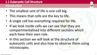
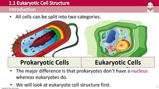
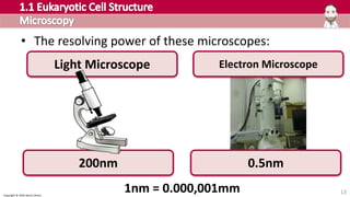
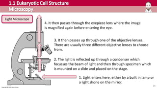
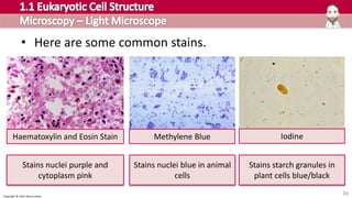
MAKING SENSE OF
Icons CC – The Pink Group
CopyrightŠ2017HenryExham
Copyright Š 2016 Henry Exham
2
• The smallest unit of life is one cell big.
• This means that cells are the key to life.
• A single cell has everything required for life.
• If we look inside cells we can see that they are
compartmentalised into different sections which
each have their own role.
• In this section we will look at the structure of
eukaryotic cells and also how to observe them using
microscopes.
Copyright Š 2016 Henry Exham
3
• All cells can be split into two categories.
Prokaryotic Cells Eukaryotic Cells
• The major difference is that prokaryotes don’t have a nucleus
whereas eukaryotes do.
• We will look at eukaryote cell structure first.
MAKING SENSE OFMAKING SENSE OF
Microscopy
Cell membrane structure
Transport across membranes
Organelles
4
Copyright Š 2016 Henry Exham
• In 1665 biology changed forever because the first
microscope was evented.
• It opened up a whole new and exciting microscopic
world which allowed scientists to study cells and very
small organisms.
• Ever since then the microscope has developed and
today it is a sophisticated piece of technology.
• You will learn about two types in this section of the
course: light microscopes and electron microscopes.
5
Copyright Š 2016 Henry Exham
a) Discuss the relative advantages of light and electron
microscopes.
b) Explain and distinguish between resolution and
magnification with reference to light microscopy
and electron microscopy.
Copyright Š 2016 Henry Exham
• In 1665 a man called Robert
Hooke built this.
• It was the first microscope.
• He looked at many things down
his microscope and drew them
in great detail.
• For example this amazing flea!
7
Copyright Š 2016 Henry Exham
• Hooke also looked at some cork
under his microscope and saw
this structure.
• He thought the compartments
looked like the small cells that
monks lived in.
• Therefore he called them cells
and that is where the term
came from.
8
Copyright Š 2016 Henry Exham
• After plenty more
research it was finally
realised that cells were
the basic unit of life.
• Schleiden and Schwann
coined the cell theory in
1839 which stated that
all living organisms are
made of one or more
cells and the cell is the
most basic unit of life.
9
Copyright Š 2016 Henry Exham
10
• Microscopes are now much
more advanced and a huge
amount has been discovered
about how cells work.
• There are two types of
microscope that you should
know about.
• They have various pros and
cons that we will review.
Light
Microscope
Electron
Microscope
Copyright Š 2016 Henry Exham
11
• The microscopes can magnify the image by this many
times.
Light Microscope Electron Microscope
X1500 X500,000
Copyright Š 2016 Henry Exham
12
• However, it is also about how
much detail they can show.
• This is measured by the
resolving power or resolution
of the microscope.
• The resolution tells you the
limit that two objects are seen
as two objects, any closer and
it would appear as one.
• This is a measure of detail. Low resolution
High resolution
Copyright Š 2016 Henry Exham
13
• The resolving power of these microscopes:
Light Microscope Electron Microscope
200nm 0.5nm
1nm = 0.000,001mm
Copyright Š 2016 Henry Exham
14
1. Light enters here, either by a built in lamp or
a light shone on the mirror.
2. The light is reflected up through a condenser which
focusses the beam of light and then through specimen which
is mounted on a slide and placed on the stage.
3. It then passes up through one of the objective lenses.
There are usually three different objective lenses to choose
from.
4. It then passes through the eyepiece lens where the image
is magnified again before entering the eye.
Light Microscope
Copyright Š 2016 Henry Exham
• To work out the total magnification you just multiply the
objective lens with the eyepiece lens.
• In order to prepare a slide for viewing on a light
microscope you need to make sure the sample is thin
enough for light to pass through.
• Most biological matter, especially when cut in a thin slice,
is transparent so usually a stain is added to highlight
certain structures.
15
Eyepiece lens magnification x Objective lens magnification = Total magnification
x10 x x40 = x400
Copyright Š 2016 Henry Exham
16
• Here are some common stains.
Haematoxylin and Eosin Stain Methylene Blue Iodine
Stains nuclei purple and
cytoplasm pink
Stains nuclei blue in animal
cells
Stains starch granules in
plant cells blue/black
Copyright Š 2016 Henry Exham
17
• Good
– Cheap, accessible and portable
– Can look at live cells with little
preparation
– For example blood samples and
sperm
• Bad
– Limited in terms of magnification
(X1500) and resolution (200nm).
Copyright Š 2016 Henry Exham
18
• The electron microscope works in a similar principle to
the light microscope, but a beam of electrons is used
instead of a beam of light.
• The electron beam has a much smaller wavelength so
can be magnified much more and has a higher
resolution.
• The beam is focused by magnets not lenses.
• There are two types the transmission electron
microscope (TEM) and the scanning electron microscope
(SEM).
Electron Microscope
Copyright Š 2016 Henry Exham
To download the whole
presentation visit
www.MrExham.com
This PowerPoint is protected under copyright.
It is designed for educational use. Either personal study or to be
presented to a class. It may be edited or duplicated for these purposes
only.
It must not be shared or distributed online in any format.
Some images used are under a separate creative commons license, these
are clearly marked.
Copyright Š 2016 Henry Exham
Brought to you by MrExham.com

Cambridge Pre-U Biology - 1.1 Eukaryotic Cell Structure

  • 1.
    MAKING SENSE OF IconsCC – The Pink Group Copyright©2017HenryExham
  • 2.
    Copyright © 2016Henry Exham 2 • The smallest unit of life is one cell big. • This means that cells are the key to life. • A single cell has everything required for life. • If we look inside cells we can see that they are compartmentalised into different sections which each have their own role. • In this section we will look at the structure of eukaryotic cells and also how to observe them using microscopes.
  • 3.
    Copyright © 2016Henry Exham 3 • All cells can be split into two categories. Prokaryotic Cells Eukaryotic Cells • The major difference is that prokaryotes don’t have a nucleus whereas eukaryotes do. • We will look at eukaryote cell structure first.
  • 4.
    MAKING SENSE OFMAKINGSENSE OF Microscopy Cell membrane structure Transport across membranes Organelles 4
  • 5.
    Copyright © 2016Henry Exham • In 1665 biology changed forever because the first microscope was evented. • It opened up a whole new and exciting microscopic world which allowed scientists to study cells and very small organisms. • Ever since then the microscope has developed and today it is a sophisticated piece of technology. • You will learn about two types in this section of the course: light microscopes and electron microscopes. 5
  • 6.
    Copyright Š 2016Henry Exham a) Discuss the relative advantages of light and electron microscopes. b) Explain and distinguish between resolution and magnification with reference to light microscopy and electron microscopy.
  • 7.
    Copyright © 2016Henry Exham • In 1665 a man called Robert Hooke built this. • It was the first microscope. • He looked at many things down his microscope and drew them in great detail. • For example this amazing flea! 7
  • 8.
    Copyright © 2016Henry Exham • Hooke also looked at some cork under his microscope and saw this structure. • He thought the compartments looked like the small cells that monks lived in. • Therefore he called them cells and that is where the term came from. 8
  • 9.
    Copyright © 2016Henry Exham • After plenty more research it was finally realised that cells were the basic unit of life. • Schleiden and Schwann coined the cell theory in 1839 which stated that all living organisms are made of one or more cells and the cell is the most basic unit of life. 9
  • 10.
    Copyright © 2016Henry Exham 10 • Microscopes are now much more advanced and a huge amount has been discovered about how cells work. • There are two types of microscope that you should know about. • They have various pros and cons that we will review. Light Microscope Electron Microscope
  • 11.
    Copyright © 2016Henry Exham 11 • The microscopes can magnify the image by this many times. Light Microscope Electron Microscope X1500 X500,000
  • 12.
    Copyright © 2016Henry Exham 12 • However, it is also about how much detail they can show. • This is measured by the resolving power or resolution of the microscope. • The resolution tells you the limit that two objects are seen as two objects, any closer and it would appear as one. • This is a measure of detail. Low resolution High resolution
  • 13.
    Copyright © 2016Henry Exham 13 • The resolving power of these microscopes: Light Microscope Electron Microscope 200nm 0.5nm 1nm = 0.000,001mm
  • 14.
    Copyright Š 2016Henry Exham 14 1. Light enters here, either by a built in lamp or a light shone on the mirror. 2. The light is reflected up through a condenser which focusses the beam of light and then through specimen which is mounted on a slide and placed on the stage. 3. It then passes up through one of the objective lenses. There are usually three different objective lenses to choose from. 4. It then passes through the eyepiece lens where the image is magnified again before entering the eye. Light Microscope
  • 15.
    Copyright © 2016Henry Exham • To work out the total magnification you just multiply the objective lens with the eyepiece lens. • In order to prepare a slide for viewing on a light microscope you need to make sure the sample is thin enough for light to pass through. • Most biological matter, especially when cut in a thin slice, is transparent so usually a stain is added to highlight certain structures. 15 Eyepiece lens magnification x Objective lens magnification = Total magnification x10 x x40 = x400
  • 16.
    Copyright © 2016Henry Exham 16 • Here are some common stains. Haematoxylin and Eosin Stain Methylene Blue Iodine Stains nuclei purple and cytoplasm pink Stains nuclei blue in animal cells Stains starch granules in plant cells blue/black
  • 17.
    Copyright © 2016Henry Exham 17 • Good – Cheap, accessible and portable – Can look at live cells with little preparation – For example blood samples and sperm • Bad – Limited in terms of magnification (X1500) and resolution (200nm).
  • 18.
    Copyright © 2016Henry Exham 18 • The electron microscope works in a similar principle to the light microscope, but a beam of electrons is used instead of a beam of light. • The electron beam has a much smaller wavelength so can be magnified much more and has a higher resolution. • The beam is focused by magnets not lenses. • There are two types the transmission electron microscope (TEM) and the scanning electron microscope (SEM). Electron Microscope
  • 19.
    Copyright Š 2016Henry Exham To download the whole presentation visit www.MrExham.com
  • 20.
    This PowerPoint isprotected under copyright. It is designed for educational use. Either personal study or to be presented to a class. It may be edited or duplicated for these purposes only. It must not be shared or distributed online in any format. Some images used are under a separate creative commons license, these are clearly marked. Copyright Š 2016 Henry Exham Brought to you by MrExham.com