Downloaded 343 times

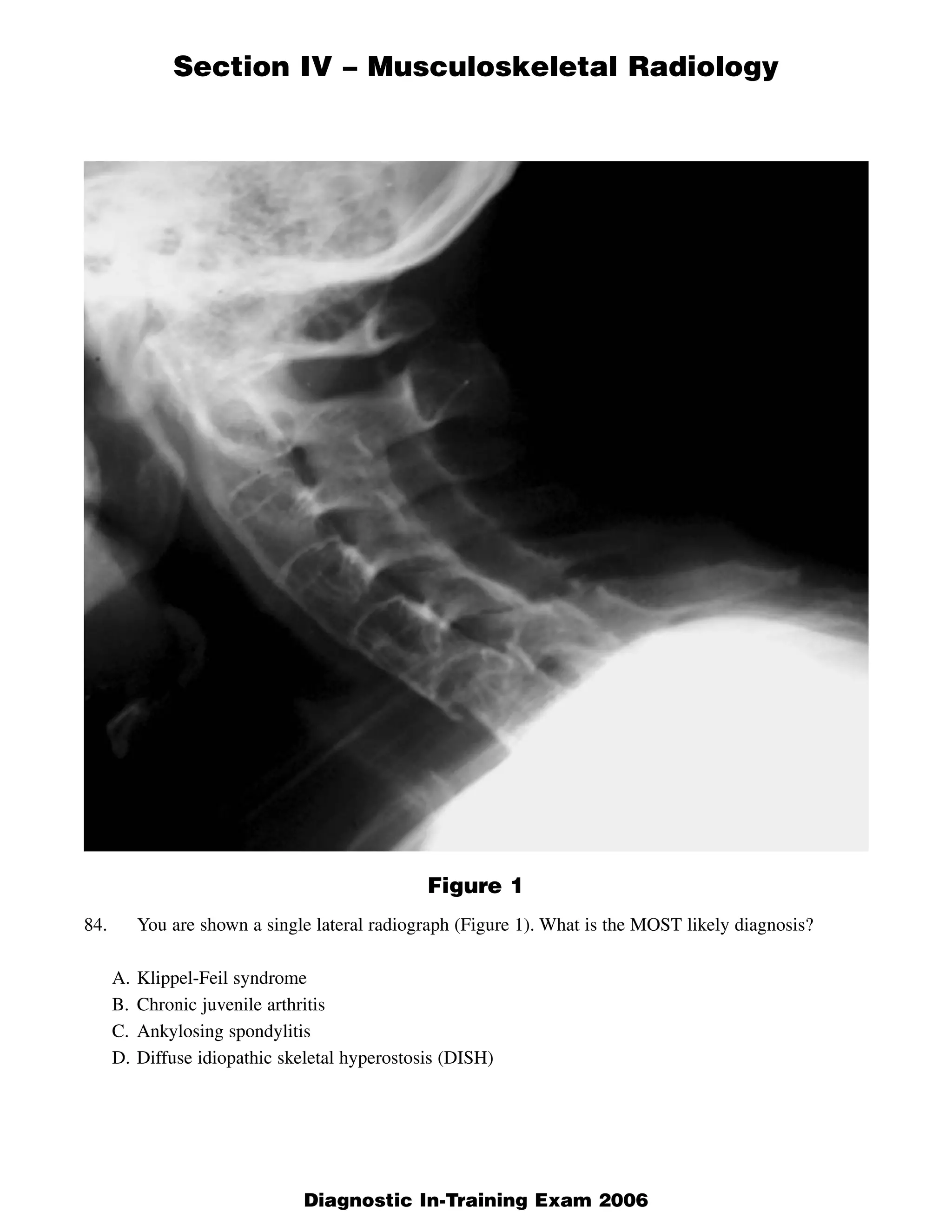
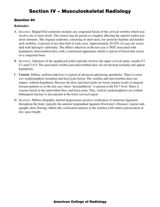
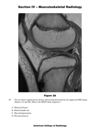
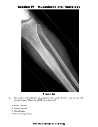
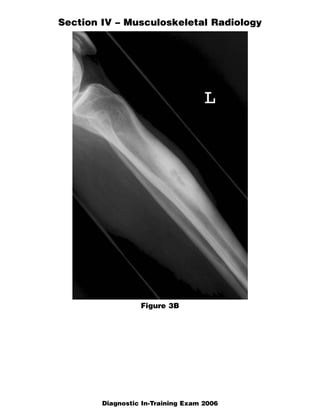
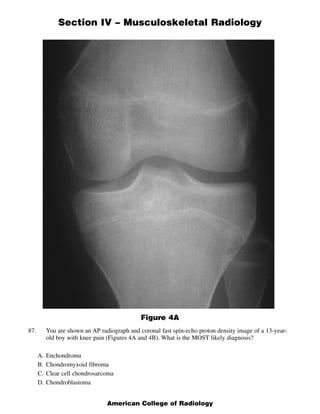
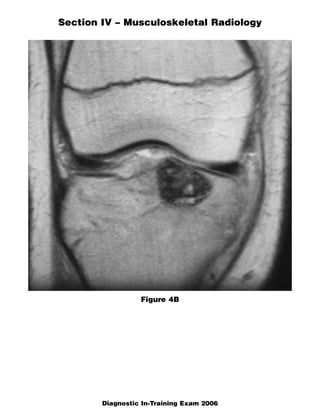

This document contains a radiology case study with 4 images (Figures 1-4) and accompanying questions. Figure 1 shows a lateral cervical spine x-ray. The diagnosis is ankylosing spondylitis based on diffuse bony ankylosis throughout the cervical spine. Figure 2 shows MRI images of the knee with a diagnosis of discoid meniscus due to excessive meniscal tissue. Figure 3 shows x-rays of the leg with a diagnosis of Brodie's abscess, seen as an elongated lytic lesion in the tibia. Figure 4 shows knee images of a 13-year-old boy with a diagnosis of chondroblastoma, seen as a well-defined lesion in the proximal tib


![ONFH[AVN HIP] -TRIPLE REGIME -A NOVAL SURGICAL CONCEPT .pptx](https://cdn.slidesharecdn.com/ss_thumbnails/onfhavnhip2026koaconcalicutdrgokuldevdrmashraf-260210064517-213ec005-thumbnail.jpg?width=640&height=640&fit=bounds)


![CTEV [ clubfoot] DR ARUN LAL ,DR MOHAMED ASHRAF travancore medical college k...](https://cdn.slidesharecdn.com/ss_thumbnails/ctevclubfootdrarunlaldrmohamedashraftravancoremedicalcollegekollamkeralaindia-260208063247-18fc466c-thumbnail.jpg?width=640&height=640&fit=bounds)


