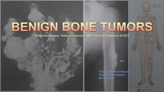
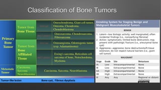
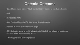
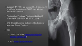
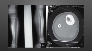
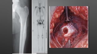
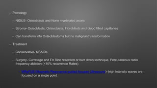
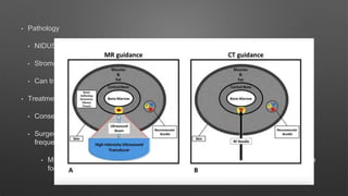
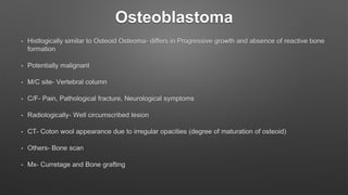
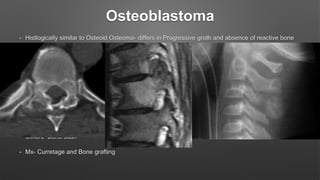
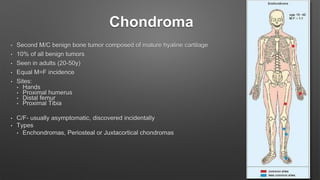
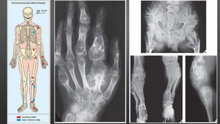
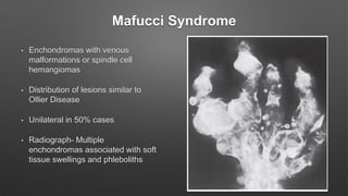
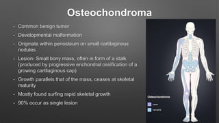
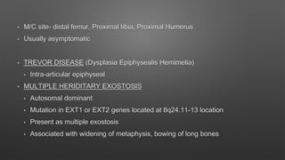
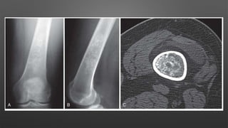
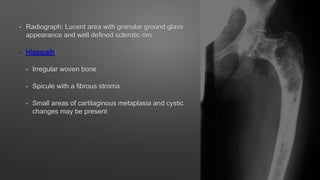
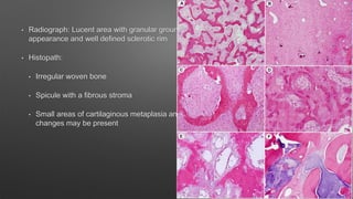
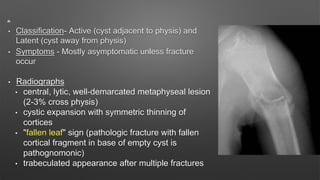
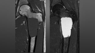
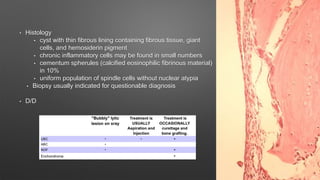
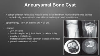
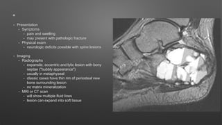
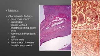
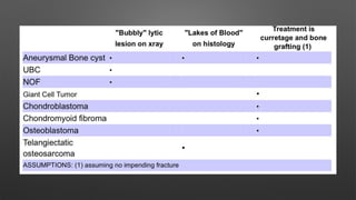
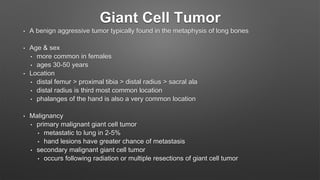
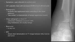
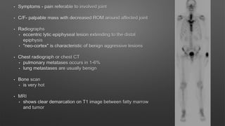
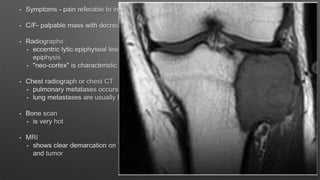
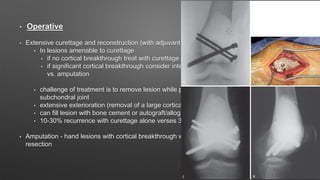
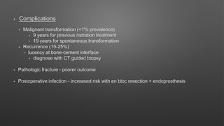
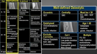
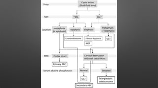
Surg Lt Cdr Anup Maurya
Resident Orthopedics
BH Lucknow
Introduction
• Usually do not behave aggressively
• Have never/ rarely been shown to metastatize
• Either asymptomatic or minimally symptomatic except when presenting with
pathological fracture
Classification of Bone Tumors
Benign Tumors
Bone forming
Osteoid Osteoma
Bone Island
Cartilage Forming
Chondroma
Osteochondroma
Fibrous Lesions
Non Ossifiying Fibroma. Cortical Desmond
Benign Fibrou Histocytoma. Fibrous Dysplasia
Osteofibrous Dysplasia Desmoplastic fibroma
Cystic Lesions
UnicameralBone Cyst. Intraosseous Ganglion
Aneurysmal Bone Cyst. Epidermis Cyst
Fatty Tumors Lipoma
Vascular Tumors Hemangioma
Osteoid Osteoma
• Osteoblastic mass called NIDUS surrounded by a zone of reactive sclerosis
• M>F
• 2nd decade of life
• Site: Proximal femur (M/C), tibia, spine (Post elements)
• No seen in bones of membranous origin
• C/F- Dull pain, worse at night, relieved with NSAIDS, not related to position or
function,, often aggravated by alcohol
• Pain aggravated by local pressure
• Suspect- Pt <30y, c/o constant back pain, spine
is stiff and scoliosis and SLRT- +ve with no
neural compression
• Radiological Findings- Radiolucent nidus (1-
1.5cm) with reactive sclerosis in cortex
• D/D- Osteoblastoma, Osteomyelitis, Brodie’s
abscess, Stress Fracture
• ∆sis-
• Tc99 bone scan- Headlight in fog and
Double Density Sign
• CT- Bulls Eye Appearance
• Pathology
• NIDUS- Osteoblasts and Nonn myelinated axons
• Stroma- Osteoblasts, Osteoclasts, Fibroblasts and blood filled capillaries
• Can transform into Osteoblastoma but no malignant transformation
• Treatment
• Conservative- NSAIDs
• Surgery- Curretage and En Bloc resection or burr down technique, Percutaneous radio
frequency ablation (<10% recurrence Rates)
• MRgFUS (Magnetic Resonance guided focuses Ultrasound)- high intensity waves are
focused on a single point
Osteoblastoma
• Histlogically similar to Osteoid Osteoma- differs in Progressive growth and absence of reactive bone
formation
• Potentially malignant
• M/C site- Vertebral column
• C/F- Pain, Pathological fracture, Neurological symptoms
• Radiologically- Well circumscribed lesion
• CT- Coton wool appearance due to irregular opacities (degree of maturation of osteoid)
• Others- Bone scan
• Mx- Curretage and Bone grafting
Osteoblastoma
• Histlogically similar to Osteoid Osteoma- differs in Progressive groth and absence of reactive bone
formation
• Potentially malignant
• M/C site- Vertebral column
• C/F- Pain, Pathological fracture, Neurological symptoms
• Radiologically- Well circumscribed lesion
• CT- Coton wool appearance due to irregular opacities (degree of maturation of osteoid)
• Others- Bone scan
• Mx- Curretage and Bone grafting
Chondroma
• Second M/C benign bone tumor composed of mature hyaline cartilage
• 10% of all benign tumors
• Seen in adults (20-50y)
• Equal M=F incidence
• Sites:
• Hands
• Proximal humerus
• Distal femur
• Proximal Tibia
• C/F- usually asymptomatic, discovered incidentally
• Types
• Enchondromas, Periosteal or Juxtacortical chondromas
• Pathology
• Result from continued growth of residual benign cartilaginous rests that are
displaced from growth plates
• Caused by failure of normal enchondral ossification
• Tumors are located in the epiphysis and adjacent parts of metaphysics and
shaft
• Deformities
• Shortening (lack of physical growth)
• Broadening
• Bowing
• Radiographic Appearnce
• Benign appearing tumors
• Intralesoinal calcifications- Stipled/ Pop corn/ Punctate
• In small bones- considerable erosion and expansion of the cortex
• Juxta cortical enchondromas are usually small (<3cm)
• Underlying cortex appears sclerotic and edges appear to be buttressed
by ring of cortical bone
• In proximal sites- deep endosteal erosion (>2/3rd of cortex) may indicate
osteochondroma
Enchondromatosis (Olliers Disease)
• Multiple enchondromas in metaphysis and diaphysis
• C/F: Knobby swelling of digits and gross disparity of
limb lengths
• Disease has strong preference for one side of body
• No hereditary or familial tendency
• Pathogeneses- Formation of ectopic nests and
failure of Chondrocytes and growth plate to mature
• Conventional radiography usually sufficient
Mafucci Syndrome
• Enchondromas with venous
malformations or spindle cell
hemangiomas
• Distribution of lesions similar to
Ollier Disease
• Unilateral in 50% cases
• Radiograph- Multiple
enchondromas associated with soft
tissue swellings and phleboliths
Osteochondroma
• Common benign tumor
• Developmental malformation
• Originate within periosteum on small cartilaginous
nodules
• Lesion- Small bony mass, often in form of a stalk
(produced by progressive enchondral ossification of a
growing cartilaginous cap)
• Growth parallels that of the mass, ceases at skeletal
maturity
• Mostly found surfing rapid skeletal growth
• 90% occur as single lesion
• M/C site- distal femur, Proximal tibia, Proximal Humerus
• Usually asymptomatic
• TREVOR DISEASE (Dysplasia Epiphysealis Hemimelia)
• Intra-articular epiphyseal
• MULTIPLE HERIDITARY EXOSTOSIS
• Autosomal dominant
• Mutation in EXT1 or EXT2 genes located at 8q24.11-13 location
• Present as multiple exostosis
• Associated with widening of metaphysis, bowing of long bones
• C/F- Palpable mass
• M>F
• Osteochondroma are of two types - Pedunculated and
Sessile
• Pedunculated - definitive stalk, directed away from the physis
of origin, continuous with normal surrounding bone
• Lesion is often covered by a cartilaginous cap- irregular
• Few mm up to 2cm thick
• Malignant change- rare (suspected when previously
quiescent lesion grows rapidly)
• Rx- En Bloc Resection if symptomatic
Fibrous Dysplasia
• Developmental anomaly of bone formation
• Hallmark- Replacement of normal bone and marrow by fibrous tissue and small woven
spicules of bone
• Can occur anywhere in bone
• McCune Albright Syndrome
• Polyostotic fibrous Dysplasia, Cutaneous pigmentation and endocrine anomalies
• Mazabraud Syndrome
• Polyostotic fibrous Dysplasia And intramuscular xanthomas
• Radiograph: Lucent area with granular ground glass
appearance and well defined sclerotic rim
• Histopath:
• Irregular woven bone
• Spicule with a fibrous stroma
• Small areas of cartilaginous metaplasia and cystic
changes may be present
• Radiograph: Lucent area with granular ground glass
appearance and well defined sclerotic rim
• Histopath:
• Irregular woven bone
• Spicule with a fibrous stroma
• Small areas of cartilaginous metaplasia and cystic
changes may be present
• Rx- Surgery is indicated in case of deformity or pathological fractures
• Intramedullary fixation in cases of impending fractures
• Cortical grafts preferred to cancellous grafts or bone graft sucstitutes because
of high rates of recurrence and slow resorption of cortical bone
• Bisphosphonates have been shown to be of some benefit
Cystic Bone Lesions
• Unicameral Bone Cyst/ Simple Bone Cysts
• Common lesions of childhood
• 85% in first 2 decades of life
• 2:1 male preponderance
• Any bone in the extremity can be affected
• M/C in proximal humerus and femur
• Prognosis- as the patient approaches skeletal maturity, UBC will often
decrease in size and may heal
• Fracture healing does not lead to cyst resolution
• Classification- Active (cyst adjacent to physis) and
Latent (cyst away from physis)
• Symptoms - Mostly asymptomatic unless fracture
occur
• Radiographs
• central, lytic, well-demarcated metaphyseal lesion
(2-3% cross physis)
• cystic expansion with symmetric thinning of
cortices
• "fallen leaf" sign (pathologic fracture with fallen
cortical fragment in base of empty cyst is
pathognomonic)
• trabeculated appearance after multiple fractures
• MRI
• very dark on T1
• very bright on T2
• gadolinium shows classic rim enhancement of a cystic lesion
• Bone scan
• is variable, but usually warm
• Histology
• cyst with thin fibrous lining containing fibrous tissue, giant
cells, and hemosiderin pigment
• chronic inflammatory cells may be found in small numbers
• cementum spherules (calcified eosinophilic fibrinous material)
in 10%
• uniform population of spindle cells without nuclear atypia
• Biopsy usually indicated for questionable diagnosis
• D/D
• Managemant
• Nonoperative
• immobilization alone
• aspiration/methylprednisolone acetate injection
• usually requires several injections, especially in very young children
• bone marrow injections have recently been reported to be effective
• Operative
• curettage and bone grafting +/- internal fixation based on tumor location
• symptomatic latent cysts that have not responded to steroid injections
• latent cysts in the proximal femur - at risk for fracture and osteonecrosis
• avoid in active lesions as communication with physis may lead to growth arrest
Aneurysmal Bone Cyst
• A benign and non-neoplastic reactive bone lesion filled with multiple blood-filled cavities
• can be locally destructive to normal bone and may extend to soft tissue
• Epidemiology - 75% of patients are < 20 yrs.
• Location
• 25% in spine
• 20% in long bones (distal femur, proximal tibia)
• usually in metaphysis
• metatarsal is the most common location in the foot
• posterior elements of pelvis
• Pathophysiology - primary and secondary forms
• primary ABC:
• driven by upregulation of the ubiquitin-specific protease USP6 (Tre2) gene on 17p13 when combined by
translocation with a promoter pairing
• secondary ABC:
• not considered a neoplasm because no known translocation has been identified
• Associated conditions
• associated with other tumors 30% of time
• giant cell tumor
• chondroblastoma
• fibrous dysplasia
• chondromyxoid fibroma
• NOF
• Prognosis
• good
• Presentation
• Symptoms
• pain and swelling
• may present with pathologic fracture
• Physical exam
• neurologic deficits possible with spine lesions
• Imaging
• Radiographs
• expansile, eccentric and lytic lesion with bony
septae ("bubbly appearance")
• usually in metaphyseal
• classic cases have thin rim of periosteal new
bone surrounding lesion
• no matrix mineralization
• MRI or CT scan
• will show multiple fluid lines
• lesion can expand into soft tissue
• Histology
• Characteristic findings
• cavernous space
• blood-filled
spaces without
endothelial lining cavity
lining
• numerous benign giant
cells
• spindle cells
• thin strands of woven
(new) bone present
• Treatment
• Nonoperative
• nonoperative fracture management - indicated until fracture has healed
• Operative
• aggressive curettage (+/- adjuvant) and bone grafting - symptomatic ABC without acute fracture
• possible adjuvants
• phenol
• argon beam
• liquid nitrogen
• outcomes
• local recurrence in up to 25% and more common in children with open physes
Giant Cell Tumor
• A benign aggressive tumor typically found in the metaphysis of long bones
• Age & sex
• more common in females
• ages 30-50 years
• Location
• distal femur > proximal tibia > distal radius > sacral ala
• distal radius is third most common location
• phalanges of the hand is also a very common location
• Malignancy
• primary malignant giant cell tumor
• metastatic to lung in 2-5%
• hand lesions have greater chance of metastasis
• secondary malignant giant cell tumor
• occurs following radiation or multiple resections of giant cell tumor
• Histology
• Type I cell
• mononucleur stromal cell that resembles interstitial fibroblasts
• has features of mesenchymal stem cells
• Type II cell
• from monocyte/macrophage family recruited from peripheral blood
• precursors of giant cells
• Type III cell
• numerous giant cells are the hallmark of this lesion
• multiple nuclei (up to 50 per cell)
• similar characteristics as osteoclasts and resorb bone
• Molecular biology
• Type II and III cells have IGF-I and IGF-II activity
• RANK pathway is important - denosumab acts on this pathway
• Nonoperative
• Radiation alone
• only indicated for inoperable or multiply recurrent lesions
• leads to 15% malignant transformation
• Medical management
• used to augment or replace surgical management
• medications
• bisphosphonates - osteclast inhibitors which may decrease the size of the tumors
• denosumab
• monoclonal antibody against RANK-ligand
• recent clinical trials suggest denosumab can decrease the size of tumor
• 90% tumor necrosis
• shows dramatic sclerosis and reconstitution of cortical bone after treatment
• Operative
• Extensive curettage and reconstruction (with adjuvant treatment)
• In lesions amenable to curettage
• if no cortical breakthrough treat with curettage and cementing
• if significant cortical breakthrough consider intercalary resection (with free fibular graft)
vs. amputation
• challenge of treatment is to remove lesion while preserving joint and providing support to
subchondral joint
• extensive exterioration (removal of a large cortical window over the lesion) is required
• can fill lesion with bone cement or autograft/allograft bone
• 10-30% recurrence with curettage alone verses 3% with adjuvant treatment
• Amputation - hand lesions with cortical breakthrough who are not amendable to intercalary
resection
• Complications
• Malignant transformation (<1% prevalence)
• 9 years for previous radiation treatment
• 19 years for spontaneous transformation
• Recurrence (15-25%)
• lucency at bone-cement interface
• diagnose with CT guided biopsy
• Pathologic fracture - poorer outcome
• Postoperative infection - increased risk with en bloc resection + endoprosthesis
Benign bone tumors
Benign bone tumors

Benign bone tumors

  • 1.
    Surg Lt CdrAnup Maurya Resident Orthopedics BH Lucknow
  • 2.
    Introduction • Usually donot behave aggressively • Have never/ rarely been shown to metastatize • Either asymptomatic or minimally symptomatic except when presenting with pathological fracture
  • 3.
  • 5.
    Benign Tumors Bone forming OsteoidOsteoma Bone Island Cartilage Forming Chondroma Osteochondroma Fibrous Lesions Non Ossifiying Fibroma. Cortical Desmond Benign Fibrou Histocytoma. Fibrous Dysplasia Osteofibrous Dysplasia Desmoplastic fibroma Cystic Lesions UnicameralBone Cyst. Intraosseous Ganglion Aneurysmal Bone Cyst. Epidermis Cyst Fatty Tumors Lipoma Vascular Tumors Hemangioma
  • 6.
    Osteoid Osteoma • Osteoblasticmass called NIDUS surrounded by a zone of reactive sclerosis • M>F • 2nd decade of life • Site: Proximal femur (M/C), tibia, spine (Post elements) • No seen in bones of membranous origin • C/F- Dull pain, worse at night, relieved with NSAIDS, not related to position or function,, often aggravated by alcohol • Pain aggravated by local pressure
  • 7.
    • Suspect- Pt<30y, c/o constant back pain, spine is stiff and scoliosis and SLRT- +ve with no neural compression • Radiological Findings- Radiolucent nidus (1- 1.5cm) with reactive sclerosis in cortex • D/D- Osteoblastoma, Osteomyelitis, Brodie’s abscess, Stress Fracture • ∆sis- • Tc99 bone scan- Headlight in fog and Double Density Sign • CT- Bulls Eye Appearance
  • 11.
    • Pathology • NIDUS-Osteoblasts and Nonn myelinated axons • Stroma- Osteoblasts, Osteoclasts, Fibroblasts and blood filled capillaries • Can transform into Osteoblastoma but no malignant transformation • Treatment • Conservative- NSAIDs • Surgery- Curretage and En Bloc resection or burr down technique, Percutaneous radio frequency ablation (<10% recurrence Rates) • MRgFUS (Magnetic Resonance guided focuses Ultrasound)- high intensity waves are focused on a single point
  • 12.
    • Pathology • NIDUS-Osteoblasts and Nonn myelinated axons • Stroma- Osteoblasts, Osteoclasts, Fibroblasts and blood filled capillaries • Can transform into Osteoblastoma but no malignant transformation • Treatment • Conservative- NSAIDs • Surgery- Curretage and En Bloc resection or burr down technique, Percutaneous radio frequency ablation (<10% recurrence Rates) • MRgFUS (Magnetic Resonance guided focuses Ultrasound)- high intensity waves are focused on a single point
  • 13.
    Osteoblastoma • Histlogically similarto Osteoid Osteoma- differs in Progressive growth and absence of reactive bone formation • Potentially malignant • M/C site- Vertebral column • C/F- Pain, Pathological fracture, Neurological symptoms • Radiologically- Well circumscribed lesion • CT- Coton wool appearance due to irregular opacities (degree of maturation of osteoid) • Others- Bone scan • Mx- Curretage and Bone grafting
  • 14.
    Osteoblastoma • Histlogically similarto Osteoid Osteoma- differs in Progressive groth and absence of reactive bone formation • Potentially malignant • M/C site- Vertebral column • C/F- Pain, Pathological fracture, Neurological symptoms • Radiologically- Well circumscribed lesion • CT- Coton wool appearance due to irregular opacities (degree of maturation of osteoid) • Others- Bone scan • Mx- Curretage and Bone grafting
  • 15.
    Chondroma • Second M/Cbenign bone tumor composed of mature hyaline cartilage • 10% of all benign tumors • Seen in adults (20-50y) • Equal M=F incidence • Sites: • Hands • Proximal humerus • Distal femur • Proximal Tibia • C/F- usually asymptomatic, discovered incidentally • Types • Enchondromas, Periosteal or Juxtacortical chondromas
  • 16.
    • Pathology • Resultfrom continued growth of residual benign cartilaginous rests that are displaced from growth plates • Caused by failure of normal enchondral ossification • Tumors are located in the epiphysis and adjacent parts of metaphysics and shaft • Deformities • Shortening (lack of physical growth) • Broadening • Bowing
  • 17.
    • Radiographic Appearnce •Benign appearing tumors • Intralesoinal calcifications- Stipled/ Pop corn/ Punctate • In small bones- considerable erosion and expansion of the cortex • Juxta cortical enchondromas are usually small (<3cm) • Underlying cortex appears sclerotic and edges appear to be buttressed by ring of cortical bone • In proximal sites- deep endosteal erosion (>2/3rd of cortex) may indicate osteochondroma
  • 19.
    Enchondromatosis (Olliers Disease) •Multiple enchondromas in metaphysis and diaphysis • C/F: Knobby swelling of digits and gross disparity of limb lengths • Disease has strong preference for one side of body • No hereditary or familial tendency • Pathogeneses- Formation of ectopic nests and failure of Chondrocytes and growth plate to mature • Conventional radiography usually sufficient
  • 21.
    Mafucci Syndrome • Enchondromaswith venous malformations or spindle cell hemangiomas • Distribution of lesions similar to Ollier Disease • Unilateral in 50% cases • Radiograph- Multiple enchondromas associated with soft tissue swellings and phleboliths
  • 22.
    Osteochondroma • Common benigntumor • Developmental malformation • Originate within periosteum on small cartilaginous nodules • Lesion- Small bony mass, often in form of a stalk (produced by progressive enchondral ossification of a growing cartilaginous cap) • Growth parallels that of the mass, ceases at skeletal maturity • Mostly found surfing rapid skeletal growth • 90% occur as single lesion
  • 23.
    • M/C site-distal femur, Proximal tibia, Proximal Humerus • Usually asymptomatic • TREVOR DISEASE (Dysplasia Epiphysealis Hemimelia) • Intra-articular epiphyseal • MULTIPLE HERIDITARY EXOSTOSIS • Autosomal dominant • Mutation in EXT1 or EXT2 genes located at 8q24.11-13 location • Present as multiple exostosis • Associated with widening of metaphysis, bowing of long bones
  • 24.
    • M/C site-distal femur, Proximal tibia, Proximal Humerus • Usually asymptomatic • TREVOR DISEASE (Dysplasia Epiphysealis Hemimelia) • Intra-articular epiphyseal • MULTIPLE HERIDITARY EXOSTOSIS • Autosomal dominant • Mutation in EXT1 or EXT2 genes located at 8q24.11-13 location • Present as multiple exostosis • Associated with widening of metaphysis, bowing of long bones
  • 25.
    • C/F- Palpablemass • M>F • Osteochondroma are of two types - Pedunculated and Sessile • Pedunculated - definitive stalk, directed away from the physis of origin, continuous with normal surrounding bone • Lesion is often covered by a cartilaginous cap- irregular • Few mm up to 2cm thick • Malignant change- rare (suspected when previously quiescent lesion grows rapidly) • Rx- En Bloc Resection if symptomatic
  • 27.
    Fibrous Dysplasia • Developmentalanomaly of bone formation • Hallmark- Replacement of normal bone and marrow by fibrous tissue and small woven spicules of bone • Can occur anywhere in bone • McCune Albright Syndrome • Polyostotic fibrous Dysplasia, Cutaneous pigmentation and endocrine anomalies • Mazabraud Syndrome • Polyostotic fibrous Dysplasia And intramuscular xanthomas
  • 28.
    • Radiograph: Lucentarea with granular ground glass appearance and well defined sclerotic rim • Histopath: • Irregular woven bone • Spicule with a fibrous stroma • Small areas of cartilaginous metaplasia and cystic changes may be present
  • 29.
    • Radiograph: Lucentarea with granular ground glass appearance and well defined sclerotic rim • Histopath: • Irregular woven bone • Spicule with a fibrous stroma • Small areas of cartilaginous metaplasia and cystic changes may be present
  • 30.
    • Rx- Surgeryis indicated in case of deformity or pathological fractures • Intramedullary fixation in cases of impending fractures • Cortical grafts preferred to cancellous grafts or bone graft sucstitutes because of high rates of recurrence and slow resorption of cortical bone • Bisphosphonates have been shown to be of some benefit
  • 31.
    Cystic Bone Lesions •Unicameral Bone Cyst/ Simple Bone Cysts • Common lesions of childhood • 85% in first 2 decades of life • 2:1 male preponderance • Any bone in the extremity can be affected • M/C in proximal humerus and femur • Prognosis- as the patient approaches skeletal maturity, UBC will often decrease in size and may heal • Fracture healing does not lead to cyst resolution
  • 32.
    • Classification- Active(cyst adjacent to physis) and Latent (cyst away from physis) • Symptoms - Mostly asymptomatic unless fracture occur • Radiographs • central, lytic, well-demarcated metaphyseal lesion (2-3% cross physis) • cystic expansion with symmetric thinning of cortices • "fallen leaf" sign (pathologic fracture with fallen cortical fragment in base of empty cyst is pathognomonic) • trabeculated appearance after multiple fractures
  • 33.
    • MRI • verydark on T1 • very bright on T2 • gadolinium shows classic rim enhancement of a cystic lesion • Bone scan • is variable, but usually warm
  • 35.
    • Histology • cystwith thin fibrous lining containing fibrous tissue, giant cells, and hemosiderin pigment • chronic inflammatory cells may be found in small numbers • cementum spherules (calcified eosinophilic fibrinous material) in 10% • uniform population of spindle cells without nuclear atypia • Biopsy usually indicated for questionable diagnosis • D/D
  • 36.
    • Managemant • Nonoperative •immobilization alone • aspiration/methylprednisolone acetate injection • usually requires several injections, especially in very young children • bone marrow injections have recently been reported to be effective • Operative • curettage and bone grafting +/- internal fixation based on tumor location • symptomatic latent cysts that have not responded to steroid injections • latent cysts in the proximal femur - at risk for fracture and osteonecrosis • avoid in active lesions as communication with physis may lead to growth arrest
  • 37.
    Aneurysmal Bone Cyst •A benign and non-neoplastic reactive bone lesion filled with multiple blood-filled cavities • can be locally destructive to normal bone and may extend to soft tissue • Epidemiology - 75% of patients are < 20 yrs. • Location • 25% in spine • 20% in long bones (distal femur, proximal tibia) • usually in metaphysis • metatarsal is the most common location in the foot • posterior elements of pelvis
  • 38.
    • Pathophysiology -primary and secondary forms • primary ABC: • driven by upregulation of the ubiquitin-specific protease USP6 (Tre2) gene on 17p13 when combined by translocation with a promoter pairing • secondary ABC: • not considered a neoplasm because no known translocation has been identified • Associated conditions • associated with other tumors 30% of time • giant cell tumor • chondroblastoma • fibrous dysplasia • chondromyxoid fibroma • NOF • Prognosis • good
  • 39.
    • Presentation • Symptoms •pain and swelling • may present with pathologic fracture • Physical exam • neurologic deficits possible with spine lesions • Imaging • Radiographs • expansile, eccentric and lytic lesion with bony septae ("bubbly appearance") • usually in metaphyseal • classic cases have thin rim of periosteal new bone surrounding lesion • no matrix mineralization • MRI or CT scan • will show multiple fluid lines • lesion can expand into soft tissue
  • 40.
    • Histology • Characteristicfindings • cavernous space • blood-filled spaces without endothelial lining cavity lining • numerous benign giant cells • spindle cells • thin strands of woven (new) bone present
  • 41.
    • Treatment • Nonoperative •nonoperative fracture management - indicated until fracture has healed • Operative • aggressive curettage (+/- adjuvant) and bone grafting - symptomatic ABC without acute fracture • possible adjuvants • phenol • argon beam • liquid nitrogen • outcomes • local recurrence in up to 25% and more common in children with open physes
  • 43.
    Giant Cell Tumor •A benign aggressive tumor typically found in the metaphysis of long bones • Age & sex • more common in females • ages 30-50 years • Location • distal femur > proximal tibia > distal radius > sacral ala • distal radius is third most common location • phalanges of the hand is also a very common location • Malignancy • primary malignant giant cell tumor • metastatic to lung in 2-5% • hand lesions have greater chance of metastasis • secondary malignant giant cell tumor • occurs following radiation or multiple resections of giant cell tumor
  • 47.
    • Histology • TypeI cell • mononucleur stromal cell that resembles interstitial fibroblasts • has features of mesenchymal stem cells • Type II cell • from monocyte/macrophage family recruited from peripheral blood • precursors of giant cells • Type III cell • numerous giant cells are the hallmark of this lesion • multiple nuclei (up to 50 per cell) • similar characteristics as osteoclasts and resorb bone • Molecular biology • Type II and III cells have IGF-I and IGF-II activity • RANK pathway is important - denosumab acts on this pathway
  • 48.
    • Nonoperative • Radiationalone • only indicated for inoperable or multiply recurrent lesions • leads to 15% malignant transformation • Medical management • used to augment or replace surgical management • medications • bisphosphonates - osteclast inhibitors which may decrease the size of the tumors • denosumab • monoclonal antibody against RANK-ligand • recent clinical trials suggest denosumab can decrease the size of tumor • 90% tumor necrosis • shows dramatic sclerosis and reconstitution of cortical bone after treatment
  • 49.
    • Operative • Extensivecurettage and reconstruction (with adjuvant treatment) • In lesions amenable to curettage • if no cortical breakthrough treat with curettage and cementing • if significant cortical breakthrough consider intercalary resection (with free fibular graft) vs. amputation • challenge of treatment is to remove lesion while preserving joint and providing support to subchondral joint • extensive exterioration (removal of a large cortical window over the lesion) is required • can fill lesion with bone cement or autograft/allograft bone • 10-30% recurrence with curettage alone verses 3% with adjuvant treatment • Amputation - hand lesions with cortical breakthrough who are not amendable to intercalary resection
  • 50.
    • Operative • Extensivecurettage and reconstruction (with adjuvant treatment) • In lesions amenable to curettage • if no cortical breakthrough treat with curettage and cementing • if significant cortical breakthrough consider intercalary resection (with free fibular graft) vs. amputation • challenge of treatment is to remove lesion while preserving joint and providing support to subchondral joint • extensive exterioration (removal of a large cortical window over the lesion) is required • can fill lesion with bone cement or autograft/allograft bone • 10-30% recurrence with curettage alone verses 3% with adjuvant treatment • Amputation - hand lesions with cortical breakthrough who are not amendable to intercalary resection
  • 51.
    • Complications • Malignanttransformation (<1% prevalence) • 9 years for previous radiation treatment • 19 years for spontaneous transformation • Recurrence (15-25%) • lucency at bone-cement interface • diagnose with CT guided biopsy • Pathologic fracture - poorer outcome • Postoperative infection - increased risk with en bloc resection + endoprosthesis