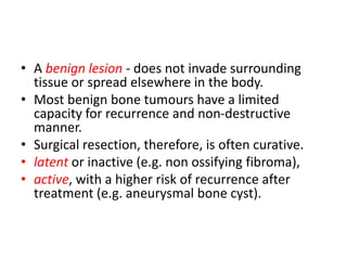
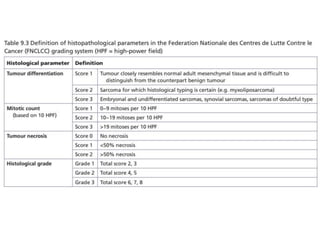
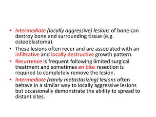
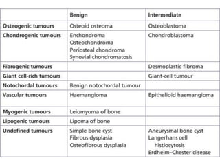
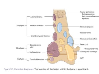
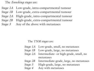
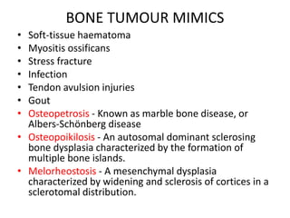
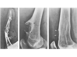
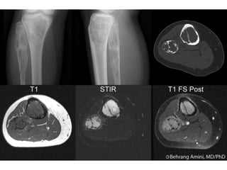
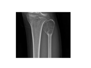
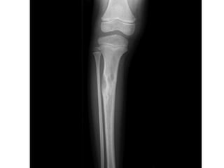
Benign bone tumors
Dr. Md. Mahbub Hasan
Resident, D-Ortho, SZMC
Tumours of bone and soft tissue are classified on
the basis of their principle cell type, and have
been classified by the World Health Organization
(WHO).
In the case of soft-tissue sarcomas, the histological
grade of the lesion is classified according to the
Federation Nationale des Centres de Lutte Contre
le Cancer (FNCLCC) system
• A benign lesion - does not invade surrounding
tissue or spread elsewhere in the body.
• Most benign bone tumours have a limited
capacity for recurrence and non-destructive
manner.
• Surgical resection, therefore, is often curative.
• latent or inactive (e.g. non ossifying fibroma),
• active, with a higher risk of recurrence after
treatment (e.g. aneurysmal bone cyst).
• Intermediate (locally aggressive) lesions of bone can
destroy bone and surrounding tissue (e.g.
osteoblastoma).
• These lesions often recur and are associated with an
infiltrative and locally destructive growth pattern.
• Recurrence is frequent following limited surgical
treatment and sometimes en bloc resection is
required to completely remove the lesion.
• Intermediate (rarely metastasizing) lesions often
behave in a similar way to locally aggressive lesions
but occasionally demonstrate the ability to spread to
distant sites.
BONE TUMOUR MIMICS
• Soft-tissue haematoma
• Myositis ossificans
• Stress fracture
• Infection
• Tendon avulsion injuries
• Gout
• Osteopetrosis - Known as marble bone disease, or
Albers-Schönberg disease
• Osteopoikilosis - An autosomal dominant sclerosing
bone dysplasia characterized by the formation of
multiple bone islands.
• Melorheostosis - A mesenchymal dysplasia
characterized by widening and sclerosis of cortices in a
sclerotomal distribution.
OSTEOID OSTEOMA
• This is a small, benign tumour formed of osteoid and woven bone
surrounded by a halo of reactive bone.
• These lesions are most common in young patients but are rare
below 5 years of age and equally rare over 30.
• They are more common in men than women.
• It is most commonly seen in the long bones, particularly the
proximal femur.
• It is rare in the trunk with the exception of the spine where it is
most often seen in the posterior arches.
• It is more often diaphyseal than metaphyseal.
• Osteoid osteomas most often present with pain, which classically is
worse at night and relieved by non-steroidal anti-inflammatories.
• When in close proximity to a joint, they can result in stiffness and
an effusion.
• In the spine, they can cause muscle spasm and scoliosis.
• Plain X-ray demonstrates an area of dense sclerosis with a
small, rounded area of osteolysis which is often obscured by
the surrounding sclerosis.
• Isotope bone scan is positive and the central nidus of the
lesion is best seen on CT scan (Figure 9.16).
• Histology demonstrates a packed mesh of thin, woven bone
with osteoblastic rimming, osteoclasts and dilated capillaries.
• The mature osteoid corresponds to the nidus, which is
surrounded by areas of dense, reactive bone with surrounding
soft tissue also showing features of inflammation.
• Without treatment, the lesion will slowly increase but over
time will regress and usually burns out over a variable period
of a number of years.
• The preferred method of treatment is by CT-guided
radiofrequency ablation, which has proven to be highly
successful in eradicating the lesion with a dramatic resolution
of symptoms.
ENCHONDROMA
• This is an intramedullary neoplasm made of well
differentiated hyaline cartilage.
• The commonest location is the tubular bones of the hand,
followed by the femur and humerus.
• Radiographically, the lesion is most commonly central with
rounded, well-defined, lobulated edges and a thin rind of
reactive sclerosis.
• It contains glandular, popcorn, ring-like opacities.
• They can reach considerable size but rarely exceed 6 cm.
• MRI demonstrates the black signal voids of internal
calcification and isotope bone scan is hot in most lesions.
• Histologically, the lesion contains lobules of cartilage
with areas of calcification.
• The chondrocytes are usually sparse with small, round,
dense nuclei.
• if there is evidence of chondroma permeating within
the marrow spaces and haversian system, this is highly
suggestive of chondrosarcoma.
• Treatment is usually not required although,
occasionally in the hand, the lesion may be removed
through curettage, particularly if there is pain or
pathological fracture.
• Serial X-rays may be helpful if there is a suspicion of a
grade 1 chondrosarcoma, as enchondromas in skeletal
maturity do not grow.
OSTEOCHONDROMA
• benign, cartilaginous neoplasm derived from an aberrant
subperiosteal nest of physeal cartilage
• common in males in adolescent years, during the final growth spurt
• most commonly occur in long bones, particularly the femur and
humerus.
• They normally present as a painless mass, bursa due to friction, or
to activity related discomfort. cause neuropathic symptoms due to
compression of a nearby nerve or may fracture producing sudden
pain.
• The lesion appears as a bony protuberance with well-defined limits,
thin outer cortex and an inner cancellous structure.
• The pathognomonic feature is that the host bone flares from the
cortex into the osteochondroma
• Some are pedunculated with a cauliflower-like summit; others have
a broad, sessile base.
• Pedunculated osteochondromas typically point away from the joint,
towards the diaphysis.
• MRI demonstrates the classic cartilaginous cap which
ranges from a few millimetres to a centimetre or more.
The cap is typically thicker in children and diminishes
with age.
• Osteochondromas tend to grow with skeletal
maturity and stop once growth stops.
• The risk of malignant transformation is rare, possibly
1%
• In asymptomatic lesions, treatment is not indicated.
• Excision may be warranted in large lesions
where local pressure effects may occur or in adults
where the risk of malignant transformation warrants
removal.
PERIOSTEAL CHONDROMA
• This is a rare, benign cartilage neoplasm occurring at the
surface of the bone.
• It is most frequently seen in children or young adults and
prefers the metaphysis of long bones, particularly the
humerus.
• It most commonly presents with pain and occasionally a
palpable lump.
• Radiographs demonstrate a superficial erosion of the bone
cortex with occasional scalloping (Figure 9.13).
• When large, the lesion demonstrates a popcorn matrix with
areas of calcification.
• Histologically, it resembles an enchondroma.
• The majority are effectively treated by curettage; though
large lesions may require more aggressive resection.
HAEMANGIOMA OF BONE
• Haemangioma are benign lesions of bone composed of
capillary blood vessels of small or large calibre.
• These are common lesions, often asymptomatic and often
identified incidentally.
• Post-mortem studies have identified them to be present in
the vertebrae of 10% of the adult population.
• Haemangiomas can be identified at any age but most
commonly present in the fifth decade.
• They are slightly more common in females than males.
• They are most commonly seen in the vertebral bodies,
followed by the craniofacial bones.
• When seen in the long bones, they most commonly affect
the metaphysis. Polyostotic disease is not uncommon.
• The majority of haemangiomas are identified incidentally. If
large, they can cause symptoms particularly in the spine,
where they may present with cord compression, pain and
neurological symptoms.
• Radiologically, they appear as a radiolucent lesion often
with coarse trabeculae.
• On MRI, they appear to contain fat, and trabeculae are
evident.
• Histology varies but typically these lesions demonstrate
thinwalled blood-filled vessels lined by a thin layer of fat.
• Treatment is often not required but, when indicated,
curettage and stabilization are often effective with a low
incidence of recurrence
SIMPLE BONE CYST
• This is a solitary, usually unilocular cystic bone cavity lined by
a fibrous membrane and filled with serous or serosanguinous
fluid.
• Males are more frequently affected and the majority occur
within the first two decades of life.
• Although they can arise at any location, the vast majority of
simple bone cysts (SBCs) occur in the proximal humerus, the
proximal femur or the proximal tibia, most commonly
affecting the metaphyseal areas close to the physis.
• In the majority of cases the lesion is asymptomatic, being
discovered incidentally.
• However, fracture through the lesion is not uncommon and
often this is the presenting feature. Occasionally, mild pain or
swelling may be present.
• X-rays demonstrate a well outlined, lytic
centrally placed, metadiaphyseal lesion
expanding and thinning the cortices
• It often abuts but does not cross the physis.
• Bone septa are often present which give the
impression of a multiloculated cyst.
• When fracture occurs, a small fragment may be
seen within the cavity, the classic ‘fallen leaf’ sign.
• MRI will demonstrate the homogeneous fluid-filled
cavity.
• Histologically, the cyst lining demonstrates
connective tissue with foci of reactive bone.
• Following fracture, there will be features of
fracture callus and new bone formation.
• Treatment is often supportive as lesions will regress
following skeletal maturity.
• Percutaneous injection of steroid has been reported
with varying success but is best reserved for ‘active’
lesions, i.e. those abutting the physis in young children.
• Curettage and bone grafting may be required in areas
at risk of fracture, and pathological fractures of the
proximal femur in particular will often require fixation
and stabilization.
• Recurrence is reported in 10–20% of cases.
• Large cysts may result in limb shortening, and avascular
necrosis of the femoral head can occur following
fracture through proximal femoral lesions.
• Spontaneous resolution following fracture has been
reported
FIBROUS DYSPLASIA
• This is a benign, medullary fibro-osseous lesion which may affect
one bone (monostotic) or a number of bones (polyostotic).
• Fibrous dysplasia can affect children and adults with equal sex and
race distribution.
• The monostotic form is considerably more frequent than the
polyostotic.
• The craniofacial bones and the femur are the most frequently
affected bones, but any bone can be affected by the monostotic
form.
• In the polyostotic form, the pelvis, femur and tibia are commonly
involved.
• Multiple sites of fibrous dysplasia may be seen in the same bone,
particularly in the monostotic form.
• The monostotic form is often discovered incidentally and is largely
asymptomatic.
• Not infrequently, pain and fracture are the presenting features.
• Bony expansion in superficial bones, deformity and
growth disturbance with deformity and lower-limb
length discrepancy can also occur.
• Fibrous dysplasia can be associated with
endocrinopathies and café-au-lait spots (coast of
Maine) in McCune–Albright syndrome, and with
intramuscular myxomas in Mazabraud syndrome.
• X-rays often demonstrate a non-aggressive, well-
circumscribed lesion with a characteristic ground-glass
matrix
• More mature lesions may show cyst formation and
secondary changes.
• The characteristic deformity resulting from proximal femoral
fibrous dysplasia is the shepherd’s crook deformity.
• There is seldom a soft-tissue component or periosteal
reaction, except in the case of fracture.
• MRI reveals a homogeneous low signal lesion on T1.
• There may be associated cystic degeneration within the
lesion.
• Histology demonstrates a characteristic mixture of benign
proliferating fibroblastic cells within islands of woven bone
which characteristically appear in a ‘Chinese letter’ formation.
• Benign giant cells are not infrequently seen and islands of
cartilage may dominate the appearance.
• Secondary ABC features are often seen.
• Treatment is often not needed.
• Correction of deformities is sometimes required but
should be undertaken with caution.
• Occasionally, in the case of fracture or impending
fracture, intralesional curettage and bone grafting may
be required and, for very large lesions, augmentation
with internal fixation may be required.
• However, recurrence following treatment is not
uncommon.
• Malignant transformation, to fibrosarcoma of bone, is
rare but more frequently seen in McCune–Albright
syndrome.
OSTEOFIBROUS DYSPLASIA
• Osteofibrous dysplasia (OFD) is a benign, non-osseous
lesion of bone typically seen in the anterior cortex of
the tibia, particularly in children or teenagers.
• It is commonly seen in children in the first decade and
can on rare occasions be polyostotic.
• It is almost exclusively seen in the tibia but can on rare
occasion affect the ipsilateral fibula or be bilateral at
presentation.
• It presents with moderate expansion of bone which is
manifest as a palpable lump or progressive bowing of
the tibia.
• It is usually painless but stress fractures and
pathological fractures can occur.
• The lesion classically presents radiographically as
intracortical osteolysis, most commonly affecting the
anterior cortex of the tibia (Figure 9.23).
• There is expansion of the cortex to accommodate the lesion
which typically has a soap bubble appearance.
• There is a narrow zone of transition between the lesion and
normal bone with a surrounding rim of sclerotic bone on
the medullary side, which may obscure the medullary
canal.
• MRI demonstrates a cortical lesion with a surrounding area
of sclerosis which demarcate the lesion from its
comparable malignant variant, adamantinoma, which
typically demonstrates a ‘moth-eaten’ margin with
medullary involvement.
• Histologically, osteofibrous dysplasia appears as irregular
fragments of woven bone rimmed by layers of lamellar
bone laid down by osteoblasts.
• The fibrous component consists of bland spindle cells with
collagen production and a matrix that may be myxoid or
fibrous.
• The histological appearance can be differentiated from fibrous
dysplasia by the absence of keratin-positive epithelial cells in
OFD, and from adamantinoma by the absence of epithelial
cell clusters.
• These three comparable lesions behave very differently in
their natural history, however: OFD tends to progress through
the first decade of life, stabilizing during the second and often
healing or resolving spontaneously thereafter.
• Treatment, therefore, is rarely indicated except in exceptional
circumstances of severe progressive deformity.
• if ever, results in metastases, which differentiates it from
adamantinoma
MULTIPLE CHONDROMAS AND
ASSOCIATED CONDITIONS
• In Ollier’s disease, multiple chondromas may be found
within the hand of one limb, or have a much wider,
hemisomic distribution, or affect the entire body with a
hemisomic prevalence.
• The disease is non-hereditary and sporadic.
• It most commonly affects the tubular bones of the
hand or foot.
• Chondromas normally present as bony swellings in
childhood which may cause deformities and limb
length discrepancy due to epiphyseal fusion anomalies.
• In Maffucci syndrome, multiple chondromas are
associated with multiple cutaneous or deep
haemangiomas.
• On X-ray, the chondromas can be very large with
consequent expansion of the bone, thinning of
the cortex or, indeed, no cortex at all (Figure 9.14).
• In Maffucci syndrome, the presence of haemangioma
may be seen on imaging by phleboliths.
• Histologically, the chondromas appear more cellular
than solitary chondromas, with more proliferative
potential.
• Transformation to a secondary sarcoma is seen in both
these conditions.
• In Ollier’s disease, this may occur in 20–30% of
patients; in Maffucci syndrome, this is much more
common and is likely to be greater than 50%.
• On X-ray, they appear as small, metaphyseal, eccentric
lytic lesions.
• They lie parallel to the long axis of the bone and, in
small bones, can cause fusiform swelling.
• The tumour often erupts out through the cortex but
causes little or no periosteal or soft-tissue reaction.
• They may be hot on isotope bone scanning and
homogeneous on MRI.
• Histologically, the tumour appears as stellate cells on a
myxoid background.
• Mitotic figures are not common though cellular atypia
may occur.
• Treatment is principally in the form of intralesional
curettage with a low risk of recurrence.
CHONDROBLASTOMA
• These are benign tumours of childhood.
• They occur most commonly at the epiphyses, usually at the ends of
long bones.
• They account for less than 1% of all bone neoplasms and are more
common in males.
• The peak age of incidence is in teenage years, rare after the age of
35 and exceptional before the age of 10.
• They typically present with pain and can occasionally cause a joint
effusion or stiffness.
• They appear as a round or oval lytic lesion ranging in size from 1 to
7 cm on X-ray.
• They appear within the epiphysis or apophysis and can cross the
physis. The cortex may be expanded but often is not breached
(Figure 9.15). MRI demonstrates a homogeneous high signal lesion.
• Histologically, they appear as ‘wet-sawdust’ with areas of chondroid
matrix, calcification and haemorrhage.
• The presence of ‘chicken-wire’ calcification is pathognomonic.
• Areas resembling an ABC may be seen in 35% of cases.
• The majority of cases can be treated with simple curettage with or
without bone grafting to the defect to support the subchondral
plate.
• Smaller lesions may be amenable to radiofrequency ablation under
CT guidance.
• Recurrence is not infrequent and can be seen in about 10% of
cases.
• Violation of the joint should be avoided to prevent contamination.
• More aggressive, en-bloc resection may be required for recurrent
disease
OSTEOBLASTOMA
• This is again a benign tumour of osteoblasts producing osteoid and woven bone.
• Similar in appearance to an osteoid osteoma, osteoblastomas are characteristically
larger than osteoid osteomas.
• These are rare lesions most commonly seen in children and teenagers, rarely seen
below the age of 8 or older than 40.
• Osteoblastoma have a predilection for the spine, again in the posterior arch, but
can occur anywhere in the skeleton.
• As with osteoid osteoma, osteoblastomas usually present with pain at the site of
the tumour.
• In the spine, they can present with nerve root compression.
• Plain X-ray demonstrates a lytic lesion, typically 2–5 cm in size, although they can
be much bigger.
• The lesion is usually central but can be eccentric or periosteal.
• Often there is a rind of sclerosis with an area of periosteal reaction if there has
been breach of
the cortex.
• Occasionally, the lesion may have features of ABC due to rapid expansion.
• The lesion is hot on bone scan and MRI often shows intense peritumoural oedema.
• Histologically, the lesion consists of large osteoblasts producing
osteoid and woven bone.
• Cytological activity may also be present.
• The majority of osteoblastomas are intraosseous at presentation
although, in advanced disease, soft tissue extension is not
uncommon.
• Reports of metastatic spread of osteoblastomas are most likely
undiagnosed osteoblast-like osteosarcomas.
• These can be differentiated by the finding of osteoid permeating
the marrow spaces and trapping the host lamellar bone.
• In the majority of cases, treatment comprises extended
intralesional curettage with radiofrequency ablation for smaller
volume lesions. Rarely, en-bloc resection may be required.
• Consideration may be given to preoperative embolization in the
case of larger lesions to reduce intraoperative haemorrhage.
EPITHELIOID HAEMANGIOMA
• Compared to haemangioma, epithelioid haemangioma are considerably more locally
aggressive.
• Patients are typically in the fourth decade, though these lesions can present at any age.
• They are more frequently seen in women. In comparison to haemangioma, these lesions are
most commonly seen in the long bones, affecting flat bones, vertebrae and tubular bones of
the extremities to a lesser extent.
• In up to 25% of cases, they may be multifocal and regional.
• Patients most frequently present with pain.
• X-ray demonstrates a well-defined, lytic, sometimes expansile, septated lesion which often
erodes the cortex and results in a soft-tissue mass. MRI demonstrates a hypointense lesion
on T1 and hyperintense lesion on T2.
• Histologically, they have a lobulated architecture which replaces marrow and infiltrates the
pre-existing bony trabeculae.
• The centre of the lesion often contains epithelioid cells formed into solid sheets while the
periphery often contains small arteriolar-like vessels lined by epithelioid cells.
• The majority of lesions require treatment, most often with curettage although occasionally
en-bloc resection is required. The prognosis is usually good with a local recurrence rate of
9%.
• Occasionally, radiotherapy may be required, particularly for inaccessible locations.

Benign bt

  • 1.
    Benign bone tumors Dr.Md. Mahbub Hasan Resident, D-Ortho, SZMC
  • 2.
    Tumours of boneand soft tissue are classified on the basis of their principle cell type, and have been classified by the World Health Organization (WHO). In the case of soft-tissue sarcomas, the histological grade of the lesion is classified according to the Federation Nationale des Centres de Lutte Contre le Cancer (FNCLCC) system
  • 3.
    • A benignlesion - does not invade surrounding tissue or spread elsewhere in the body. • Most benign bone tumours have a limited capacity for recurrence and non-destructive manner. • Surgical resection, therefore, is often curative. • latent or inactive (e.g. non ossifying fibroma), • active, with a higher risk of recurrence after treatment (e.g. aneurysmal bone cyst).
  • 5.
    • Intermediate (locallyaggressive) lesions of bone can destroy bone and surrounding tissue (e.g. osteoblastoma). • These lesions often recur and are associated with an infiltrative and locally destructive growth pattern. • Recurrence is frequent following limited surgical treatment and sometimes en bloc resection is required to completely remove the lesion. • Intermediate (rarely metastasizing) lesions often behave in a similar way to locally aggressive lesions but occasionally demonstrate the ability to spread to distant sites.
  • 9.
    BONE TUMOUR MIMICS •Soft-tissue haematoma • Myositis ossificans • Stress fracture • Infection • Tendon avulsion injuries • Gout • Osteopetrosis - Known as marble bone disease, or Albers-Schönberg disease • Osteopoikilosis - An autosomal dominant sclerosing bone dysplasia characterized by the formation of multiple bone islands. • Melorheostosis - A mesenchymal dysplasia characterized by widening and sclerosis of cortices in a sclerotomal distribution.
  • 10.
    OSTEOID OSTEOMA • Thisis a small, benign tumour formed of osteoid and woven bone surrounded by a halo of reactive bone. • These lesions are most common in young patients but are rare below 5 years of age and equally rare over 30. • They are more common in men than women. • It is most commonly seen in the long bones, particularly the proximal femur. • It is rare in the trunk with the exception of the spine where it is most often seen in the posterior arches. • It is more often diaphyseal than metaphyseal. • Osteoid osteomas most often present with pain, which classically is worse at night and relieved by non-steroidal anti-inflammatories. • When in close proximity to a joint, they can result in stiffness and an effusion. • In the spine, they can cause muscle spasm and scoliosis.
  • 11.
    • Plain X-raydemonstrates an area of dense sclerosis with a small, rounded area of osteolysis which is often obscured by the surrounding sclerosis. • Isotope bone scan is positive and the central nidus of the lesion is best seen on CT scan (Figure 9.16). • Histology demonstrates a packed mesh of thin, woven bone with osteoblastic rimming, osteoclasts and dilated capillaries. • The mature osteoid corresponds to the nidus, which is surrounded by areas of dense, reactive bone with surrounding soft tissue also showing features of inflammation. • Without treatment, the lesion will slowly increase but over time will regress and usually burns out over a variable period of a number of years. • The preferred method of treatment is by CT-guided radiofrequency ablation, which has proven to be highly successful in eradicating the lesion with a dramatic resolution of symptoms.
  • 13.
    ENCHONDROMA • This isan intramedullary neoplasm made of well differentiated hyaline cartilage. • The commonest location is the tubular bones of the hand, followed by the femur and humerus. • Radiographically, the lesion is most commonly central with rounded, well-defined, lobulated edges and a thin rind of reactive sclerosis. • It contains glandular, popcorn, ring-like opacities. • They can reach considerable size but rarely exceed 6 cm. • MRI demonstrates the black signal voids of internal calcification and isotope bone scan is hot in most lesions.
  • 14.
    • Histologically, thelesion contains lobules of cartilage with areas of calcification. • The chondrocytes are usually sparse with small, round, dense nuclei. • if there is evidence of chondroma permeating within the marrow spaces and haversian system, this is highly suggestive of chondrosarcoma. • Treatment is usually not required although, occasionally in the hand, the lesion may be removed through curettage, particularly if there is pain or pathological fracture. • Serial X-rays may be helpful if there is a suspicion of a grade 1 chondrosarcoma, as enchondromas in skeletal maturity do not grow.
  • 16.
    OSTEOCHONDROMA • benign, cartilaginousneoplasm derived from an aberrant subperiosteal nest of physeal cartilage • common in males in adolescent years, during the final growth spurt • most commonly occur in long bones, particularly the femur and humerus. • They normally present as a painless mass, bursa due to friction, or to activity related discomfort. cause neuropathic symptoms due to compression of a nearby nerve or may fracture producing sudden pain. • The lesion appears as a bony protuberance with well-defined limits, thin outer cortex and an inner cancellous structure. • The pathognomonic feature is that the host bone flares from the cortex into the osteochondroma • Some are pedunculated with a cauliflower-like summit; others have a broad, sessile base. • Pedunculated osteochondromas typically point away from the joint, towards the diaphysis.
  • 17.
    • MRI demonstratesthe classic cartilaginous cap which ranges from a few millimetres to a centimetre or more. The cap is typically thicker in children and diminishes with age. • Osteochondromas tend to grow with skeletal maturity and stop once growth stops. • The risk of malignant transformation is rare, possibly 1% • In asymptomatic lesions, treatment is not indicated. • Excision may be warranted in large lesions where local pressure effects may occur or in adults where the risk of malignant transformation warrants removal.
  • 19.
    PERIOSTEAL CHONDROMA • Thisis a rare, benign cartilage neoplasm occurring at the surface of the bone. • It is most frequently seen in children or young adults and prefers the metaphysis of long bones, particularly the humerus. • It most commonly presents with pain and occasionally a palpable lump. • Radiographs demonstrate a superficial erosion of the bone cortex with occasional scalloping (Figure 9.13). • When large, the lesion demonstrates a popcorn matrix with areas of calcification. • Histologically, it resembles an enchondroma. • The majority are effectively treated by curettage; though large lesions may require more aggressive resection.
  • 21.
    HAEMANGIOMA OF BONE •Haemangioma are benign lesions of bone composed of capillary blood vessels of small or large calibre. • These are common lesions, often asymptomatic and often identified incidentally. • Post-mortem studies have identified them to be present in the vertebrae of 10% of the adult population. • Haemangiomas can be identified at any age but most commonly present in the fifth decade. • They are slightly more common in females than males. • They are most commonly seen in the vertebral bodies, followed by the craniofacial bones. • When seen in the long bones, they most commonly affect the metaphysis. Polyostotic disease is not uncommon.
  • 22.
    • The majorityof haemangiomas are identified incidentally. If large, they can cause symptoms particularly in the spine, where they may present with cord compression, pain and neurological symptoms. • Radiologically, they appear as a radiolucent lesion often with coarse trabeculae. • On MRI, they appear to contain fat, and trabeculae are evident. • Histology varies but typically these lesions demonstrate thinwalled blood-filled vessels lined by a thin layer of fat. • Treatment is often not required but, when indicated, curettage and stabilization are often effective with a low incidence of recurrence
  • 24.
    SIMPLE BONE CYST •This is a solitary, usually unilocular cystic bone cavity lined by a fibrous membrane and filled with serous or serosanguinous fluid. • Males are more frequently affected and the majority occur within the first two decades of life. • Although they can arise at any location, the vast majority of simple bone cysts (SBCs) occur in the proximal humerus, the proximal femur or the proximal tibia, most commonly affecting the metaphyseal areas close to the physis. • In the majority of cases the lesion is asymptomatic, being discovered incidentally. • However, fracture through the lesion is not uncommon and often this is the presenting feature. Occasionally, mild pain or swelling may be present.
  • 25.
    • X-rays demonstratea well outlined, lytic centrally placed, metadiaphyseal lesion expanding and thinning the cortices • It often abuts but does not cross the physis. • Bone septa are often present which give the impression of a multiloculated cyst. • When fracture occurs, a small fragment may be seen within the cavity, the classic ‘fallen leaf’ sign. • MRI will demonstrate the homogeneous fluid-filled cavity. • Histologically, the cyst lining demonstrates connective tissue with foci of reactive bone. • Following fracture, there will be features of fracture callus and new bone formation.
  • 26.
    • Treatment isoften supportive as lesions will regress following skeletal maturity. • Percutaneous injection of steroid has been reported with varying success but is best reserved for ‘active’ lesions, i.e. those abutting the physis in young children. • Curettage and bone grafting may be required in areas at risk of fracture, and pathological fractures of the proximal femur in particular will often require fixation and stabilization. • Recurrence is reported in 10–20% of cases. • Large cysts may result in limb shortening, and avascular necrosis of the femoral head can occur following fracture through proximal femoral lesions. • Spontaneous resolution following fracture has been reported
  • 28.
    FIBROUS DYSPLASIA • Thisis a benign, medullary fibro-osseous lesion which may affect one bone (monostotic) or a number of bones (polyostotic). • Fibrous dysplasia can affect children and adults with equal sex and race distribution. • The monostotic form is considerably more frequent than the polyostotic. • The craniofacial bones and the femur are the most frequently affected bones, but any bone can be affected by the monostotic form. • In the polyostotic form, the pelvis, femur and tibia are commonly involved. • Multiple sites of fibrous dysplasia may be seen in the same bone, particularly in the monostotic form. • The monostotic form is often discovered incidentally and is largely asymptomatic. • Not infrequently, pain and fracture are the presenting features.
  • 29.
    • Bony expansionin superficial bones, deformity and growth disturbance with deformity and lower-limb length discrepancy can also occur. • Fibrous dysplasia can be associated with endocrinopathies and café-au-lait spots (coast of Maine) in McCune–Albright syndrome, and with intramuscular myxomas in Mazabraud syndrome. • X-rays often demonstrate a non-aggressive, well- circumscribed lesion with a characteristic ground-glass matrix • More mature lesions may show cyst formation and secondary changes.
  • 30.
    • The characteristicdeformity resulting from proximal femoral fibrous dysplasia is the shepherd’s crook deformity. • There is seldom a soft-tissue component or periosteal reaction, except in the case of fracture. • MRI reveals a homogeneous low signal lesion on T1. • There may be associated cystic degeneration within the lesion. • Histology demonstrates a characteristic mixture of benign proliferating fibroblastic cells within islands of woven bone which characteristically appear in a ‘Chinese letter’ formation. • Benign giant cells are not infrequently seen and islands of cartilage may dominate the appearance. • Secondary ABC features are often seen.
  • 31.
    • Treatment isoften not needed. • Correction of deformities is sometimes required but should be undertaken with caution. • Occasionally, in the case of fracture or impending fracture, intralesional curettage and bone grafting may be required and, for very large lesions, augmentation with internal fixation may be required. • However, recurrence following treatment is not uncommon. • Malignant transformation, to fibrosarcoma of bone, is rare but more frequently seen in McCune–Albright syndrome.
  • 33.
    OSTEOFIBROUS DYSPLASIA • Osteofibrousdysplasia (OFD) is a benign, non-osseous lesion of bone typically seen in the anterior cortex of the tibia, particularly in children or teenagers. • It is commonly seen in children in the first decade and can on rare occasions be polyostotic. • It is almost exclusively seen in the tibia but can on rare occasion affect the ipsilateral fibula or be bilateral at presentation. • It presents with moderate expansion of bone which is manifest as a palpable lump or progressive bowing of the tibia. • It is usually painless but stress fractures and pathological fractures can occur.
  • 34.
    • The lesionclassically presents radiographically as intracortical osteolysis, most commonly affecting the anterior cortex of the tibia (Figure 9.23). • There is expansion of the cortex to accommodate the lesion which typically has a soap bubble appearance. • There is a narrow zone of transition between the lesion and normal bone with a surrounding rim of sclerotic bone on the medullary side, which may obscure the medullary canal. • MRI demonstrates a cortical lesion with a surrounding area of sclerosis which demarcate the lesion from its comparable malignant variant, adamantinoma, which typically demonstrates a ‘moth-eaten’ margin with medullary involvement. • Histologically, osteofibrous dysplasia appears as irregular fragments of woven bone rimmed by layers of lamellar bone laid down by osteoblasts.
  • 35.
    • The fibrouscomponent consists of bland spindle cells with collagen production and a matrix that may be myxoid or fibrous. • The histological appearance can be differentiated from fibrous dysplasia by the absence of keratin-positive epithelial cells in OFD, and from adamantinoma by the absence of epithelial cell clusters. • These three comparable lesions behave very differently in their natural history, however: OFD tends to progress through the first decade of life, stabilizing during the second and often healing or resolving spontaneously thereafter. • Treatment, therefore, is rarely indicated except in exceptional circumstances of severe progressive deformity. • if ever, results in metastases, which differentiates it from adamantinoma
  • 37.
    MULTIPLE CHONDROMAS AND ASSOCIATEDCONDITIONS • In Ollier’s disease, multiple chondromas may be found within the hand of one limb, or have a much wider, hemisomic distribution, or affect the entire body with a hemisomic prevalence. • The disease is non-hereditary and sporadic. • It most commonly affects the tubular bones of the hand or foot. • Chondromas normally present as bony swellings in childhood which may cause deformities and limb length discrepancy due to epiphyseal fusion anomalies.
  • 39.
    • In Maffuccisyndrome, multiple chondromas are associated with multiple cutaneous or deep haemangiomas. • On X-ray, the chondromas can be very large with consequent expansion of the bone, thinning of the cortex or, indeed, no cortex at all (Figure 9.14). • In Maffucci syndrome, the presence of haemangioma may be seen on imaging by phleboliths. • Histologically, the chondromas appear more cellular than solitary chondromas, with more proliferative potential. • Transformation to a secondary sarcoma is seen in both these conditions. • In Ollier’s disease, this may occur in 20–30% of patients; in Maffucci syndrome, this is much more common and is likely to be greater than 50%.
  • 40.
    • On X-ray,they appear as small, metaphyseal, eccentric lytic lesions. • They lie parallel to the long axis of the bone and, in small bones, can cause fusiform swelling. • The tumour often erupts out through the cortex but causes little or no periosteal or soft-tissue reaction. • They may be hot on isotope bone scanning and homogeneous on MRI. • Histologically, the tumour appears as stellate cells on a myxoid background. • Mitotic figures are not common though cellular atypia may occur. • Treatment is principally in the form of intralesional curettage with a low risk of recurrence.
  • 44.
    CHONDROBLASTOMA • These arebenign tumours of childhood. • They occur most commonly at the epiphyses, usually at the ends of long bones. • They account for less than 1% of all bone neoplasms and are more common in males. • The peak age of incidence is in teenage years, rare after the age of 35 and exceptional before the age of 10. • They typically present with pain and can occasionally cause a joint effusion or stiffness. • They appear as a round or oval lytic lesion ranging in size from 1 to 7 cm on X-ray. • They appear within the epiphysis or apophysis and can cross the physis. The cortex may be expanded but often is not breached (Figure 9.15). MRI demonstrates a homogeneous high signal lesion.
  • 45.
    • Histologically, theyappear as ‘wet-sawdust’ with areas of chondroid matrix, calcification and haemorrhage. • The presence of ‘chicken-wire’ calcification is pathognomonic. • Areas resembling an ABC may be seen in 35% of cases. • The majority of cases can be treated with simple curettage with or without bone grafting to the defect to support the subchondral plate. • Smaller lesions may be amenable to radiofrequency ablation under CT guidance. • Recurrence is not infrequent and can be seen in about 10% of cases. • Violation of the joint should be avoided to prevent contamination. • More aggressive, en-bloc resection may be required for recurrent disease
  • 46.
    OSTEOBLASTOMA • This isagain a benign tumour of osteoblasts producing osteoid and woven bone. • Similar in appearance to an osteoid osteoma, osteoblastomas are characteristically larger than osteoid osteomas. • These are rare lesions most commonly seen in children and teenagers, rarely seen below the age of 8 or older than 40. • Osteoblastoma have a predilection for the spine, again in the posterior arch, but can occur anywhere in the skeleton. • As with osteoid osteoma, osteoblastomas usually present with pain at the site of the tumour. • In the spine, they can present with nerve root compression. • Plain X-ray demonstrates a lytic lesion, typically 2–5 cm in size, although they can be much bigger. • The lesion is usually central but can be eccentric or periosteal. • Often there is a rind of sclerosis with an area of periosteal reaction if there has been breach of the cortex. • Occasionally, the lesion may have features of ABC due to rapid expansion. • The lesion is hot on bone scan and MRI often shows intense peritumoural oedema.
  • 47.
    • Histologically, thelesion consists of large osteoblasts producing osteoid and woven bone. • Cytological activity may also be present. • The majority of osteoblastomas are intraosseous at presentation although, in advanced disease, soft tissue extension is not uncommon. • Reports of metastatic spread of osteoblastomas are most likely undiagnosed osteoblast-like osteosarcomas. • These can be differentiated by the finding of osteoid permeating the marrow spaces and trapping the host lamellar bone. • In the majority of cases, treatment comprises extended intralesional curettage with radiofrequency ablation for smaller volume lesions. Rarely, en-bloc resection may be required. • Consideration may be given to preoperative embolization in the case of larger lesions to reduce intraoperative haemorrhage.
  • 48.
    EPITHELIOID HAEMANGIOMA • Comparedto haemangioma, epithelioid haemangioma are considerably more locally aggressive. • Patients are typically in the fourth decade, though these lesions can present at any age. • They are more frequently seen in women. In comparison to haemangioma, these lesions are most commonly seen in the long bones, affecting flat bones, vertebrae and tubular bones of the extremities to a lesser extent. • In up to 25% of cases, they may be multifocal and regional. • Patients most frequently present with pain. • X-ray demonstrates a well-defined, lytic, sometimes expansile, septated lesion which often erodes the cortex and results in a soft-tissue mass. MRI demonstrates a hypointense lesion on T1 and hyperintense lesion on T2. • Histologically, they have a lobulated architecture which replaces marrow and infiltrates the pre-existing bony trabeculae. • The centre of the lesion often contains epithelioid cells formed into solid sheets while the periphery often contains small arteriolar-like vessels lined by epithelioid cells. • The majority of lesions require treatment, most often with curettage although occasionally en-bloc resection is required. The prognosis is usually good with a local recurrence rate of 9%. • Occasionally, radiotherapy may be required, particularly for inaccessible locations.