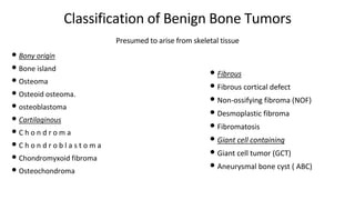
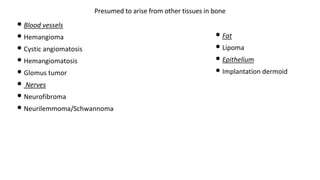
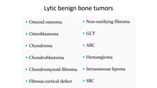
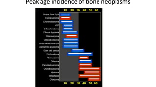
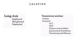
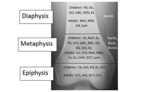
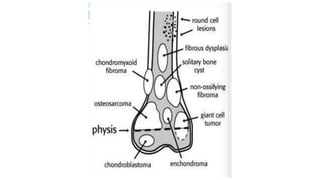
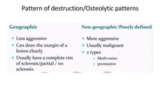
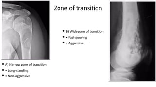
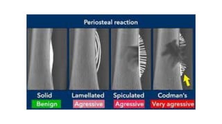
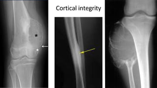
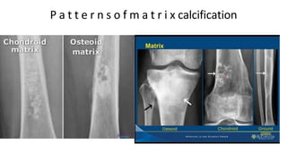
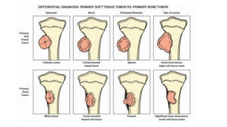
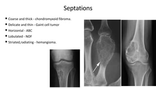
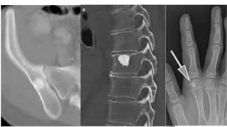
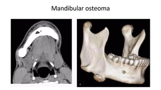
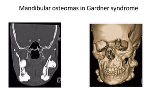
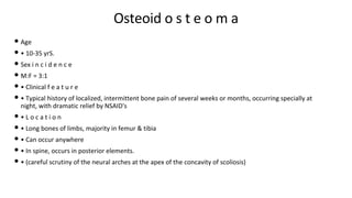
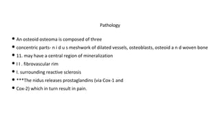
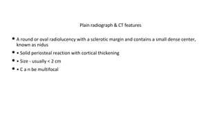
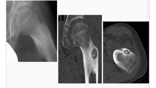
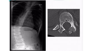
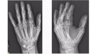
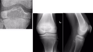
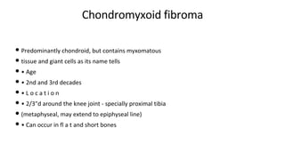
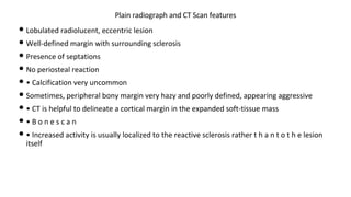
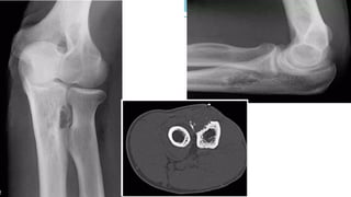
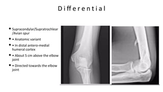
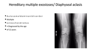
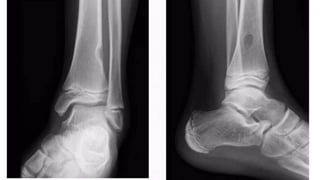
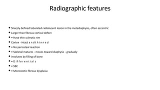
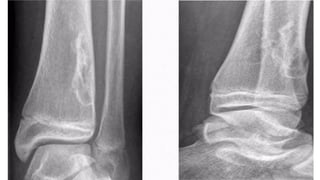
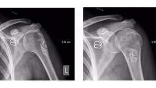
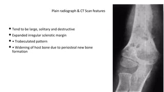
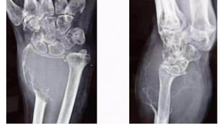
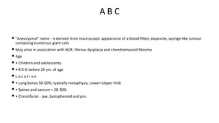
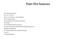
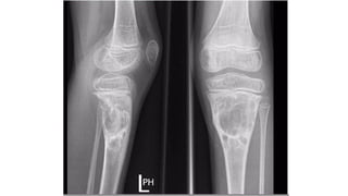
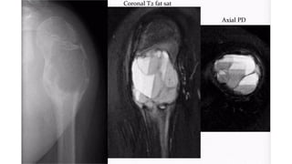
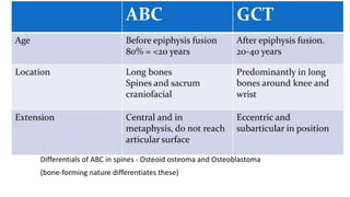
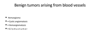
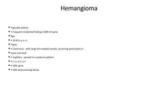
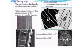
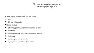
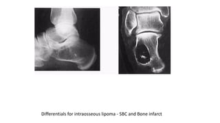
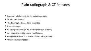
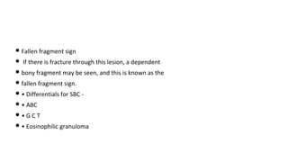
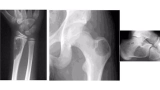
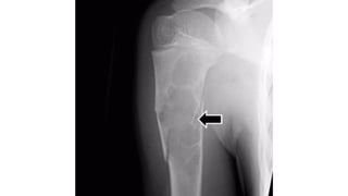
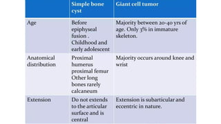
BENIGN BONE LESIONS X RAY
• PRESENTER : T SRIHARSHA
• MODERATOR : DR PRUDVEESH
Epidemiology of bone tumors
Incidence : Benign>Malignant
It is likely that benign lesions are underestimated because they often are asymptomatic and not
clinically recognized.
Classification of Benign Bone Tumors
• Bony origin
• Bone island
• Osteoma
• Osteoid osteoma.
• osteoblastoma
• Cartilaginous
• C h o n d r o m a
• C h o n d r o b l a s t o m a
• Chondromyxoid fibroma
• Osteochondroma
Presumed to arise from skeletal tissue
• Fibrous
• Fibrous cortical defect
• Non-ossifying fibroma (NOF)
• Desmoplastic fibroma
• Fibromatosis
• Giant cell containing
• Giant cell tumor (GCT)
• Aneurysmal bone cyst ( ABC)
• Blood vessels
• Hemangioma
• Cystic angiomatosis
• Hemangiomatosis
• Glomus tumor
• Nerves
• Neurofibroma
• Neurilemmoma/Schwannoma
Presumed to arise from other tissues in bone
• Fat
• Lipoma
• Epithelium
• Implantation dermoid
• Presumed to arise from joints
• Intraosseous ganglion
• Pigmented villonodular synovitis ( PVNS)
• Synovial chondromatosis
• Lipoma arborescens
• No known origin
• Solitary bone cyst (SBC)
• Non-neoplastic tumors
• Brodie's abscess
• Hydatid
• Hematoma
• Infarction
• Histiocytosis
Lytic benign bone tumors
Diagnostic modality of choice
• Clinical feature
• May be less useful As many lesions present with non-specific features of pain, swelling or pathological
fracture.
• Sex incidence is of little diagnostic value in primary bone tumors .
• Plain radiograph
• Mainstay of imaging modality
• Views: Antero-posterior, lateral and oblique (at least 2 views 90* to each other)
• • CT Scan
• Can define matrix calcification , Fracture , Soft tissue extension and endosteal scalloping.
• To assess bones like spine, pelvis, scapula and mandible.
• Nidus in osteoid osteoma - CT is max we can do for osteoid osteoma.
• MRI
• Gives idea about varieties of tissues present
• To assess chondroid matrix, fluid fluid levels, bone marrow involvement, cortical breach and
joint involvement.
• Intraosseous edema and extraosseous inflammatory
• response may be demonstrated
• Pre-op planning / post-op evaluation
• Angiography
• To see tumor vessels
• To see encasement of major adjacent vessels
• To distinguish between tumors, e.g. Osteosarcoma is highly vascular and chondrosarcoma is
hypovascular
• Bone scintigraphy
• Compound - technetium labelled diphosphonate
• Phases
• Blood pool phase
• Vascular lesion e.g. ABC, avascular Isesion e.g. Cartilage tumors
• Delayed phase
• • Helps to identify multiplicity of lesions
• • To see distant metastases
• • Patient is symptomatic (e.g. bone pain), but no lesion is identified in plain radiograph or CT
Scan, then Bone scintigraphy can be helpful.
Peak age incidence of bone neoplasms
L o c a t i o n
Behavior and morphology of a lesion
• Pattern of destruction
• Zone of transition
• Cortical integrity
• Matrix calcification
• Periosteal reaction.
• Soft tissue vs bone
• Septations
Pattern of destruction/Osteolytic patterns
Zone of transition
• A) Narrow zone of transition
• • Long-standing
• • Non-aggressive
• B) Wide zone of transition
• • Fast-growing
• • Aggressive
P e r i o s t e a l r e a c t i o n
Cortical integrity
P a t t e r n s o f m a t r i x calcification
Septations
• Coarse and thick - chondromyxoid fibroma.
• Delicate and thin - Gaint cell tumor
• Horizontal - ABC
• Lobulated - NOF
• Striated,radiating - hemangioma.
Benign bone tumors of bony origin
• Bone island
• Osteoma
• Osteoidosteoma
• Osteoblastoma
Bone island / Enostosis
• Common benign bone lesion
• • Usually seen as an incidental finding
• Constitute a small focus of compact bone within a cancellous bone
• One of the " don't touch lesions"
• Age - Any age
• • May grow in size up to the age of skeletal maturity
• • L o c a t i o n
• • Can occur anywhere in the skeleton
• • some predilection for pelvis, long bones, spine and ribs.
Plain radiographic findings
• Single or multiple
• Round or oval
• Always medullary in location
• Has radiating thorn-like spicules with narrow zone of t r a n s i t i o n
• Size - usually < 15mm, can be large up to 4 cm
• • No periosteal reaction, no cortical expansion
• In diaphyseal bone, the long axis of a bone island typically parallels the long axis of the
involved bone. In the metaphysis, and other regions, these are typically more spherical.
O s t e o m a
• Seen almost exclusively in bones formed in membrane
• • Slow growing, usually asymptomatic, found incidentally
• • L o c a t i o n - Paranasal sinus , Skull v a u l t , Mandible
• • Size
• • Varies, some > 2 . 5 cm
• • Types
• Ivory/dense
• • Lacks haversian system
• Spongy
• • Resembles normal bone
• Ivory osteomas appear very radio-dense
• Spongy osteomas may demonstrate central marrow
• Complications
• • Growth within PNS c a n cause mucocele
• • Growth from inner table of skull may produce raised intracranial pressure
• • Association - "Gardner syndrome"
• Multiple osteomas of skull, mandible and long bones
• > Familial adenomatous polyposis
• Soft-tissue tumors of connective tissue origin
Radiographic features
Ivory osteoma
Mandibular osteoma
Mandibular osteomas in Gardner syndrome
Osteoid o s t e o m a
• Age
• • 10-35 yrS.
• Sex i n c i d e n c e
• M:F = 3:1
• • Clinical f e a t u r e
• • Typical history of localized, intermittent bone pain of several weeks or months, occurring specially at
night, with dramatic relief by NSAID's
• • L o c a t i o n
• • Long bones of limbs, majority in femur & tibia
• • Can occur anywhere
• • In spine, occurs in posterior elements.
• • (careful scrutiny of the neural arches at the apex of the concavity of scoliosis)
• An osteoid osteoma is composed of three
• concentric parts- n i d u s meshwork of dilated vessels, osteoblasts, osteoid a n d woven bone
• 11. may have a central region of mineralization
• I I . fibrovascular rim
• I. surrounding reactive sclerosis
• ***The nidus releases prostaglandins (via Cox-1 and
• Cox-2) which in turn result in pain.
Pathology
• A round or oval radiolucency with a sclerotic margin and contains a small dense center,
known as nidus
• • Solid periosteal reaction with cortical thickening
• • Size - usually < 2 cm
• • C a n be multifocal
Plain radiograph & CT features
• Most important means in cases of medullary osteoid
• osteoma, where radiographs remain normal
• Also important in any young patients with bone pain
• and normal radiographs
• • Findings
• • Intense focal area of increased activity surrounded by less intense activity from the reactive
sclerosis.
• • Evident in blood-pool image and persists in delayed
• image.
B o n e s c a n
• Osteoblastoma (>2cm)
• Stress fracture - that c a u s e s c h r o n i c cortical
• thickening
• • Chronic osteomyelitis
• • Area of radiolucency more irregular
• • Sequestra are irregular or linear in shape,
• differentiating from central nidus of osteoid
• o s t e o m a
Differentials
O s t e o b l a s t o m a
• Similar in histological characteristics as osteoid osteoma but usually larger
• Less c o m m o n t h a n osteoid o s t e o m a
• Age
• • 80% of patients under the age of 30
• In 2nd and 3rd decades
• Clinical feature
• • Long insidious history of pain, often worsening at night
• Not relieved by NSAID's
• Limitation of movement
• • L o c a t i o n
• • Spine ( posterior element), flat bones
• • Metaphysis and distal diaphysis of long bones.
• • Pathology
• Larger than osteoid osteoma
• • Irregular in shape, friable and hemorrhagic
• • Abundant osteoid tissue and many thin-walled capillaries
• S i z e = 2 - 1 0 c m
• Radiolucent lesion
• Margin - irregular but sharply demarcated
• Surrounding sclerosis - varies but can be profound
• Can cause cortical expansion and thinning
• Calcification o r ossification o f osteoid tissue -
• punctate or amorphous
• Sometimes, lesion can be aggressive with soft tissue
• masses containing calcification and ossification
• • B o n e s c a n
• • Active both in blood-pool and delayed phase
Plain radiograph and CT features
Cartilage-forming benign bone
t u m o r s
• C h o n d r o m a
• Chondroblastoma
• Chondromyxoid
• fi b r o m a
• Osteochondroma
Chondroma/Enchondroma
• Relatively common benign tumor
• • Single/multifocal
• • Usually central medullary
• • Age
• Childhood to adulthood, peak incidence = 10-30 years
• • Clinical feature
• Rarely symptomatic
• • Patient may admit localized hard swelling for many years
• • May be complicated by pathological fracture
• • L o c a t i o n
• • 50% in hands - commonly in phalanges, less commonly in metacarpals
• • 10% in small bones of feet
• • 20% - long bones
• • 20% - flat bones - pelvis, scapula and vertebral bodies.
• Cartilaginous tissue is not radiopaque
• • Radiolucent lesion
• Expansion and thinning of overlying cortex
• No periosteal reaction in the absence of fracture
• Z o n e o f t r a n s i t i o n is n a r r o w a n d s c l e r o t i c
• Endosteal scalloping +
• Flecks o f c a l c i fi c a t i o n w i t h i n t h e lesion
• usually purely lytic in phalanges, in other
• locations, enchondromas are expansile, with
• characteristic "rings and arcs" calcifications.
Plain radiograph & CT scan features
• T1
• • Low to intermediate signal
• T 2
• • typically of background intense high signal
• (high water content of hyaline cartilage matrix)
• • they can be focal regions of signal drop out where calcification
• p r e s e n t
• • P o s t - c o n t r a s t
• • Variable e n h a n c e m e n t
MRI
Special type- Juxtacortical chondroma
• R a r e
• • Usually in young adults and
• related to the cortex of a long
• bone and phalanges
• Radiologically -
• • Well-defined soft tissue mass with
• c a l c i fi c a t i o n
• • Bordered by thin, incomplete shell
• of overlying bone
• • Pressure erosion causes scalloping
• of underlying cortex
• • Va r i a b l e s c l e r o t i c r e a c t i o n
• Ollier disease
• • non-hereditary, sporadic, skeletal disorder characterized by multiple enchondromas that
are principally located in the metaphyseal regions.
• Maffucci's syndrome
• • Rare association of dyschondroplasia with
• cavernous hemangiomas in soft tissues
A s s o c i a t i o n s
• Implantation dermoid cyst - in hands
• B . Medullary bone infarct
• A . Curvilinear peripheral calcification in the infarct
Differentials
C h o n d r o b l a s t o m a
• R a r e
• • Characteristically occur in epiphyses of apophyses
• of long bones in young patients
• Age
• • < 2 o y r s
• Sex prevalence
• • Slight male predominant
• • L o c a t i o n
• • Most tumors occur in epiphyses of long bones, specially around
• the hips, knees or shoulders
• • Also occur in apophyses
• Well-defined oval lucent lesion
• • S i z e = 1-10 c m
• Position- eccentric in the epiphyses
• Margin- thin sclerotic rim
• • Cortical expansion = +-
• Periosteal r e a c t i o n = +-
• Internal calcification = about quarter of cases = stippled calcification
Plain radiograph and CT Scan features
• Ideal for evaluating transphyseal or transcortical
• expansion
• • To see bone marrow & soft tissue edema
• •Bone s c a n
• • Increased activity in blood-pool phase
• Differentials
• • Osteomyelitis - Brodie's abscess (same location)
• • Giant cell tumor - older age group
MRI
Chondromyxoid fibroma
• Predominantly chondroid, but contains myxomatous
• tissue and giant cells as its name tells
• • Age
• • 2nd and 3rd decades
• • L o c a t i o n
• • 2/3"d around the knee joint - specially proximal tibia
• (metaphyseal, may extend to epiphyseal line)
• • Can occur in fl a t and short bones
• Lobulated radiolucent, eccentric lesion
• Well-defined margin with surrounding sclerosis
• Presence of septations
• No periosteal reaction
• • Calcification very uncommon
• Sometimes, peripheral bony margin very hazy and poorly defined, appearing aggressive
• • CT is helpful to delineate a cortical margin in the expanded soft-tissue mass
• • B o n e s c a n
• • Increased activity is usually localized to the reactive sclerosis rather t h a n t o t h e lesion
itself
Plain radiograph and CT Scan features
D i ff e r e n t i a l s
• NOF - younger age group
• • Chondroblastoma - younger age group
• • ABC = ill-defined endosteal margin
O s t e o c h o n d r o m a
• Relatively common
• Osseous outgrowth arising from bony cortex
• • Single/multiple
• • Capped by cartilage
• In long bones, grows away from the metaphyses to the diaphysis,
• i.e. away from the joint - typical characteristic
• Age
• • Develop during childhood
• • Can present at any age
• • L o c a t i o n
• • Lower limb =50%
• • Upper limb = 10-20%
• • Flat bones like scapula and pelvis
• • Types
• • Sessile - usually in pelvis
• • pedunculated
• In long bones, in the metaphysis & grows away from the joint
• Size - varies, can be as long as 10 cm
• Pedunculated type - cortex of lesion continuous with the underlying bone and medullary cavity
merging through the cortical defect
• • Cartilage cap best seen in CT
• As age increases - calcification increases ( punctate or
• curvilinear)
• Growth ceases after skeletal maturation
• Sessile type in flat bones may resemble cauliflower
Plain radiograph & CT scan features
D i ff e r e n t i a l
• Supracondylar/Supratrochlear
/Avian spur
• • Anatomic variant
• • In distal antero-medial
humeral cortex
• • About 5 cm above the elbow
joint
• • Directed towards the elbow
joint
Hereditary multiple exostoses/ Diaphyseal aclasis
• A u t o s o m a l d o m i n a n t d i s o r d e r
• Multiple
• o s t e o c h o n d r o m a s
• • Diagnosed by the age
• of 12 years
Malignant transformation of cartilage
forming tumors
• T u m o r s that c a n t r a n s f o r m
• • C h o n d r o m a
• • O s t e o c h o n d r o m a
• Common if location - in long and flat bones
• More common in diaphyseal aclasis
• When to suspect transformation??
• • Recent increase in pain
• • Alteration of radiological appearances on serial films
• • Late development of pathological fracture
• • Cortical d e s t r u c t i o n
• • S o f t t i s s u e m a s s
• Benign bone
Benign bone tumors from skeletal
t i s s u e - F i b r o u s t u m o r s
• Fibrous cortical defect
• Non-ossifying fibroma
• Desmoplastic fibroma
• Fibromatosis
F i b r o u s c o r t i c a l d e f e c t
• Extremely common
• Usually discovered by chance
• Histologically identical to NOF
• • Age
• • Typically occur in children
• • 2-15 y e a r s
• • L o c a t i o n
• • Occurs in distal femur, ends of tibia
• • Metaphysis or diametaphyseal regions
• • Less frequent in upper limbs
• Sharply defined lucent intracortical lesion
• They are blister-like cortical expansion
• Size = small than 2-3 c m
• May appear only lucent lesion when on frontal view
• • Have fine sclerotic margin
• Increasing skeletal maturity = increasing sclerosis =
• migrates to diaphysis = involutes
• No periosteal reaction
• Not reach medullary cavity
Radiographic features
• N O F
• • O s t e o i d o s t e o m a
• • Stress fracture
D i ff e r e n t i a l s
Non-ossifying fibroma (NOF)
• Most common benign fibrous lesion
• Similar to fibrous cortical defect, but much larger and
• occurs in slightly older age
• they spontaneously heal, being gradually filled in by
• b o n e
• Age
• • 10-20 yrs.
• Location
• • Vast majority around the knee joint.
• • Femur being the commonest
Radiographic features
• Sharply defined lobulated radiolucent lesion in the metadiaphysis, often eccentric
• Larger than fibrous cortical defect
• • Have thin sclerotic rim
• Cortex - intact a n d t h i n n e d
• • No periosteal reaction
• • Skeletal matures - moves toward diaphysis - gradually
• involutes by filling of bone
• • D i f f e r e n t i a l s
• • SBC
• • Monostotic fibrous dysplasia
Desmoplastic fibroma
• Extremely rare
• • Exhibits dense fibrous tissue
• • Do not metastasize but locally aggressive
• Age - usually in young adults
• Clinical feature
• • Constant pain
• • Lesion is often t e n d e r
• • L o c a t i o n
• • Metadiaphysis of long bones
• • Pelvis and spine
• • Abundant collagenous stroma
• • Fibroblasts, myofibroblasts & mesenchymal cells
• Tend to be large, solitary and destructive
• Expanded irregular sclerotic margin
• • Trabeculated pattern
• • Widening of host bone due to periosteal new bone
formation
Plain radiograph & CT Scan features
Giant cell containing
• GCT
• • ABC
• • Brown tumor of hyperparathyroidism
• • C h o n d r o b l a s t o m a
• • Chondromyxoid fibroma.
GCT O s t e o c l a s t o m a s
• Name coming from its histological picture
• • GCT containing richly vascular tissue with spindle cells and numerous giant cells
• • It neither forms bone nor cartilage
• • Locally aggressive
• • Can recur after excision
• • Age
• • 20-40 y e a r s
• • 3% develop in immature skeleton
• • L o c a t i o n
• •around the knee: distal femur and proximal tibia: 50-65%
• • distal radius: 10-15%
• • sacrum: 4-10%
• • vertebral body: 5-10%
• A lucent lesion typically situated beneath the articular surface
• E c c e n t r i c
• Narrow z o n e o f transition
• 'soap-bubble' pattern of calcification
• No sclerotic margin
• • overlying cortex is thinned, expanded or deficient
• • Soft-tissue mass may be present
• • No calcification or ossification until complicated by f r a c t u r e
• • CT Scan & MRI
• • To delineate soft tissue extension
• • B o n e s c a n
• • Increased activity in blood-pool phase
Plain film & CT features
A B C
• "Aneurysmal" name - is derived from macroscopic appearance of a blood-filled, expansile, sponge-like tumour
containing numerous giant cells
• May arise in association with NOF, fibrous dysplasia and chondromyxoid fibroma
• Age
• • Children and adolescents.
• • 8 0 % before 20 yrs. of age
• L o c a t i o n
• • Long bones 50-60%, typically metaphysis, Lower>Upper limb
• • Spines and sacrum = 20-30%
• • Craniofacial - jaw, basisphenoid and pns
Plain film features
• An expansile lytic lesion
• S i z e = 2 - 2 0 c m
• Z o n e o f transition - can be ill-defined
• Thin sclerotic margin
• • Cortical breach and soft tissue extension
• CT & MRI
• • To delineate soft tissue extension
• • Shows characteristic fluid-fluid levels (representing areas of
• blood of variable ages)
• ***Other benign lesions with fluid-fluid levels
• • G C T
• • Chondroblastoma
Differentials of ABC in spines - Osteoid osteoma and Osteoblastoma
(bone-forming nature differentiates these)
Benign tumors arising from blood vessels
• Hemangioma
• • Cystic angiomatosis
• • Hemangiomatosis
• • G l o m u s t u m o r
Hemangioma
• Typically solitary
• • Frequent incidental finding in MRI of spine
• Age
• • 10-45 y e a r s
• Types
• • Cavernous - with large thin-walled vessels, occurring particularly in
• spine and skull
• • Capillary - spread in a sunburst pattern
• • L o c a t i o n
• • 50% spine
• • 50% skull and long bones
Plain fi l m a n d CT f e a t u r e s
• Osteolytic lesion with a trabecular pattern
• • No expansion in spine
• • Can cause cortical expansion in skull and long bones
• with thin rim o f sclerosis
• • In long bones, osteolytic lesion may have soap-bubble
• appearance with stippled radiodensity
• • Can affect posterior elements of vertebra
Glomus tumor/Glomangioma/
Hemangiopericytoma
• Rare, highly differentiated vascular tumor
• • Age
• • 4th and 5th decades
• Clinical feature
• • Extremely painful, tender and sensitive to cold
• L o c a t i o n
• • Terminal phalanx, particularly subungual portion
• • Pathology
• • branching vascular channels
• • aggregates of specialized glomus cells
Plain film f e a t u r e s
• Lucent lesion with extremely sharp margin
• • May show osseous erosion or thinning of adjacent
• cortical b o n e
• • B o n e S c a n
• • Increased activity in early phase and blood-pool phases
Benign tumor arising from fat -
Intraosseous Lipoma
• Extremely rare
• • Age
• • Wide range = 5-85 years
• • Usually detected in 4th-5th decades
• • L o c a t i o n
• • Lower limb = 75% ( metaphysis)
• • Calcaneum>femur >tibia>fibula
• • Upper limb
• • Skull and mandible
• • Spine & pelvis
• • Ribs
Plain radiograph feature
• Expansile radiolucent lesion with sharply defined
• sclerotic margin
• • No periosteal new bone formation
• Within calcaneum, lipoma has characteristic
• appearance, osteolytic lesion with a central focus
• o f o s s i fi c a t i o n
• CT & MRI
• • Can be homogenous fat content
• • Fatty lesion with central necroses, central
• calcifications o r o s s i fi c a t i o n s
• • Fatty lesion with multiple central necroses, central
• calcifications or ossifications
Differentials for intraosseous lipoma - SBC and Bone infarct
Benign tumor of unknown origin
• Always unilocular
• • Found incidentally, more than half present due to a pathological
• f r a c t u r e
• Age
• • Sex • 1st and 2nd decades
• C a n o c c u r a f t e r s k e l e t a l i n c i d e n c e
• • M:F = 2-3:1
• m a t u r a t i o n
• • L o c a t i o n
• Typically intramedullary in long bones
• In childhood, near growth plate, then migrates to diaphysis, then involutes
• Proximal humerus> Proximal femur>Other long bones
• Calcaneum & Talus ( after skeletal maturation)
• • Pathology
• • Cyst contains clear liquid unless there has been contamination by bleeding
• following a fracture.
• • Cyst is lined by a thin layer of connective tissue.
Simple bone cyst / Unicameral bone cyst/Solitary bone cyst
Plain radiograph & CT features
• A central radiolucent lesion in metadiaphysis is
• c h a r a c t e r i s t i c
• • Cortex may be thinned and expanded
• Sclerotic margin
• • A serpiginous margin (by prominent ridges of bone)
• may cause the cyst to appear multilocular.
• • No periosteal reaction unless a fracture has occured
• • No internal calcification
• Fallen fragment sign
• If there is fracture through this lesion, a dependent
• bony fragment may be seen, and this is known as the
• fallen fragment sign.
• • Differentials for SBC -
• • ABC
• • G C T
• • Eosinophilic granuloma
Summary
• Age
• L o c a t i o n
• Multiplicity
• Shape
• Size
• X-ray appearance - lytic/dense
• Z o n e o f transition
• Sclerosis
• • Periosteal reaction
• Matrix calcification
• Soft tissue involvement
Thank you
• References
• Yochum and Rowe’s essentials of skeletal radiology.
• David Sutton’s textbook of radiology and imaging
• Next presentation : Journal presentation by Dr ramudu on 17-12-2024.

benign bone lesions x ray radiology guide.pdf

  • 1.
    BENIGN BONE LESIONSX RAY • PRESENTER : T SRIHARSHA • MODERATOR : DR PRUDVEESH
  • 2.
    Epidemiology of bonetumors Incidence : Benign>Malignant It is likely that benign lesions are underestimated because they often are asymptomatic and not clinically recognized.
  • 3.
    Classification of BenignBone Tumors • Bony origin • Bone island • Osteoma • Osteoid osteoma. • osteoblastoma • Cartilaginous • C h o n d r o m a • C h o n d r o b l a s t o m a • Chondromyxoid fibroma • Osteochondroma Presumed to arise from skeletal tissue • Fibrous • Fibrous cortical defect • Non-ossifying fibroma (NOF) • Desmoplastic fibroma • Fibromatosis • Giant cell containing • Giant cell tumor (GCT) • Aneurysmal bone cyst ( ABC)
  • 4.
    • Blood vessels •Hemangioma • Cystic angiomatosis • Hemangiomatosis • Glomus tumor • Nerves • Neurofibroma • Neurilemmoma/Schwannoma Presumed to arise from other tissues in bone • Fat • Lipoma • Epithelium • Implantation dermoid
  • 5.
    • Presumed toarise from joints • Intraosseous ganglion • Pigmented villonodular synovitis ( PVNS) • Synovial chondromatosis • Lipoma arborescens • No known origin • Solitary bone cyst (SBC) • Non-neoplastic tumors • Brodie's abscess • Hydatid • Hematoma • Infarction • Histiocytosis
  • 6.
  • 7.
    Diagnostic modality ofchoice • Clinical feature • May be less useful As many lesions present with non-specific features of pain, swelling or pathological fracture. • Sex incidence is of little diagnostic value in primary bone tumors . • Plain radiograph • Mainstay of imaging modality • Views: Antero-posterior, lateral and oblique (at least 2 views 90* to each other) • • CT Scan • Can define matrix calcification , Fracture , Soft tissue extension and endosteal scalloping. • To assess bones like spine, pelvis, scapula and mandible. • Nidus in osteoid osteoma - CT is max we can do for osteoid osteoma.
  • 8.
    • MRI • Givesidea about varieties of tissues present • To assess chondroid matrix, fluid fluid levels, bone marrow involvement, cortical breach and joint involvement. • Intraosseous edema and extraosseous inflammatory • response may be demonstrated • Pre-op planning / post-op evaluation • Angiography • To see tumor vessels • To see encasement of major adjacent vessels • To distinguish between tumors, e.g. Osteosarcoma is highly vascular and chondrosarcoma is hypovascular
  • 9.
    • Bone scintigraphy •Compound - technetium labelled diphosphonate • Phases • Blood pool phase • Vascular lesion e.g. ABC, avascular Isesion e.g. Cartilage tumors • Delayed phase • • Helps to identify multiplicity of lesions • • To see distant metastases • • Patient is symptomatic (e.g. bone pain), but no lesion is identified in plain radiograph or CT Scan, then Bone scintigraphy can be helpful.
  • 11.
    Peak age incidenceof bone neoplasms
  • 12.
    L o ca t i o n
  • 15.
    Behavior and morphologyof a lesion • Pattern of destruction • Zone of transition • Cortical integrity • Matrix calcification • Periosteal reaction. • Soft tissue vs bone • Septations
  • 16.
  • 18.
    Zone of transition •A) Narrow zone of transition • • Long-standing • • Non-aggressive • B) Wide zone of transition • • Fast-growing • • Aggressive
  • 19.
    P e ri o s t e a l r e a c t i o n
  • 21.
  • 22.
    P a tt e r n s o f m a t r i x calcification
  • 24.
    Septations • Coarse andthick - chondromyxoid fibroma. • Delicate and thin - Gaint cell tumor • Horizontal - ABC • Lobulated - NOF • Striated,radiating - hemangioma.
  • 25.
    Benign bone tumorsof bony origin • Bone island • Osteoma • Osteoidosteoma • Osteoblastoma
  • 26.
    Bone island /Enostosis • Common benign bone lesion • • Usually seen as an incidental finding • Constitute a small focus of compact bone within a cancellous bone • One of the " don't touch lesions" • Age - Any age • • May grow in size up to the age of skeletal maturity • • L o c a t i o n • • Can occur anywhere in the skeleton • • some predilection for pelvis, long bones, spine and ribs.
  • 27.
    Plain radiographic findings •Single or multiple • Round or oval • Always medullary in location • Has radiating thorn-like spicules with narrow zone of t r a n s i t i o n • Size - usually < 15mm, can be large up to 4 cm • • No periosteal reaction, no cortical expansion • In diaphyseal bone, the long axis of a bone island typically parallels the long axis of the involved bone. In the metaphysis, and other regions, these are typically more spherical.
  • 29.
    O s te o m a • Seen almost exclusively in bones formed in membrane • • Slow growing, usually asymptomatic, found incidentally • • L o c a t i o n - Paranasal sinus , Skull v a u l t , Mandible • • Size • • Varies, some > 2 . 5 cm • • Types • Ivory/dense • • Lacks haversian system • Spongy • • Resembles normal bone
  • 30.
    • Ivory osteomasappear very radio-dense • Spongy osteomas may demonstrate central marrow • Complications • • Growth within PNS c a n cause mucocele • • Growth from inner table of skull may produce raised intracranial pressure • • Association - "Gardner syndrome" • Multiple osteomas of skull, mandible and long bones • > Familial adenomatous polyposis • Soft-tissue tumors of connective tissue origin Radiographic features
  • 31.
  • 32.
  • 33.
    Mandibular osteomas inGardner syndrome
  • 34.
    Osteoid o st e o m a • Age • • 10-35 yrS. • Sex i n c i d e n c e • M:F = 3:1 • • Clinical f e a t u r e • • Typical history of localized, intermittent bone pain of several weeks or months, occurring specially at night, with dramatic relief by NSAID's • • L o c a t i o n • • Long bones of limbs, majority in femur & tibia • • Can occur anywhere • • In spine, occurs in posterior elements. • • (careful scrutiny of the neural arches at the apex of the concavity of scoliosis)
  • 35.
    • An osteoidosteoma is composed of three • concentric parts- n i d u s meshwork of dilated vessels, osteoblasts, osteoid a n d woven bone • 11. may have a central region of mineralization • I I . fibrovascular rim • I. surrounding reactive sclerosis • ***The nidus releases prostaglandins (via Cox-1 and • Cox-2) which in turn result in pain. Pathology
  • 36.
    • A roundor oval radiolucency with a sclerotic margin and contains a small dense center, known as nidus • • Solid periosteal reaction with cortical thickening • • Size - usually < 2 cm • • C a n be multifocal Plain radiograph & CT features
  • 37.
    • Most importantmeans in cases of medullary osteoid • osteoma, where radiographs remain normal • Also important in any young patients with bone pain • and normal radiographs • • Findings • • Intense focal area of increased activity surrounded by less intense activity from the reactive sclerosis. • • Evident in blood-pool image and persists in delayed • image. B o n e s c a n
  • 39.
    • Osteoblastoma (>2cm) •Stress fracture - that c a u s e s c h r o n i c cortical • thickening • • Chronic osteomyelitis • • Area of radiolucency more irregular • • Sequestra are irregular or linear in shape, • differentiating from central nidus of osteoid • o s t e o m a Differentials
  • 40.
    O s te o b l a s t o m a • Similar in histological characteristics as osteoid osteoma but usually larger • Less c o m m o n t h a n osteoid o s t e o m a • Age • • 80% of patients under the age of 30 • In 2nd and 3rd decades • Clinical feature • • Long insidious history of pain, often worsening at night • Not relieved by NSAID's • Limitation of movement • • L o c a t i o n • • Spine ( posterior element), flat bones • • Metaphysis and distal diaphysis of long bones. • • Pathology • Larger than osteoid osteoma • • Irregular in shape, friable and hemorrhagic • • Abundant osteoid tissue and many thin-walled capillaries
  • 41.
    • S iz e = 2 - 1 0 c m • Radiolucent lesion • Margin - irregular but sharply demarcated • Surrounding sclerosis - varies but can be profound • Can cause cortical expansion and thinning • Calcification o r ossification o f osteoid tissue - • punctate or amorphous • Sometimes, lesion can be aggressive with soft tissue • masses containing calcification and ossification • • B o n e s c a n • • Active both in blood-pool and delayed phase Plain radiograph and CT features
  • 44.
    Cartilage-forming benign bone tu m o r s • C h o n d r o m a • Chondroblastoma • Chondromyxoid • fi b r o m a • Osteochondroma
  • 45.
    Chondroma/Enchondroma • Relatively commonbenign tumor • • Single/multifocal • • Usually central medullary • • Age • Childhood to adulthood, peak incidence = 10-30 years • • Clinical feature • Rarely symptomatic • • Patient may admit localized hard swelling for many years • • May be complicated by pathological fracture • • L o c a t i o n • • 50% in hands - commonly in phalanges, less commonly in metacarpals • • 10% in small bones of feet • • 20% - long bones • • 20% - flat bones - pelvis, scapula and vertebral bodies.
  • 46.
    • Cartilaginous tissueis not radiopaque • • Radiolucent lesion • Expansion and thinning of overlying cortex • No periosteal reaction in the absence of fracture • Z o n e o f t r a n s i t i o n is n a r r o w a n d s c l e r o t i c • Endosteal scalloping + • Flecks o f c a l c i fi c a t i o n w i t h i n t h e lesion • usually purely lytic in phalanges, in other • locations, enchondromas are expansile, with • characteristic "rings and arcs" calcifications. Plain radiograph & CT scan features
  • 47.
    • T1 • •Low to intermediate signal • T 2 • • typically of background intense high signal • (high water content of hyaline cartilage matrix) • • they can be focal regions of signal drop out where calcification • p r e s e n t • • P o s t - c o n t r a s t • • Variable e n h a n c e m e n t MRI
  • 49.
    Special type- Juxtacorticalchondroma • R a r e • • Usually in young adults and • related to the cortex of a long • bone and phalanges • Radiologically - • • Well-defined soft tissue mass with • c a l c i fi c a t i o n • • Bordered by thin, incomplete shell • of overlying bone • • Pressure erosion causes scalloping • of underlying cortex • • Va r i a b l e s c l e r o t i c r e a c t i o n
  • 50.
    • Ollier disease •• non-hereditary, sporadic, skeletal disorder characterized by multiple enchondromas that are principally located in the metaphyseal regions. • Maffucci's syndrome • • Rare association of dyschondroplasia with • cavernous hemangiomas in soft tissues A s s o c i a t i o n s
  • 51.
    • Implantation dermoidcyst - in hands • B . Medullary bone infarct • A . Curvilinear peripheral calcification in the infarct Differentials
  • 52.
    C h on d r o b l a s t o m a • R a r e • • Characteristically occur in epiphyses of apophyses • of long bones in young patients • Age • • < 2 o y r s • Sex prevalence • • Slight male predominant • • L o c a t i o n • • Most tumors occur in epiphyses of long bones, specially around • the hips, knees or shoulders • • Also occur in apophyses
  • 53.
    • Well-defined ovallucent lesion • • S i z e = 1-10 c m • Position- eccentric in the epiphyses • Margin- thin sclerotic rim • • Cortical expansion = +- • Periosteal r e a c t i o n = +- • Internal calcification = about quarter of cases = stippled calcification Plain radiograph and CT Scan features
  • 54.
    • Ideal forevaluating transphyseal or transcortical • expansion • • To see bone marrow & soft tissue edema • •Bone s c a n • • Increased activity in blood-pool phase • Differentials • • Osteomyelitis - Brodie's abscess (same location) • • Giant cell tumor - older age group MRI
  • 56.
    Chondromyxoid fibroma • Predominantlychondroid, but contains myxomatous • tissue and giant cells as its name tells • • Age • • 2nd and 3rd decades • • L o c a t i o n • • 2/3"d around the knee joint - specially proximal tibia • (metaphyseal, may extend to epiphyseal line) • • Can occur in fl a t and short bones
  • 57.
    • Lobulated radiolucent,eccentric lesion • Well-defined margin with surrounding sclerosis • Presence of septations • No periosteal reaction • • Calcification very uncommon • Sometimes, peripheral bony margin very hazy and poorly defined, appearing aggressive • • CT is helpful to delineate a cortical margin in the expanded soft-tissue mass • • B o n e s c a n • • Increased activity is usually localized to the reactive sclerosis rather t h a n t o t h e lesion itself Plain radiograph and CT Scan features
  • 59.
    D i ffe r e n t i a l s • NOF - younger age group • • Chondroblastoma - younger age group • • ABC = ill-defined endosteal margin
  • 60.
    O s te o c h o n d r o m a • Relatively common • Osseous outgrowth arising from bony cortex • • Single/multiple • • Capped by cartilage • In long bones, grows away from the metaphyses to the diaphysis, • i.e. away from the joint - typical characteristic • Age • • Develop during childhood • • Can present at any age • • L o c a t i o n • • Lower limb =50% • • Upper limb = 10-20% • • Flat bones like scapula and pelvis • • Types • • Sessile - usually in pelvis • • pedunculated
  • 61.
    • In longbones, in the metaphysis & grows away from the joint • Size - varies, can be as long as 10 cm • Pedunculated type - cortex of lesion continuous with the underlying bone and medullary cavity merging through the cortical defect • • Cartilage cap best seen in CT • As age increases - calcification increases ( punctate or • curvilinear) • Growth ceases after skeletal maturation • Sessile type in flat bones may resemble cauliflower Plain radiograph & CT scan features
  • 63.
    D i ffe r e n t i a l • Supracondylar/Supratrochlear /Avian spur • • Anatomic variant • • In distal antero-medial humeral cortex • • About 5 cm above the elbow joint • • Directed towards the elbow joint
  • 64.
    Hereditary multiple exostoses/Diaphyseal aclasis • A u t o s o m a l d o m i n a n t d i s o r d e r • Multiple • o s t e o c h o n d r o m a s • • Diagnosed by the age • of 12 years
  • 65.
    Malignant transformation ofcartilage forming tumors • T u m o r s that c a n t r a n s f o r m • • C h o n d r o m a • • O s t e o c h o n d r o m a • Common if location - in long and flat bones • More common in diaphyseal aclasis • When to suspect transformation?? • • Recent increase in pain • • Alteration of radiological appearances on serial films • • Late development of pathological fracture • • Cortical d e s t r u c t i o n • • S o f t t i s s u e m a s s • Benign bone
  • 66.
    Benign bone tumorsfrom skeletal t i s s u e - F i b r o u s t u m o r s • Fibrous cortical defect • Non-ossifying fibroma • Desmoplastic fibroma • Fibromatosis
  • 67.
    F i br o u s c o r t i c a l d e f e c t • Extremely common • Usually discovered by chance • Histologically identical to NOF • • Age • • Typically occur in children • • 2-15 y e a r s • • L o c a t i o n • • Occurs in distal femur, ends of tibia • • Metaphysis or diametaphyseal regions • • Less frequent in upper limbs
  • 68.
    • Sharply definedlucent intracortical lesion • They are blister-like cortical expansion • Size = small than 2-3 c m • May appear only lucent lesion when on frontal view • • Have fine sclerotic margin • Increasing skeletal maturity = increasing sclerosis = • migrates to diaphysis = involutes • No periosteal reaction • Not reach medullary cavity Radiographic features
  • 70.
    • N OF • • O s t e o i d o s t e o m a • • Stress fracture D i ff e r e n t i a l s
  • 71.
    Non-ossifying fibroma (NOF) •Most common benign fibrous lesion • Similar to fibrous cortical defect, but much larger and • occurs in slightly older age • they spontaneously heal, being gradually filled in by • b o n e • Age • • 10-20 yrs. • Location • • Vast majority around the knee joint. • • Femur being the commonest
  • 72.
    Radiographic features • Sharplydefined lobulated radiolucent lesion in the metadiaphysis, often eccentric • Larger than fibrous cortical defect • • Have thin sclerotic rim • Cortex - intact a n d t h i n n e d • • No periosteal reaction • • Skeletal matures - moves toward diaphysis - gradually • involutes by filling of bone • • D i f f e r e n t i a l s • • SBC • • Monostotic fibrous dysplasia
  • 75.
    Desmoplastic fibroma • Extremelyrare • • Exhibits dense fibrous tissue • • Do not metastasize but locally aggressive • Age - usually in young adults • Clinical feature • • Constant pain • • Lesion is often t e n d e r • • L o c a t i o n • • Metadiaphysis of long bones • • Pelvis and spine • • Abundant collagenous stroma • • Fibroblasts, myofibroblasts & mesenchymal cells
  • 76.
    • Tend tobe large, solitary and destructive • Expanded irregular sclerotic margin • • Trabeculated pattern • • Widening of host bone due to periosteal new bone formation Plain radiograph & CT Scan features
  • 77.
    Giant cell containing •GCT • • ABC • • Brown tumor of hyperparathyroidism • • C h o n d r o b l a s t o m a • • Chondromyxoid fibroma.
  • 78.
    GCT O st e o c l a s t o m a s • Name coming from its histological picture • • GCT containing richly vascular tissue with spindle cells and numerous giant cells • • It neither forms bone nor cartilage • • Locally aggressive • • Can recur after excision • • Age • • 20-40 y e a r s • • 3% develop in immature skeleton • • L o c a t i o n • •around the knee: distal femur and proximal tibia: 50-65% • • distal radius: 10-15% • • sacrum: 4-10% • • vertebral body: 5-10%
  • 79.
    • A lucentlesion typically situated beneath the articular surface • E c c e n t r i c • Narrow z o n e o f transition • 'soap-bubble' pattern of calcification • No sclerotic margin • • overlying cortex is thinned, expanded or deficient • • Soft-tissue mass may be present • • No calcification or ossification until complicated by f r a c t u r e • • CT Scan & MRI • • To delineate soft tissue extension • • B o n e s c a n • • Increased activity in blood-pool phase Plain film & CT features
  • 81.
    A B C •"Aneurysmal" name - is derived from macroscopic appearance of a blood-filled, expansile, sponge-like tumour containing numerous giant cells • May arise in association with NOF, fibrous dysplasia and chondromyxoid fibroma • Age • • Children and adolescents. • • 8 0 % before 20 yrs. of age • L o c a t i o n • • Long bones 50-60%, typically metaphysis, Lower>Upper limb • • Spines and sacrum = 20-30% • • Craniofacial - jaw, basisphenoid and pns
  • 82.
    Plain film features •An expansile lytic lesion • S i z e = 2 - 2 0 c m • Z o n e o f transition - can be ill-defined • Thin sclerotic margin • • Cortical breach and soft tissue extension • CT & MRI • • To delineate soft tissue extension • • Shows characteristic fluid-fluid levels (representing areas of • blood of variable ages) • ***Other benign lesions with fluid-fluid levels • • G C T • • Chondroblastoma
  • 85.
    Differentials of ABCin spines - Osteoid osteoma and Osteoblastoma (bone-forming nature differentiates these)
  • 86.
    Benign tumors arisingfrom blood vessels • Hemangioma • • Cystic angiomatosis • • Hemangiomatosis • • G l o m u s t u m o r
  • 87.
    Hemangioma • Typically solitary •• Frequent incidental finding in MRI of spine • Age • • 10-45 y e a r s • Types • • Cavernous - with large thin-walled vessels, occurring particularly in • spine and skull • • Capillary - spread in a sunburst pattern • • L o c a t i o n • • 50% spine • • 50% skull and long bones
  • 88.
    Plain fi lm a n d CT f e a t u r e s • Osteolytic lesion with a trabecular pattern • • No expansion in spine • • Can cause cortical expansion in skull and long bones • with thin rim o f sclerosis • • In long bones, osteolytic lesion may have soap-bubble • appearance with stippled radiodensity • • Can affect posterior elements of vertebra
  • 91.
    Glomus tumor/Glomangioma/ Hemangiopericytoma • Rare,highly differentiated vascular tumor • • Age • • 4th and 5th decades • Clinical feature • • Extremely painful, tender and sensitive to cold • L o c a t i o n • • Terminal phalanx, particularly subungual portion • • Pathology • • branching vascular channels • • aggregates of specialized glomus cells
  • 92.
    Plain film fe a t u r e s • Lucent lesion with extremely sharp margin • • May show osseous erosion or thinning of adjacent • cortical b o n e • • B o n e S c a n • • Increased activity in early phase and blood-pool phases
  • 93.
    Benign tumor arisingfrom fat - Intraosseous Lipoma • Extremely rare • • Age • • Wide range = 5-85 years • • Usually detected in 4th-5th decades • • L o c a t i o n • • Lower limb = 75% ( metaphysis) • • Calcaneum>femur >tibia>fibula • • Upper limb • • Skull and mandible • • Spine & pelvis • • Ribs
  • 94.
    Plain radiograph feature •Expansile radiolucent lesion with sharply defined • sclerotic margin • • No periosteal new bone formation • Within calcaneum, lipoma has characteristic • appearance, osteolytic lesion with a central focus • o f o s s i fi c a t i o n • CT & MRI • • Can be homogenous fat content • • Fatty lesion with central necroses, central • calcifications o r o s s i fi c a t i o n s • • Fatty lesion with multiple central necroses, central • calcifications or ossifications
  • 95.
    Differentials for intraosseouslipoma - SBC and Bone infarct
  • 96.
    Benign tumor ofunknown origin • Always unilocular • • Found incidentally, more than half present due to a pathological • f r a c t u r e • Age • • Sex • 1st and 2nd decades • C a n o c c u r a f t e r s k e l e t a l i n c i d e n c e • • M:F = 2-3:1 • m a t u r a t i o n • • L o c a t i o n • Typically intramedullary in long bones • In childhood, near growth plate, then migrates to diaphysis, then involutes • Proximal humerus> Proximal femur>Other long bones • Calcaneum & Talus ( after skeletal maturation) • • Pathology • • Cyst contains clear liquid unless there has been contamination by bleeding • following a fracture. • • Cyst is lined by a thin layer of connective tissue. Simple bone cyst / Unicameral bone cyst/Solitary bone cyst
  • 97.
    Plain radiograph &CT features • A central radiolucent lesion in metadiaphysis is • c h a r a c t e r i s t i c • • Cortex may be thinned and expanded • Sclerotic margin • • A serpiginous margin (by prominent ridges of bone) • may cause the cyst to appear multilocular. • • No periosteal reaction unless a fracture has occured • • No internal calcification
  • 98.
    • Fallen fragmentsign • If there is fracture through this lesion, a dependent • bony fragment may be seen, and this is known as the • fallen fragment sign. • • Differentials for SBC - • • ABC • • G C T • • Eosinophilic granuloma
  • 103.
    Summary • Age • Lo c a t i o n • Multiplicity • Shape • Size • X-ray appearance - lytic/dense • Z o n e o f transition • Sclerosis • • Periosteal reaction • Matrix calcification • Soft tissue involvement
  • 104.
    Thank you • References •Yochum and Rowe’s essentials of skeletal radiology. • David Sutton’s textbook of radiology and imaging • Next presentation : Journal presentation by Dr ramudu on 17-12-2024.