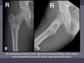
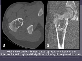
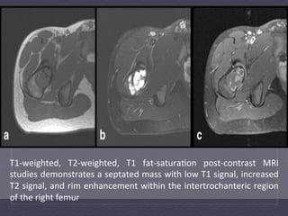
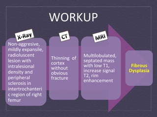
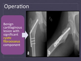
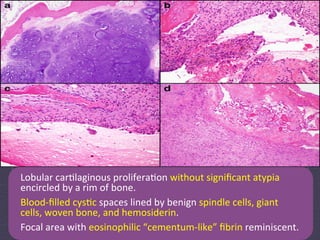
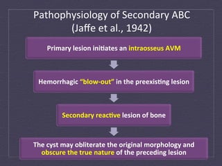
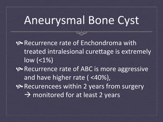
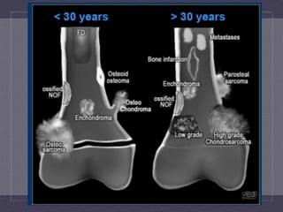
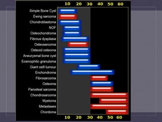
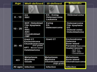
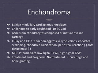
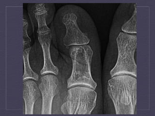
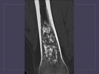
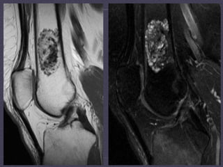
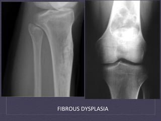
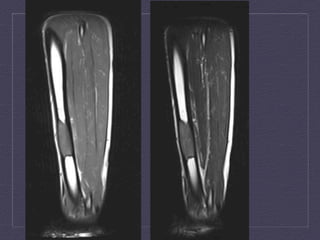
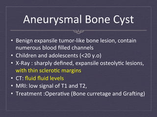
Case	
  Report:	
  
Enchondroma	
  in	
  	
  
Secondary	
  Aneurysmal	
  Bone	
  Cyst	
  	
  
Skeletal	
  Radiol.	
  2011	
  Nov;41(11):1475-­‐8.	
  Epub	
  2012	
  May	
  26	


Introduc:on	
  
•  Enchondroma	
  are	
  common	
  benign	
  car:laginous	
  tumors	
  
•  Most	
  common	
  in	
  metaphyseal	
  regions	
  
•  Most	
   common	
   sites:	
   Metacarpals,	
   Metatarsals,	
   Phalanges,	
  
   Proximal	
  humerus,	
  and	
  Femur	
  
•  X-­‐Ray:	
   well-­‐defined	
   ly:c	
   lesions,	
   punctate	
   intralesional	
  
   lesions	
   (giant	
   cell	
   tumor	
   of	
   bone,	
   fibrous	
   dysplasia,	
  
   chondrosarcoma,	
  and	
  osteonecrosis	
  
•  Histology:	
   lobulated	
   tumor	
   composed	
   of	
   hyaline	
   car:lage	
  
   that	
   lacks	
   significant	
   cellularity	
   and	
   atypia,	
   lamellar	
   bones	
  
   surround	
  the	
  lobules	
  

                     Cys:c	
  change	
  in	
  enchondroma	
  is	
  unusual	
  case	
  
Clinical	
  History	
  
Male,	
  13	
  years	
  old	


                                                                          Medical	
  Evalua:on	
  
                                                   Antalgic	
  gait	


                          Exacerbated	
  on	
  daily	
  
                          exercises	
  training	
  

      Right	
  groin	
  pain	
  
      radiated	
  down	
  the	
  
      front	
  of	
  thigh	
  
AP	
  and	
  lateral	
  views	
  of	
  the	
  right	
  hip	
  demonstra:ng	
  a	
  ly:c	
  lesion	
  
        with	
  peripheral	
  sclerosis	
  of	
  the	
  right	
  proximal	
  femur	
  
Axial	
  and	
  coronal	
  CT	
  demonstrates	
  septated,	
  ly:c	
  lesion	
  in	
  the	
  
intertrochanteric	
  region	
  with	
  significant	
  thinning	
  of	
  the	
  posterior	
  cortex	
  
T1-­‐weighted,	
   T2-­‐weighted,	
   T1	
   fat-­‐satura:on	
   post-­‐contrast	
   MRI	
  
studies	
  demonstrates	
  a	
  septated	
  mass	
  with	
  low	
  T1	
  signal,	
  increased	
  
T2	
  signal,	
  and	
  rim	
  enhancement	
  within	
  the	
  intertrochanteric	
  region	
  
of	
  the	
  right	
  femur	
  
WORKUP	
  
Non-­‐aggresive,	
  
mildly	
  expansile,	
  
radioluscent	
                                            Mul:lobulated,	
  
                                 Thinning	
  	
  of	
     septated	
  mass	
  
lesion	
  with	
                 cortex	
  
intralesional	
                  without	
                with	
  low	
  T1,	
      Fibrous	
  
density	
  and	
                 obvious	
                increase	
  signal	
     Dysplasia	
  
peripheral	
                     fracture	
               T2,	
  rim	
  
                                                          enhancement	
  
sclerosis	
  in	
  
intertrochanteri
c	
  region	
  of	
  right	
  
femur	
  
Opera:on	
  

Benign	
  
car:laginous	
  
lesion	
  with	
  
significant	
  
cys:c	
  
fibroosseus	
  
component	
  
Lobular	
  car:laginous	
  prolifera:on	
  without	
  significant	
  atypia	
  
encircled	
  by	
  a	
  rim	
  of	
  bone.	
  
Blood-­‐filled	
  cys:c	
  spaces	
  lined	
  by	
  benign	
  spindle	
  cells,	
  giant	
  
cells,	
  woven	
  bone,	
  and	
  hemosiderin.	
  
Focal	
  area	
  with	
  eosinophilic	
  “cementum-­‐like”	
  fibrin	
  reminiscent.	
  
Discussion	
  

 Aneurysmal	
  cys:c	
  changes	
  occur	
  secondary	
  
   to	
  number	
  of	
  pathologic	
  lesions	
  à	
  never	
  
   been	
  reported	
  in	
  Enchondroma	
  
Fibrous	
  Dysplasia	
  
  Radiographic	
  in	
  this	
  case:	
  Fibrous	
  Dysplasia	
  
  X-­‐Ray:	
  Radiolucent	
  lesion	
  with	
  ‘ground-­‐glass’	
  
    opacifica:on	
  
  Histology:	
  prolifera:ng	
  fibroblasts	
  with	
  osteoid	
  
    trabeculae	
  
  FD	
  with	
  massive	
  chondroid	
  differen:a:on	
  à	
  
    Fibrocar:laginous	
  Dysplasia	
  (FCD)	
  
  Rare	
  occasions:	
  Calcifica:on	
  may	
  be	
  so	
  extensive	
  
    à	
  mimicking	
  Enchondroma	
  or	
  Chondrosarcoma	
  
Aneurysmal	
  Bone	
  Cyst	
  
 Aneurysmal	
  Bone	
  Cyst	
  (ABC)	
  are	
  benign	
  
   osteoly:c	
  lesions	
  
 Blood-­‐filled	
  spaces	
  separated	
  by	
  connec:ve	
  
   :ssue	
  septae	
  
 Common	
  sites:	
  Long	
  bones	
  of	
  lower	
  
   extremity,	
  and	
  posterior	
  of	
  spine	
  
 Primary	
  and	
  secondary	
  ABC	
  
Pathophysiology	
  of	
  Secondary	
  ABC	
  	
  
       (Jaffe	
  et	
  al.,	
  1942)	
  
      Primary	
  lesion	
  ini6ates	
  an	
  intraosseus	
  AVM	


  Hemorrhagic	
  “blow-­‐out”	
  in	
  the	
  preexis6ng	
  lesion	


             Secondary	
  reac6ve	
  lesion	
  of	
  bone	


The	
  cyst	
  may	
  obliterate	
  the	
  original	
  morphology	
  and	
  
  obscure	
  the	
  true	
  nature	
  of	
  the	
  preceding	
  lesion	
  
Aneurysmal	
  Bone	
  Cyst	
  
 Recurrence	
  rate	
  of	
  Enchondroma	
  with	
  
   treated	
  intralesional	
  cure`age	
  is	
  extremely	
  
   low	
  (<1%)	
  
 Recurrence	
  rate	
  of	
  ABC	
  is	
  more	
  aggressive	
  
   and	
  have	
  higher	
  rate	
  (	
  <40%),	
  	
  
 Recurencees	
  within	
  2	
  years	
  from	
  surgery	
  
   à	
  monitored	
  for	
  at	
  least	
  2	
  years	
  
Thank You
BUBBLY	
  LYTIC	
  BONE	
  LESIONS	
  
•       F	
  :	
  Fibrous	
  Dysplasia	
  or	
  Fibrous	
  Cor:cal	
  Defect	
  
•       O:	
  Osteoblastoma	
  
•       G	
  :	
  Giant	
  cell	
  tumour	
  (GCT)	
  
•       M	
  :	
  Metastases	
  
•       A	
  :	
  Aneurysmal	
  Bone	
  Cyst	
  (ABC)	
  
•       C	
  :	
  Chondroblastoma	
  or	
  Chondromyxoid	
  fibroma	
  
•       H	
  :Hyperparathyroidism	
  (Brown	
  Tumor)	
  	
  
•       I	
  :	
  infec:on	
  (Osteomyeli:s)	
  
•       N	
  :	
  Non-­‐ossifying	
  Fibroma	
  
•       E	
  :Enchondroma	
  or	
  Eosinophilic	
  Granuloma	
  
•       S	
  :	
  Simple	
  Bone	
  Cyst	
  
AJR	
  	
  August	
  2009	
  vol	
  193	
  no.	
  2	
  W79-­‐W94	
  	
  
Distribu:on	
  of	
  Lesions	
  
        Enchondroma	
                                      Fibrous	
  Dysplasia	
                                          ABC	
                                          GCT	


Layout	
  and	
  distribuLon:	
  Frank	
  Gaillard	
  2009,	
  Line	
  drawing	
  of	
  skeleton:	
  Patrick	
  Lynch	
  2006,	
  CreaLve	
  Common	
  NC-­‐SA-­‐BY	
  
Enchondroma	
  
  Benign	
  medullary	
  car:laginous	
  neoplasm	
  
  Childhood	
  to	
  early	
  adulthood	
  (10-­‐30	
  y.o)	
  
  Arise	
  from	
  chondrocytes	
  composed	
  of	
  mature	
  hyaline	
  
    car:lage	
  
  X-­‐Ray	
  and	
  CT:	
  1-­‐2	
  cm	
  non-­‐aggressive	
  ly:c	
  lesions,	
  endosteal	
  
    scalloping,	
  chondroid	
  calcifica:on,	
  periosteal	
  reac:on	
  (-­‐),sof	
  
    :ssue	
  mass	
  (-­‐)	
  
  MRI:	
  Intermediate-­‐low	
  signal	
  T1WI,	
  high	
  signal	
  T2WI	
  
  Treatment	
  and	
  Prognosis:	
  No	
  treatment	
  à	
  cure`age	
  and	
  
    bone	
  grafing	
  
Fibrous	
  Dysplasia	
  
•  Benign	
  tumour-­‐like	
  congenital	
  process	
  
•  Defect	
  in	
  osteoblas:c	
  differen:a:on	
  and	
  matura:on,	
  with	
  
   progressive	
  replacement	
  of	
  normal	
  bone	
  with	
  immature	
  woven	
  
   bone	
  
•  Children	
  and	
  yound	
  adults,	
  <	
  30	
  y.o	
  
•  X-­‐Ray	
  and	
  CT:	
  Well	
  circumscribed	
  cys:c	
  or	
  sclero:c	
  lesion,	
  
   Ground-­‐glass	
  opacity,	
  Endosteal	
  scalloping,	
  intact	
  bone	
  
•  MRI:	
  Heterogenous	
  Intermediate	
  signal	
  T1WI,	
  heterogenous	
  
   low	
  signal	
  T2WI	
  
•  Treatment:	
  no	
  treatment	
  à	
  surgical	
  decompression	
  
FIBROUS	
  DYSPLASIA	
  
Aneurysmal	
  Bone	
  Cyst	
  
•  Benign	
  expansile	
  tumor-­‐like	
  bone	
  lesion,	
  contain	
  
   numerous	
  blood	
  filled	
  channels	
  
•  Children	
  and	
  adolescents	
  (<20	
  y.o)	
  
•  X-­‐Ray	
  :	
  sharply	
  defined,	
  expansile	
  osteoly:c	
  lesions,	
  
   with	
  thin	
  sclero:c	
  margins	
  
•  CT:	
  fluid	
  fluid	
  levels	
  
•  MRI:	
  low	
  signal	
  of	
  T1	
  and	
  T2,	
  
•  Treatment	
  :Opera:ve	
  (Bone	
  curretage	
  and	
  Grafing)	
  
Giant	
  Cell	
  Tumor	
  
  Benign,	
  metaepiphysis	
  of	
  long	
  bones	
  	
  
  Early	
  adulthood	
  (	
  20-­‐	
  50	
  y.o)	
  
  Single	
  lesions	
  (knee,	
  distal	
  radius,	
  sacrum)	
  
  X-­‐Ray	
  and	
  CT	
  :	
  occurs	
  only	
  with	
  a	
  	
  closed	
  epiphyseal	
  
    plate,	
  ar:cular	
  surface,	
  well	
  defined	
  with	
  non-­‐
    sclero:c	
  margin,	
  periosteal	
  reac:on	
  (10-­‐30%	
  cases),	
  
    no	
  calcifica:on	
  
  MRI:	
  low	
  signal	
  in	
  T1WI	
  and	
  T2WI	
  
  Angiography:	
  hypervascular	
  tumor	
  with	
  the	
  rest	
  
    being	
  hipo/avascular	
  
ENCHONDROMA	
  VS	
  LOW	
  GRADE	
  CHONDROSARCOMA	
  
Unicameral	
  (Simple)	
  
	
  Bone	
  Cyst	


Fallen-­‐Fragment	
  sign	
  

Enchondroma with Secondary Aneurysmal Bone Cyst

  • 1.
    Case  Report:   Enchondroma  in     Secondary  Aneurysmal  Bone  Cyst     Skeletal  Radiol.  2011  Nov;41(11):1475-­‐8.  Epub  2012  May  26    
  • 2.
    Introduc:on   •  Enchondroma  are  common  benign  car:laginous  tumors   •  Most  common  in  metaphyseal  regions   •  Most   common   sites:   Metacarpals,   Metatarsals,   Phalanges,   Proximal  humerus,  and  Femur   •  X-­‐Ray:   well-­‐defined   ly:c   lesions,   punctate   intralesional   lesions   (giant   cell   tumor   of   bone,   fibrous   dysplasia,   chondrosarcoma,  and  osteonecrosis   •  Histology:   lobulated   tumor   composed   of   hyaline   car:lage   that   lacks   significant   cellularity   and   atypia,   lamellar   bones   surround  the  lobules   Cys:c  change  in  enchondroma  is  unusual  case  
  • 3.
    Clinical  History   Male,  13  years  old   Medical  Evalua:on   Antalgic  gait   Exacerbated  on  daily   exercises  training   Right  groin  pain   radiated  down  the   front  of  thigh  
  • 4.
    AP  and  lateral  views  of  the  right  hip  demonstra:ng  a  ly:c  lesion   with  peripheral  sclerosis  of  the  right  proximal  femur  
  • 5.
    Axial  and  coronal  CT  demonstrates  septated,  ly:c  lesion  in  the   intertrochanteric  region  with  significant  thinning  of  the  posterior  cortex  
  • 6.
    T1-­‐weighted,   T2-­‐weighted,   T1   fat-­‐satura:on   post-­‐contrast   MRI   studies  demonstrates  a  septated  mass  with  low  T1  signal,  increased   T2  signal,  and  rim  enhancement  within  the  intertrochanteric  region   of  the  right  femur  
  • 7.
    WORKUP   Non-­‐aggresive,   mildly  expansile,   radioluscent   Mul:lobulated,   Thinning    of   septated  mass   lesion  with   cortex   intralesional   without   with  low  T1,   Fibrous   density  and   obvious   increase  signal   Dysplasia   peripheral   fracture   T2,  rim   enhancement   sclerosis  in   intertrochanteri c  region  of  right   femur  
  • 8.
    Opera:on   Benign   car:laginous   lesion  with   significant   cys:c   fibroosseus   component  
  • 9.
    Lobular  car:laginous  prolifera:on  without  significant  atypia   encircled  by  a  rim  of  bone.   Blood-­‐filled  cys:c  spaces  lined  by  benign  spindle  cells,  giant   cells,  woven  bone,  and  hemosiderin.   Focal  area  with  eosinophilic  “cementum-­‐like”  fibrin  reminiscent.  
  • 10.
    Discussion    Aneurysmal  cys:c  changes  occur  secondary   to  number  of  pathologic  lesions  à  never   been  reported  in  Enchondroma  
  • 11.
    Fibrous  Dysplasia    Radiographic  in  this  case:  Fibrous  Dysplasia     X-­‐Ray:  Radiolucent  lesion  with  ‘ground-­‐glass’   opacifica:on     Histology:  prolifera:ng  fibroblasts  with  osteoid   trabeculae     FD  with  massive  chondroid  differen:a:on  à   Fibrocar:laginous  Dysplasia  (FCD)     Rare  occasions:  Calcifica:on  may  be  so  extensive   à  mimicking  Enchondroma  or  Chondrosarcoma  
  • 12.
    Aneurysmal  Bone  Cyst    Aneurysmal  Bone  Cyst  (ABC)  are  benign   osteoly:c  lesions    Blood-­‐filled  spaces  separated  by  connec:ve   :ssue  septae    Common  sites:  Long  bones  of  lower   extremity,  and  posterior  of  spine    Primary  and  secondary  ABC  
  • 13.
    Pathophysiology  of  Secondary  ABC     (Jaffe  et  al.,  1942)   Primary  lesion  ini6ates  an  intraosseus  AVM   Hemorrhagic  “blow-­‐out”  in  the  preexis6ng  lesion   Secondary  reac6ve  lesion  of  bone   The  cyst  may  obliterate  the  original  morphology  and   obscure  the  true  nature  of  the  preceding  lesion  
  • 14.
    Aneurysmal  Bone  Cyst    Recurrence  rate  of  Enchondroma  with   treated  intralesional  cure`age  is  extremely   low  (<1%)    Recurrence  rate  of  ABC  is  more  aggressive   and  have  higher  rate  (  <40%),      Recurencees  within  2  years  from  surgery   à  monitored  for  at  least  2  years  
  • 15.
  • 21.
    BUBBLY  LYTIC  BONE  LESIONS   •  F  :  Fibrous  Dysplasia  or  Fibrous  Cor:cal  Defect   •  O:  Osteoblastoma   •  G  :  Giant  cell  tumour  (GCT)   •  M  :  Metastases   •  A  :  Aneurysmal  Bone  Cyst  (ABC)   •  C  :  Chondroblastoma  or  Chondromyxoid  fibroma   •  H  :Hyperparathyroidism  (Brown  Tumor)     •  I  :  infec:on  (Osteomyeli:s)   •  N  :  Non-­‐ossifying  Fibroma   •  E  :Enchondroma  or  Eosinophilic  Granuloma   •  S  :  Simple  Bone  Cyst   AJR    August  2009  vol  193  no.  2  W79-­‐W94    
  • 22.
    Distribu:on  of  Lesions   Enchondroma   Fibrous  Dysplasia   ABC   GCT   Layout  and  distribuLon:  Frank  Gaillard  2009,  Line  drawing  of  skeleton:  Patrick  Lynch  2006,  CreaLve  Common  NC-­‐SA-­‐BY  
  • 23.
    Enchondroma     Benign  medullary  car:laginous  neoplasm     Childhood  to  early  adulthood  (10-­‐30  y.o)     Arise  from  chondrocytes  composed  of  mature  hyaline   car:lage     X-­‐Ray  and  CT:  1-­‐2  cm  non-­‐aggressive  ly:c  lesions,  endosteal   scalloping,  chondroid  calcifica:on,  periosteal  reac:on  (-­‐),sof   :ssue  mass  (-­‐)     MRI:  Intermediate-­‐low  signal  T1WI,  high  signal  T2WI     Treatment  and  Prognosis:  No  treatment  à  cure`age  and   bone  grafing  
  • 27.
    Fibrous  Dysplasia   • Benign  tumour-­‐like  congenital  process   •  Defect  in  osteoblas:c  differen:a:on  and  matura:on,  with   progressive  replacement  of  normal  bone  with  immature  woven   bone   •  Children  and  yound  adults,  <  30  y.o   •  X-­‐Ray  and  CT:  Well  circumscribed  cys:c  or  sclero:c  lesion,   Ground-­‐glass  opacity,  Endosteal  scalloping,  intact  bone   •  MRI:  Heterogenous  Intermediate  signal  T1WI,  heterogenous   low  signal  T2WI   •  Treatment:  no  treatment  à  surgical  decompression  
  • 28.
  • 31.
    Aneurysmal  Bone  Cyst   •  Benign  expansile  tumor-­‐like  bone  lesion,  contain   numerous  blood  filled  channels   •  Children  and  adolescents  (<20  y.o)   •  X-­‐Ray  :  sharply  defined,  expansile  osteoly:c  lesions,   with  thin  sclero:c  margins   •  CT:  fluid  fluid  levels   •  MRI:  low  signal  of  T1  and  T2,   •  Treatment  :Opera:ve  (Bone  curretage  and  Grafing)  
  • 37.
    Giant  Cell  Tumor     Benign,  metaepiphysis  of  long  bones       Early  adulthood  (  20-­‐  50  y.o)     Single  lesions  (knee,  distal  radius,  sacrum)     X-­‐Ray  and  CT  :  occurs  only  with  a    closed  epiphyseal   plate,  ar:cular  surface,  well  defined  with  non-­‐ sclero:c  margin,  periosteal  reac:on  (10-­‐30%  cases),   no  calcifica:on     MRI:  low  signal  in  T1WI  and  T2WI     Angiography:  hypervascular  tumor  with  the  rest   being  hipo/avascular  
  • 39.
    ENCHONDROMA  VS  LOW  GRADE  CHONDROSARCOMA  
  • 41.
    Unicameral  (Simple)    Bone  Cyst     Fallen-­‐Fragment  sign