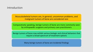
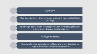
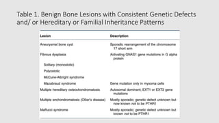
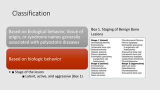
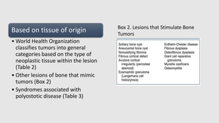
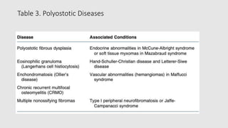
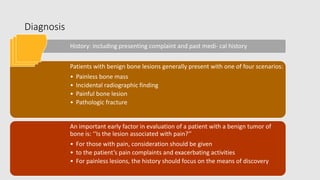
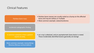
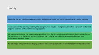
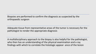
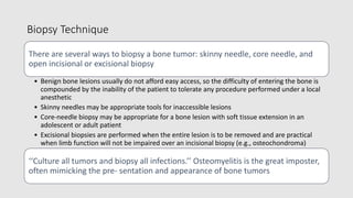
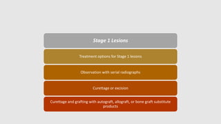
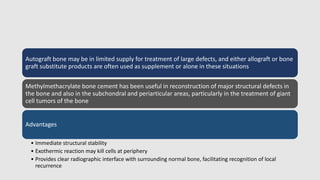
Benign Tumors of Bone
Introduction
Musculoskeletal tumors are, in general, uncommon problems, and
malignant tumors of bone are considered rare
Comparatively speaking, benign tumors of bone are more commonly seen
by orthopaedic surgeons and primary care practitioners
Benign tumors of bone may exhibit various biologic and clinical behaviors that
require a broad spectrum of treatment options
Many benign tumors of bone are incidental findings
Etiology
Most bone tumors, either benign or malignant, have no identifiable
etiology
Few benign bone lesions are associated with preexisting conditions
or have a hereditary or familial pattern
Pathophysiology
Unknown for most benign tumors of bone and is best described for
congenital and inherited syndromes (Table 1)
Table 1. Benign Bone Lesions with Consistent Genetic Defects
and/ or Hereditary or Familial Inheritance Patterns
Classification
Based on biological behavior, tissue of
origin, or syndrome names generally
associated with polyostotic diseases
Based on biologic behavior
โ€ข โ–  Stage of the lesion
โ–  Latent, active, and aggressive (Box 1)
Box 1. Staging of Benign Bone
Lesions
Based on tissue of origin
โ€ข World Health Organization
classifies tumors into general
categories based on the type of
neoplastic tissue within the lesion
(Table 2)
โ€ข Other lesions of bone that mimic
tumors (Box 2)
โ€ข Syndromes associated with
polyostotic disease (Table 3)
Box 2. Lesions that Stimulate Bone
Tumors
Table 2 World Health Organization Benign Bone Tumor
Categories
Table 3. Polyostotic Diseases
Diagnosis
History: including presenting complaint and past medi- cal history
Patients with benign bone lesions generally present with one of four scenarios:
โ€ข Painless bone mass
โ€ข Incidental radiographic finding
โ€ข Painful bone lesion
โ€ข Pathologic fracture
An important early factor in evaluation of a patient with a benign tumor of
bone is: โ€˜โ€˜Is the lesion associated with pain?โ€™โ€™
โ€ข For those with pain, consideration should be given
โ€ข to the patientโ€™s pain complaints and exacerbating activities
โ€ข For painless lesions, the history should focus on the means of discovery
Clinical Features
โ€ข Painless bone masses are usually noted as a bump on the affected
bone and may be solitary or multiple
โ€ข Most common example: osteochondroma
Painless bone mass
Incidental radiographic finding
โ€ข an x-ray is obtained, and an asymptomatic bony lesion is noted.
These incidentally identified lesions generally are benign
A common scenario is that a patient
injures an area,
Most common example: nonossifying
fibroma (fibrous cortical defect)
Painful bone lesion
โ€ข โ–  When a patient presents with pain as the primary complaint and an underlying
tumor of bone is identified, a more thorough evaluation is warranted to rule out
an aggressive benign tumor that is associated with structural compromise of the
bone or even a malignancy
โ€ข โ–  Differential diagnosis in this situation may be extensive
Pathologic Fracture
โ€ข โ–  Most children with a pathologic fracture through an underlying benign tumor of
bone can be managed conservatively initially to allow for fracture healing, and
then a biopsy may be performed in a delayed fashion if appropriate
Key question in determining whether there is cause for
concern: Was pain present preceding the time of the
fracture?
โ€ข โ–  Preceding pain: Possible aggressive benign or
malignant lesion: may warrant biopsy/surgical
intervention
No preceding pain: Very likely a benign, inactive lesion
โ€ข โ–  Common examples: nonossifying fibroma, simple bone
cyst
Physical Examination
โ€ข General physical examination
โ€ข Look for signs of multifocal disease (cafe ฬ-au-lait spots, axillary freckling,
multiple bony masses, soft tissue myxomas, soft tissue hemangiomas, short
stature, limb deformities), generalized lymphadenopathy
โ€ข Examine for possible sites of referred pain
โ€ข Focused examination concentrating on the area of concern
โ€ข Specific findings to assess are the presence of a visible mass, limb length
discrepancy, overlying skin changes, increased tactile temperature, presence
of a soft tissue mass, tenderness on palpation of the area, compromised joint
range of motion, surrounding muscle atrophy, neurovascular status of the
limb and any lymphadenopathy
Radiographic Evaluation
โ€ข At a minimum, two orthogonal view x-rays of
the affected area should be obtained, an AP
and a lateral view
โ€ข Oblique views may also be of assistance
โ€ข โ– In the case of an inactive lesion that is an
incidental radiographic finding, observation
and serial x-rays are often all that is
warranted. This regimen affords the
opportunity to observe the natural history of
the process
X-ray films are a
necessary part of the
work-up of any patient
with a musculoskeletal
complaint
โ–  Consider proximal x-ray evaluation of an area that can be associated with a referred pain
pattern (e.g., hip pa- thology presenting as knee pain, spine pathology presenting as hip pain)
โ–  Size of the lesion on initial evaluation is generally of limited help in establishing a diagnosis
โ–  Systematic review of the x-ray is essential
The soft tissues should be evaluated for mineralization or other significant findings
The bone then should be evaluated with attention to location of the lesion, what the lesion is
doing to the bone, what the bone is doing in response to the lesion, any other characteristic
findings that may suggest a given diagnosis, especially matrix mineralization
Dense sclerotic margin around the tumor is a characteristic sign of a benign tumor of bone
(Fig. 1).
Mature periosteal reaction. If the periosteum has had an opportunity to react to the
expansion of the bone by forming mature bone, a benign process is favored
Lack of sclerotic rim suggests more rapid growth of the underlying lesion (Fig. 2).
Soft tissue extension: Any extension of the lesion into the surrounding soft tissues is an
ominous sign and sug- gests a rapidly growing processโ€”a malignancy, an infection, or an
aggressive benign process
Onion-skinning, sequential periosteal elevation, is also indicative of a rapidly growing
process; it can be seen in both benign and malignant tumors
Codmanโ€™s triangles are a result of rapid periosteal elevation indicative of a rapidly growing
process such as an infection or malignancy
Specific Radiographic Findings (Table 4)
Table 4. Radiographic Types of Responses to Bone Lesions
Fig 1. Benign nonossifying fibroma of femur.
Note sclerotic margins
Fig 2. Lytic epiphyseal and metaphyseal lesion
of distal femur without sclerotic margins,
consistent with giant cell tumor of bone
โ–  Location of the lesion within the bone is important in the formulation
of the differential diagnosis.
โ€ข Majority of lesions occur within the metaphysis.
โ€ข Neoplasms that arise in the epiphysis are giant cell tumor, chondroblastoma, and clear cell
chondrosarcoma. In addition, juxta-articular cysts such as those from pigmented villonodular
synovitis and degenerative geodes are often located in the epiphysis of adult bones.
Aneurysmal bone cysts rarely occur in the epiphysis. Brodieโ€™s abscesses often are located in
the epiphysis of children, and tuberculosis may cause epiphyseal โ€˜โ€˜kissing cystsโ€™โ€™ in adults (Fig
3 and Box 3)
โ–  Benign-appearing diaphyseal lesions tend to be eo- sinophilic
granuloma, fibrous dysplasia, โ€˜โ€˜inactiveโ€™โ€™ positioned simple bone cysts,
and osteomyelitis
Fig 3. PGCAT lesions. Epiphyseal lesions have a
relatively narrow differential diagnosis that can be
recalled using the mnemonic PGCAT, which
includes such common epiphyseal lesions as PVNS
(pigmented villonodular synovitis), GCT (giant cell
tumor of bone), chondroblastoma, ABC
(aneurysmal bone cyst), and Tb (tuberculosis)
Box 3. Differential Diagnosis of Epiphyseal Lesions: PGCAT
Other Imaging
Studies
Technetium-99m bone scan may be helpful
in identifying other sites of skeletal
involvement
Computed tomography (CT) scans are
generally helpful in assessing specific bone
lesions such as osteoid osteoma (to identify
the nidus) and enchondroma (to determine
the extent of endosteal scalloping)
Magnetic resonance imaging (MRI) useful to delineate any associated soft tissue
masses and the intraosseous extent of the process. It also helps to distinguish
between tissues of similar densities but different histologies. In certain situations,
specific findings may be helpful:
โ€ข Fluid โ€“ fluid levels are associated with aneurysmal bone cyst
โ€ข Perilesional edema is often seen with osteomyelitis, osteoid osteoma, eosinophilic granuloma
(Langerhans cell histiocytosis), and chondroblastoma and may be extensive
โ€ข Peripheral enhancement alone (without central en- hancement) is seen with simple cysts,
degenerative geodes, or intraosseous synovial cysts.
โ€ข Fat, especially surrounding a central area of ossification (the cockade sign), is diagnostic of
intraosseous lipoma
Biopsy
Should be the last step in the evaluation of a benign bone tumor and performed only after careful planning.
There is always the remote possibility that benign tumor may be a malignancy, therefore a properly performed
biopsy is essential for future limb salvage options
It is recommended that the surgeon who would perform the ultimate limb-sparing surgical procedure also be
the one to perform the biopsy so that the biopsy track may be placed in the appropriate location for a
definitive orthopaedic surgery
If a radiologist is to perform the biopsy, guidance for needle placement is recommended from the orthopedist
Biopsies are performed to confirm the diagnosis as suspected by the
orthopaedic surgeon
Adequate tissue from representative areas of the tumor is necessary for the
pathologist to render the appropriate diagnosis
A multidisciplinary approach to the biopsy is also helpful for the pathologist,
who then has an understanding of the physical examination and x-ray
findings with which to correlate the histologic appear- ance of the lesion
Biopsy Technique
There are several ways to biopsy a bone tumor: skinny needle, core needle, and
open incisional or excisional biopsy
โ€ข Benign bone lesions usually do not afford easy access, so the difficulty of entering the bone is
compounded by the inability of the patient to tolerate any procedure performed under a local
anesthetic
โ€ข Skinny needles may be appropriate tools for inaccessible lesions
โ€ข Core-needle biopsy may be appropriate for a bone lesion with soft tissue extension in an
adolescent or adult patient
โ€ข Excisional biopsies are performed when the entire lesion is to be removed and are practical
when limb function will not be impaired over an incisional biopsy (e.g., osteochondroma)
โ€˜โ€˜Culture all tumors and biopsy all infections.โ€™โ€™ Osteomyelitis is the great imposter,
often mimicking the pre- sentation and appearance of bone tumors
Surgical Management
General Principles
Treatment options for benign tumors of bone are matched to the aggressiveness of the lesion.
Specific treatment considerations may apply to individual tumor types and the stage of the lesion (see Box
1)
Other variables affecting treatment choices
Clinical picture, radiographic evaluation, physicianโ€™s judgment and experience, patient and/or family
goals/desires
Stage 1 Lesions
Treatment options for Stage 1 lesions
Observation with serial radiographs
Curettage or excision
Curettage and grafting with autograft, allograft, or bone graft substitute
products
Stage 1 lesions identified as an incidental finding: Observation is
appropriate in most cases.
โ€ข Serial radiographic and clinical evaluation
โ€ข The potential for these lesions to remain stable or
โ€ข resolve on their own is greater than for a lesion asso- ciated with symptoms or a more
active radiographic appearance.
Stage 1 lesions in high-stress areas (proximal femur) and those that are
symptomatic or cause structural compromise to the bone should be
considered for surgical intervention
โ€ข Generally, curettage and grafting with or without stabilization is elected in these cases.
โ€ข Need for stabilization is determined by the presence or absence of fracture, anatomic site,
risk of subsequent fracture, activity level, and ability to comply with postoperative
restrictions.
Stage
2
Lesions
โ€ข Concerns: Stage 2 lesions often pose a greater risk of pathologic fracture
based on the location and size of the lesion as well as activity and age of
the patient
โ€ข Standard of care: Generally treated with an intralesional procedure (with
the exception of juxtacortical chondroma), with the defect filled with
bone graft, bone graft substitute materials, or cementation
โ€ข Specific exceptions
โ€ข Juxtacortical chondroma: Recurrence rate is less following en bloc
excision
โ€ข Osteoid osteoma: Radiofrequency ablation has generally supplanted
curettage: medical management with nonsteroidal anti-inflammatory
agents is also an option
Stage 3 Lesions
โ€ข Concerns: Stage 3 lesions continue their growth unless treated. More aggressive
and have a greater risk of pathologic fracture, local recurrence and in some cases
(giant cell tumors and chondroblastomas) a risk of metastatic disease
โ€ข Standard of care: Generally treated with an extended curettage with or without
adjuvant agents (phenol, laser, liquid nitrogen) and with or without stabilization
โ€ข Defect may be filled with bone graft or bone graft
โ€ข substitute products, bone cement, or structural grafts
โ€ข Indications for stabilization same as for stage 1 and 2 lesions (above)
โ€ข Exceptions
โ€ข Expendable bone or multiply recurrent tumors with extensive bone destruction:
wide en bloc excision
โ€ข In the case of wide excision, reconstruction com- mences in a similar fashion to
that following removal of a sarcoma, including a megaprosthesis or allograft
prosthetic composite reconstruction
Reconstruction
Removal of large, active, or aggressive lesions may result in a large structural defect that
warrants additional con- sideration for reconstructive options
None of the commercially available bone graft substitute materials are approved by the
Food and Drug Administration for reconstruction of structural de- fects in bone
A combination of an osteoinductive agent, an osteoconductive product, and a source of
osteoprogenitor cells (bone marrow aspirate) recreates the elements contained in an
autograft
Autograft bone may be in limited supply for treatment of large defects, and either allograft or bone
graft substitute products are often used as supplement or alone in these situations
Methylmethacrylate bone cement has been useful in reconstruction of major structural defects in
the bone and also in the subchondral and periarticular areas, particularly in the treatment of giant
cell tumors of the bone
Advantages
โ€ข Immediate structural stability
โ€ข Exothermic reaction may kill cells at periphery
โ€ข Provides clear radiographic interface with surrounding normal bone, facilitating recognition of local
recurrence
Results and Outcomes
Results of treatment for benign tumors of bone are gen- erally favorable, barring
any complications of a surgical procedure or pathologic fracture through a lesion
Local recurrence rates vary according to the underlying diagnosis.
Many assessment tools are available for functional outcome, including the
Musculoskeletal Tumor Society Rating Scale, The Hospital for Special Surgery Knee
and Harris Hip scores, as well as the Health Satisfaction Questionnaire (SF-36).
Postoperative Management and Follow-Up
Variables important in determining postoperative management
(including activity, weight bearing)
Surgical procedure performed, upper versus lower
extremity, specific location, size of defect, type of reconstruction,
presence or absence of stabilization device, underlying disease
process, medical comorbidities, and compliance
Stage 3 (aggressive) lesions merit a closer follow-up, much like a
sarcoma, particularly for the first 2 years postoperatively, to
evaluate for local recurrence and metastatic foci of disease
Benign bone lesions with a high risk of local recurrence
โ€ข Giant cell tumor of bone: near 50% recurrence if treated with simple curettage and bone
grafting and as low as 3% to 17% when adjuvant agents, extended curettage, and cementation
are used
โ€ข Aneurysmal bone cysts: Risk of recurrence for secondary aneurysmal bone cyst depends on
the accompanying lesion. Primary aneurysmal bone cysts have a local recurrence rate as high
as 32%.
โ€ข Chondroblastomas: 5% local recurrence risk
Benign bone tumors with risk of pulmonary metastases: giant cell tumor,
chondroblastoma
THANK YOU

Benign Bone Tumor.pptx

  • 1.
  • 2.
    Introduction Musculoskeletal tumors are,in general, uncommon problems, and malignant tumors of bone are considered rare Comparatively speaking, benign tumors of bone are more commonly seen by orthopaedic surgeons and primary care practitioners Benign tumors of bone may exhibit various biologic and clinical behaviors that require a broad spectrum of treatment options Many benign tumors of bone are incidental findings
  • 3.
    Etiology Most bone tumors,either benign or malignant, have no identifiable etiology Few benign bone lesions are associated with preexisting conditions or have a hereditary or familial pattern Pathophysiology Unknown for most benign tumors of bone and is best described for congenital and inherited syndromes (Table 1)
  • 4.
    Table 1. BenignBone Lesions with Consistent Genetic Defects and/ or Hereditary or Familial Inheritance Patterns
  • 5.
    Classification Based on biologicalbehavior, tissue of origin, or syndrome names generally associated with polyostotic diseases Based on biologic behavior โ€ข โ–  Stage of the lesion โ–  Latent, active, and aggressive (Box 1) Box 1. Staging of Benign Bone Lesions
  • 6.
    Based on tissueof origin โ€ข World Health Organization classifies tumors into general categories based on the type of neoplastic tissue within the lesion (Table 2) โ€ข Other lesions of bone that mimic tumors (Box 2) โ€ข Syndromes associated with polyostotic disease (Table 3) Box 2. Lesions that Stimulate Bone Tumors
  • 7.
    Table 2 WorldHealth Organization Benign Bone Tumor Categories
  • 8.
  • 9.
    Diagnosis History: including presentingcomplaint and past medi- cal history Patients with benign bone lesions generally present with one of four scenarios: โ€ข Painless bone mass โ€ข Incidental radiographic finding โ€ข Painful bone lesion โ€ข Pathologic fracture An important early factor in evaluation of a patient with a benign tumor of bone is: โ€˜โ€˜Is the lesion associated with pain?โ€™โ€™ โ€ข For those with pain, consideration should be given โ€ข to the patientโ€™s pain complaints and exacerbating activities โ€ข For painless lesions, the history should focus on the means of discovery
  • 10.
    Clinical Features โ€ข Painlessbone masses are usually noted as a bump on the affected bone and may be solitary or multiple โ€ข Most common example: osteochondroma Painless bone mass Incidental radiographic finding โ€ข an x-ray is obtained, and an asymptomatic bony lesion is noted. These incidentally identified lesions generally are benign A common scenario is that a patient injures an area, Most common example: nonossifying fibroma (fibrous cortical defect)
  • 11.
    Painful bone lesion โ€ขโ–  When a patient presents with pain as the primary complaint and an underlying tumor of bone is identified, a more thorough evaluation is warranted to rule out an aggressive benign tumor that is associated with structural compromise of the bone or even a malignancy โ€ข โ–  Differential diagnosis in this situation may be extensive Pathologic Fracture โ€ข โ–  Most children with a pathologic fracture through an underlying benign tumor of bone can be managed conservatively initially to allow for fracture healing, and then a biopsy may be performed in a delayed fashion if appropriate
  • 12.
    Key question indetermining whether there is cause for concern: Was pain present preceding the time of the fracture? โ€ข โ–  Preceding pain: Possible aggressive benign or malignant lesion: may warrant biopsy/surgical intervention No preceding pain: Very likely a benign, inactive lesion โ€ข โ–  Common examples: nonossifying fibroma, simple bone cyst
  • 13.
    Physical Examination โ€ข Generalphysical examination โ€ข Look for signs of multifocal disease (cafe ฬ-au-lait spots, axillary freckling, multiple bony masses, soft tissue myxomas, soft tissue hemangiomas, short stature, limb deformities), generalized lymphadenopathy โ€ข Examine for possible sites of referred pain โ€ข Focused examination concentrating on the area of concern โ€ข Specific findings to assess are the presence of a visible mass, limb length discrepancy, overlying skin changes, increased tactile temperature, presence of a soft tissue mass, tenderness on palpation of the area, compromised joint range of motion, surrounding muscle atrophy, neurovascular status of the limb and any lymphadenopathy
  • 14.
    Radiographic Evaluation โ€ข Ata minimum, two orthogonal view x-rays of the affected area should be obtained, an AP and a lateral view โ€ข Oblique views may also be of assistance โ€ข โ– In the case of an inactive lesion that is an incidental radiographic finding, observation and serial x-rays are often all that is warranted. This regimen affords the opportunity to observe the natural history of the process X-ray films are a necessary part of the work-up of any patient with a musculoskeletal complaint
  • 15.
    โ–  Consider proximalx-ray evaluation of an area that can be associated with a referred pain pattern (e.g., hip pa- thology presenting as knee pain, spine pathology presenting as hip pain) โ–  Size of the lesion on initial evaluation is generally of limited help in establishing a diagnosis โ–  Systematic review of the x-ray is essential The soft tissues should be evaluated for mineralization or other significant findings The bone then should be evaluated with attention to location of the lesion, what the lesion is doing to the bone, what the bone is doing in response to the lesion, any other characteristic findings that may suggest a given diagnosis, especially matrix mineralization
  • 16.
    Dense sclerotic marginaround the tumor is a characteristic sign of a benign tumor of bone (Fig. 1). Mature periosteal reaction. If the periosteum has had an opportunity to react to the expansion of the bone by forming mature bone, a benign process is favored Lack of sclerotic rim suggests more rapid growth of the underlying lesion (Fig. 2). Soft tissue extension: Any extension of the lesion into the surrounding soft tissues is an ominous sign and sug- gests a rapidly growing processโ€”a malignancy, an infection, or an aggressive benign process Onion-skinning, sequential periosteal elevation, is also indicative of a rapidly growing process; it can be seen in both benign and malignant tumors Codmanโ€™s triangles are a result of rapid periosteal elevation indicative of a rapidly growing process such as an infection or malignancy Specific Radiographic Findings (Table 4)
  • 17.
    Table 4. RadiographicTypes of Responses to Bone Lesions
  • 18.
    Fig 1. Benignnonossifying fibroma of femur. Note sclerotic margins Fig 2. Lytic epiphyseal and metaphyseal lesion of distal femur without sclerotic margins, consistent with giant cell tumor of bone
  • 19.
    โ–  Location ofthe lesion within the bone is important in the formulation of the differential diagnosis. โ€ข Majority of lesions occur within the metaphysis. โ€ข Neoplasms that arise in the epiphysis are giant cell tumor, chondroblastoma, and clear cell chondrosarcoma. In addition, juxta-articular cysts such as those from pigmented villonodular synovitis and degenerative geodes are often located in the epiphysis of adult bones. Aneurysmal bone cysts rarely occur in the epiphysis. Brodieโ€™s abscesses often are located in the epiphysis of children, and tuberculosis may cause epiphyseal โ€˜โ€˜kissing cystsโ€™โ€™ in adults (Fig 3 and Box 3) โ–  Benign-appearing diaphyseal lesions tend to be eo- sinophilic granuloma, fibrous dysplasia, โ€˜โ€˜inactiveโ€™โ€™ positioned simple bone cysts, and osteomyelitis
  • 20.
    Fig 3. PGCATlesions. Epiphyseal lesions have a relatively narrow differential diagnosis that can be recalled using the mnemonic PGCAT, which includes such common epiphyseal lesions as PVNS (pigmented villonodular synovitis), GCT (giant cell tumor of bone), chondroblastoma, ABC (aneurysmal bone cyst), and Tb (tuberculosis)
  • 21.
    Box 3. DifferentialDiagnosis of Epiphyseal Lesions: PGCAT
  • 22.
    Other Imaging Studies Technetium-99m bonescan may be helpful in identifying other sites of skeletal involvement Computed tomography (CT) scans are generally helpful in assessing specific bone lesions such as osteoid osteoma (to identify the nidus) and enchondroma (to determine the extent of endosteal scalloping)
  • 23.
    Magnetic resonance imaging(MRI) useful to delineate any associated soft tissue masses and the intraosseous extent of the process. It also helps to distinguish between tissues of similar densities but different histologies. In certain situations, specific findings may be helpful: โ€ข Fluid โ€“ fluid levels are associated with aneurysmal bone cyst โ€ข Perilesional edema is often seen with osteomyelitis, osteoid osteoma, eosinophilic granuloma (Langerhans cell histiocytosis), and chondroblastoma and may be extensive โ€ข Peripheral enhancement alone (without central en- hancement) is seen with simple cysts, degenerative geodes, or intraosseous synovial cysts. โ€ข Fat, especially surrounding a central area of ossification (the cockade sign), is diagnostic of intraosseous lipoma
  • 24.
    Biopsy Should be thelast step in the evaluation of a benign bone tumor and performed only after careful planning. There is always the remote possibility that benign tumor may be a malignancy, therefore a properly performed biopsy is essential for future limb salvage options It is recommended that the surgeon who would perform the ultimate limb-sparing surgical procedure also be the one to perform the biopsy so that the biopsy track may be placed in the appropriate location for a definitive orthopaedic surgery If a radiologist is to perform the biopsy, guidance for needle placement is recommended from the orthopedist
  • 25.
    Biopsies are performedto confirm the diagnosis as suspected by the orthopaedic surgeon Adequate tissue from representative areas of the tumor is necessary for the pathologist to render the appropriate diagnosis A multidisciplinary approach to the biopsy is also helpful for the pathologist, who then has an understanding of the physical examination and x-ray findings with which to correlate the histologic appear- ance of the lesion
  • 26.
    Biopsy Technique There areseveral ways to biopsy a bone tumor: skinny needle, core needle, and open incisional or excisional biopsy โ€ข Benign bone lesions usually do not afford easy access, so the difficulty of entering the bone is compounded by the inability of the patient to tolerate any procedure performed under a local anesthetic โ€ข Skinny needles may be appropriate tools for inaccessible lesions โ€ข Core-needle biopsy may be appropriate for a bone lesion with soft tissue extension in an adolescent or adult patient โ€ข Excisional biopsies are performed when the entire lesion is to be removed and are practical when limb function will not be impaired over an incisional biopsy (e.g., osteochondroma) โ€˜โ€˜Culture all tumors and biopsy all infections.โ€™โ€™ Osteomyelitis is the great imposter, often mimicking the pre- sentation and appearance of bone tumors
  • 27.
    Surgical Management General Principles Treatmentoptions for benign tumors of bone are matched to the aggressiveness of the lesion. Specific treatment considerations may apply to individual tumor types and the stage of the lesion (see Box 1) Other variables affecting treatment choices Clinical picture, radiographic evaluation, physicianโ€™s judgment and experience, patient and/or family goals/desires
  • 28.
    Stage 1 Lesions Treatmentoptions for Stage 1 lesions Observation with serial radiographs Curettage or excision Curettage and grafting with autograft, allograft, or bone graft substitute products
  • 29.
    Stage 1 lesionsidentified as an incidental finding: Observation is appropriate in most cases. โ€ข Serial radiographic and clinical evaluation โ€ข The potential for these lesions to remain stable or โ€ข resolve on their own is greater than for a lesion asso- ciated with symptoms or a more active radiographic appearance. Stage 1 lesions in high-stress areas (proximal femur) and those that are symptomatic or cause structural compromise to the bone should be considered for surgical intervention โ€ข Generally, curettage and grafting with or without stabilization is elected in these cases. โ€ข Need for stabilization is determined by the presence or absence of fracture, anatomic site, risk of subsequent fracture, activity level, and ability to comply with postoperative restrictions.
  • 30.
    Stage 2 Lesions โ€ข Concerns: Stage2 lesions often pose a greater risk of pathologic fracture based on the location and size of the lesion as well as activity and age of the patient โ€ข Standard of care: Generally treated with an intralesional procedure (with the exception of juxtacortical chondroma), with the defect filled with bone graft, bone graft substitute materials, or cementation โ€ข Specific exceptions โ€ข Juxtacortical chondroma: Recurrence rate is less following en bloc excision โ€ข Osteoid osteoma: Radiofrequency ablation has generally supplanted curettage: medical management with nonsteroidal anti-inflammatory agents is also an option
  • 31.
    Stage 3 Lesions โ€ขConcerns: Stage 3 lesions continue their growth unless treated. More aggressive and have a greater risk of pathologic fracture, local recurrence and in some cases (giant cell tumors and chondroblastomas) a risk of metastatic disease โ€ข Standard of care: Generally treated with an extended curettage with or without adjuvant agents (phenol, laser, liquid nitrogen) and with or without stabilization โ€ข Defect may be filled with bone graft or bone graft โ€ข substitute products, bone cement, or structural grafts โ€ข Indications for stabilization same as for stage 1 and 2 lesions (above) โ€ข Exceptions โ€ข Expendable bone or multiply recurrent tumors with extensive bone destruction: wide en bloc excision โ€ข In the case of wide excision, reconstruction com- mences in a similar fashion to that following removal of a sarcoma, including a megaprosthesis or allograft prosthetic composite reconstruction
  • 32.
    Reconstruction Removal of large,active, or aggressive lesions may result in a large structural defect that warrants additional con- sideration for reconstructive options None of the commercially available bone graft substitute materials are approved by the Food and Drug Administration for reconstruction of structural de- fects in bone A combination of an osteoinductive agent, an osteoconductive product, and a source of osteoprogenitor cells (bone marrow aspirate) recreates the elements contained in an autograft
  • 33.
    Autograft bone maybe in limited supply for treatment of large defects, and either allograft or bone graft substitute products are often used as supplement or alone in these situations Methylmethacrylate bone cement has been useful in reconstruction of major structural defects in the bone and also in the subchondral and periarticular areas, particularly in the treatment of giant cell tumors of the bone Advantages โ€ข Immediate structural stability โ€ข Exothermic reaction may kill cells at periphery โ€ข Provides clear radiographic interface with surrounding normal bone, facilitating recognition of local recurrence
  • 34.
    Results and Outcomes Resultsof treatment for benign tumors of bone are gen- erally favorable, barring any complications of a surgical procedure or pathologic fracture through a lesion Local recurrence rates vary according to the underlying diagnosis. Many assessment tools are available for functional outcome, including the Musculoskeletal Tumor Society Rating Scale, The Hospital for Special Surgery Knee and Harris Hip scores, as well as the Health Satisfaction Questionnaire (SF-36).
  • 35.
    Postoperative Management andFollow-Up Variables important in determining postoperative management (including activity, weight bearing) Surgical procedure performed, upper versus lower extremity, specific location, size of defect, type of reconstruction, presence or absence of stabilization device, underlying disease process, medical comorbidities, and compliance Stage 3 (aggressive) lesions merit a closer follow-up, much like a sarcoma, particularly for the first 2 years postoperatively, to evaluate for local recurrence and metastatic foci of disease
  • 36.
    Benign bone lesionswith a high risk of local recurrence โ€ข Giant cell tumor of bone: near 50% recurrence if treated with simple curettage and bone grafting and as low as 3% to 17% when adjuvant agents, extended curettage, and cementation are used โ€ข Aneurysmal bone cysts: Risk of recurrence for secondary aneurysmal bone cyst depends on the accompanying lesion. Primary aneurysmal bone cysts have a local recurrence rate as high as 32%. โ€ข Chondroblastomas: 5% local recurrence risk Benign bone tumors with risk of pulmonary metastases: giant cell tumor, chondroblastoma
  • 37.

Editor's Notes

  • #21ย In the figure, illustrates common pediatric epiphyseal lesions. Chondroblastoma is represented by the purely epiphyseal radiolucency without sclerotic borders. Aneurysmal bone cyst is represented by the septated, expansile lesion centered in the meta-epiphysis. Osteomyelitis, of which tuberculosis is only a representative example, may manifest itself as a metaphyseal radiolucency eroding across into the epiphysis, as shown on the pediatric figure. The femur on the right, without open growth plates, illustrates the common adult epiphyseal lesions. Synovial processes, of which PVNS is only a representative example, are relatively common. Degenerative geodes and intraosseous ganglions are more common examples of such processes. Giant cell tumor of bone has its epicenter in the metaphysis but classically erodes down to subchondral bone within the epiphysis.