Downloaded 215 times
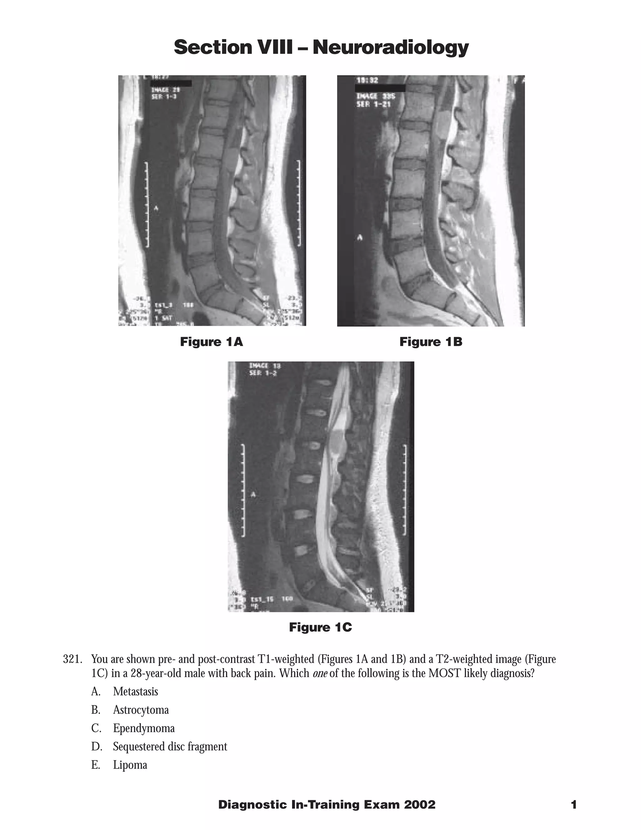

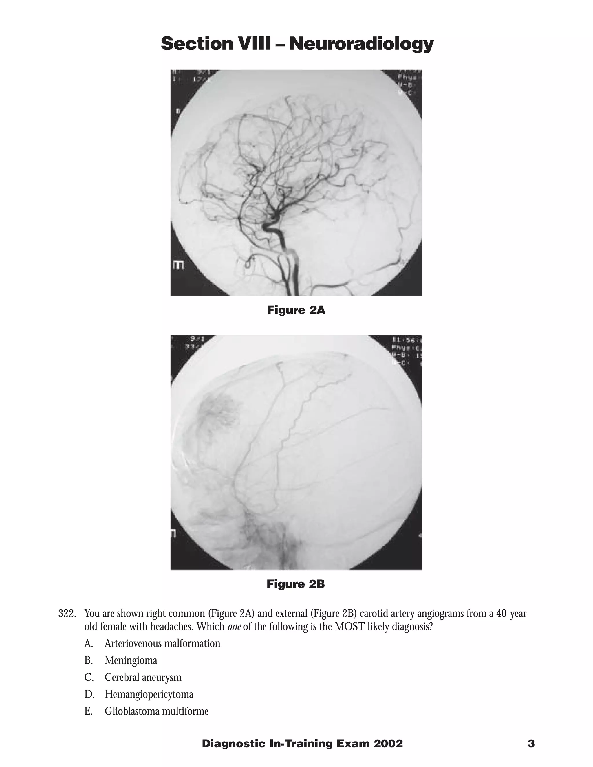

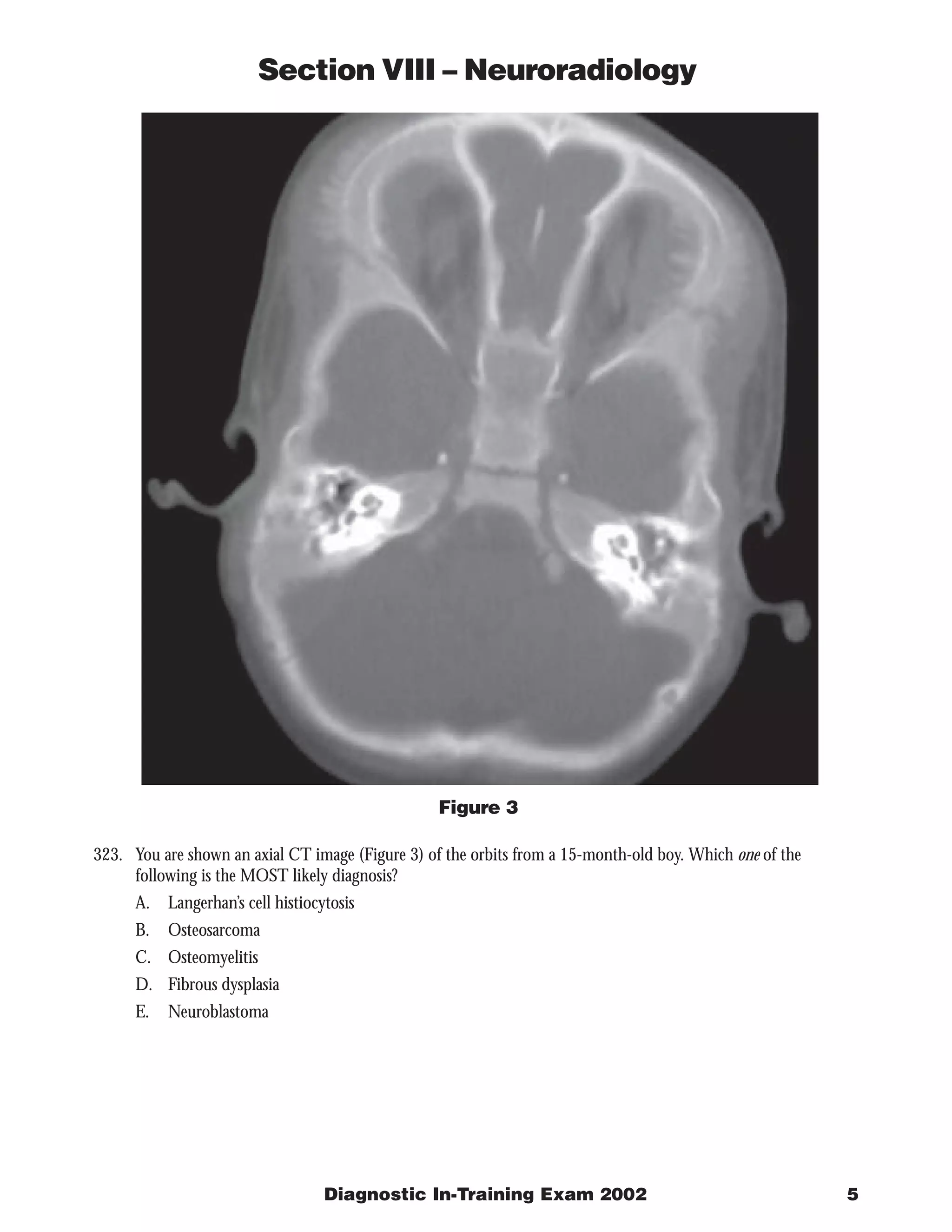


The document describes 3 radiology cases: 1) Images of the lumbar spine show a well-defined intradural mass at the filum terminale that is isointense on T1 and T2 with enhancement, most consistent with myxopapillary ependymoma. 2) Cerebral angiography shows a dural-based enhancing mass supplied by the external carotid artery, consistent with meningioma. 3) CT of the orbits in a 15-month-old boy shows spiculated bone formation in both orbits, suggestive of neuroblastoma metastases.