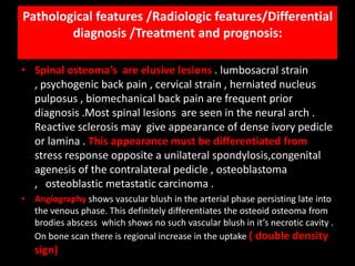
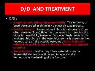
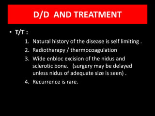
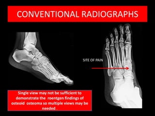
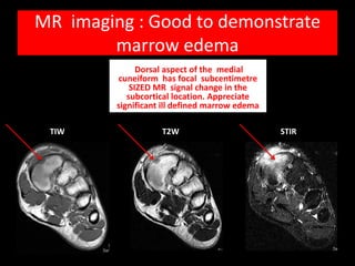
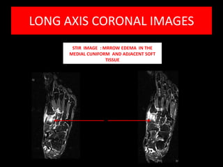
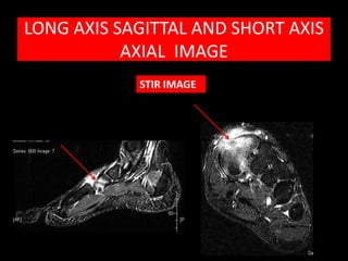
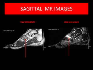
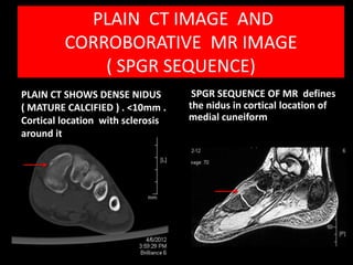
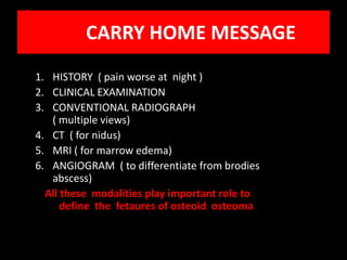
Osteoid osteoma is a benign bone tumor most common in young patients, typically causing pain worsened at night. Radiographs show a small lucent nidus surrounded by reactive sclerosis. CT precisely defines the calcified nidus. MRI demonstrates surrounding bone marrow edema. Treatment involves surgical excision or thermocoagulation of the nidus, providing effective pain relief. A multimodality imaging approach is useful to characterize the lesion and differentiate it from other causes of bone pain.
A classification of bone tumours. Modified after Revised WHO Classification –Schajowicz (1994)
Osteoblastoma
Are larger: > 2 cm.
Periosteal reaction may be more prominent than encountered in osteoid osteomas
A classification of bone tumours. Modified after Revised WHO Classification –Schajowicz (1994)
Osteoblastoma
Are larger: > 2 cm.
Periosteal reaction may be more prominent than encountered in osteoid osteomas
Giant osteoid osteoma of tibial shaft: A rare case reportApollo Hospitals
Giant osteoid osteoma of the tibial shaft is a rare entity.
Though this tumor is seen commonly in axial skeleton, so far
no conclusive report has been published on its periosteal
involvement of tibial shaft diaphysis.
ADACTYLY IN FETUS
PORENCEPHALIC CYST IN FETUS
SEPTO-OPTIC DYSPLASIA IN FETUS
MUSCLE HERNIA IN ADULT
FETAL REDUCTION
AGENESIS OF CORPUS CALLOSUM
FLAT FETAL FACIAL PROFILE
Anti ulcer drugs and their Advance pharmacology ||
Anti-ulcer drugs are medications used to prevent and treat ulcers in the stomach and upper part of the small intestine (duodenal ulcers). These ulcers are often caused by an imbalance between stomach acid and the mucosal lining, which protects the stomach lining.
||Scope: Overview of various classes of anti-ulcer drugs, their mechanisms of action, indications, side effects, and clinical considerations.
MANAGEMENT OF ATRIOVENTRICULAR CONDUCTION BLOCK.pdfJim Jacob Roy
Cardiac conduction defects can occur due to various causes.
Atrioventricular conduction blocks ( AV blocks ) are classified into 3 types.
This document describes the acute management of AV block.
Explore natural remedies for syphilis treatment in Singapore. Discover alternative therapies, herbal remedies, and lifestyle changes that may complement conventional treatments. Learn about holistic approaches to managing syphilis symptoms and supporting overall health.
Lung Cancer: Artificial Intelligence, Synergetics, Complex System Analysis, S...Oleg Kshivets
RESULTS: Overall life span (LS) was 2252.1±1742.5 days and cumulative 5-year survival (5YS) reached 73.2%, 10 years – 64.8%, 20 years – 42.5%. 513 LCP lived more than 5 years (LS=3124.6±1525.6 days), 148 LCP – more than 10 years (LS=5054.4±1504.1 days).199 LCP died because of LC (LS=562.7±374.5 days). 5YS of LCP after bi/lobectomies was significantly superior in comparison with LCP after pneumonectomies (78.1% vs.63.7%, P=0.00001 by log-rank test). AT significantly improved 5YS (66.3% vs. 34.8%) (P=0.00000 by log-rank test) only for LCP with N1-2. Cox modeling displayed that 5YS of LCP significantly depended on: phase transition (PT) early-invasive LC in terms of synergetics, PT N0—N12, cell ratio factors (ratio between cancer cells- CC and blood cells subpopulations), G1-3, histology, glucose, AT, blood cell circuit, prothrombin index, heparin tolerance, recalcification time (P=0.000-0.038). Neural networks, genetic algorithm selection and bootstrap simulation revealed relationships between 5YS and PT early-invasive LC (rank=1), PT N0—N12 (rank=2), thrombocytes/CC (3), erythrocytes/CC (4), eosinophils/CC (5), healthy cells/CC (6), lymphocytes/CC (7), segmented neutrophils/CC (8), stick neutrophils/CC (9), monocytes/CC (10); leucocytes/CC (11). Correct prediction of 5YS was 100% by neural networks computing (area under ROC curve=1.0; error=0.0).
CONCLUSIONS: 5YS of LCP after radical procedures significantly depended on: 1) PT early-invasive cancer; 2) PT N0--N12; 3) cell ratio factors; 4) blood cell circuit; 5) biochemical factors; 6) hemostasis system; 7) AT; 8) LC characteristics; 9) LC cell dynamics; 10) surgery type: lobectomy/pneumonectomy; 11) anthropometric data. Optimal diagnosis and treatment strategies for LC are: 1) screening and early detection of LC; 2) availability of experienced thoracic surgeons because of complexity of radical procedures; 3) aggressive en block surgery and adequate lymph node dissection for completeness; 4) precise prediction; 5) adjuvant chemoimmunoradiotherapy for LCP with unfavorable prognosis.
Ethanol (CH3CH2OH), or beverage alcohol, is a two-carbon alcohol
that is rapidly distributed in the body and brain. Ethanol alters many
neurochemical systems and has rewarding and addictive properties. It
is the oldest recreational drug and likely contributes to more morbidity,
mortality, and public health costs than all illicit drugs combined. The
5th edition of the Diagnostic and Statistical Manual of Mental Disorders
(DSM-5) integrates alcohol abuse and alcohol dependence into a single
disorder called alcohol use disorder (AUD), with mild, moderate,
and severe subclassifications (American Psychiatric Association, 2013).
In the DSM-5, all types of substance abuse and dependence have been
combined into a single substance use disorder (SUD) on a continuum
from mild to severe. A diagnosis of AUD requires that at least two of
the 11 DSM-5 behaviors be present within a 12-month period (mild
AUD: 2–3 criteria; moderate AUD: 4–5 criteria; severe AUD: 6–11 criteria).
The four main behavioral effects of AUD are impaired control over
drinking, negative social consequences, risky use, and altered physiological
effects (tolerance, withdrawal). This chapter presents an overview
of the prevalence and harmful consequences of AUD in the U.S.,
the systemic nature of the disease, neurocircuitry and stages of AUD,
comorbidities, fetal alcohol spectrum disorders, genetic risk factors, and
pharmacotherapies for AUD.
These simplified slides by Dr. Sidra Arshad present an overview of the non-respiratory functions of the respiratory tract.
Learning objectives:
1. Enlist the non-respiratory functions of the respiratory tract
2. Briefly explain how these functions are carried out
3. Discuss the significance of dead space
4. Differentiate between minute ventilation and alveolar ventilation
5. Describe the cough and sneeze reflexes
Study Resources:
1. Chapter 39, Guyton and Hall Textbook of Medical Physiology, 14th edition
2. Chapter 34, Ganong’s Review of Medical Physiology, 26th edition
3. Chapter 17, Human Physiology by Lauralee Sherwood, 9th edition
4. Non-respiratory functions of the lungs https://academic.oup.com/bjaed/article/13/3/98/278874
Factory Supply Best Quality Pmk Oil CAS 28578–16–7 PMK Powder in Stockrebeccabio
Factory Supply Best Quality Pmk Oil CAS 28578–16–7 PMK Powder in Stock
Telegram: bmksupplier
signal: +85264872720
threema: TUD4A6YC
You can contact me on Telegram or Threema
Communicate promptly and reply
Free of customs clearance, Double Clearance 100% pass delivery to USA, Canada, Spain, Germany, Netherland, Poland, Italy, Sweden, UK, Czech Republic, Australia, Mexico, Russia, Ukraine, Kazakhstan.Door to door service
Hot Selling Organic intermediates
Pulmonary Thromboembolism - etilogy, types, medical- Surgical and nursing man...VarunMahajani
Disruption of blood supply to lung alveoli due to blockage of one or more pulmonary blood vessels is called as Pulmonary thromboembolism. In this presentation we will discuss its causes, types and its management in depth.
Report Back from SGO 2024: What’s the Latest in Cervical Cancer?bkling
Are you curious about what’s new in cervical cancer research or unsure what the findings mean? Join Dr. Emily Ko, a gynecologic oncologist at Penn Medicine, to learn about the latest updates from the Society of Gynecologic Oncology (SGO) 2024 Annual Meeting on Women’s Cancer. Dr. Ko will discuss what the research presented at the conference means for you and answer your questions about the new developments.
Title: Sense of Smell
Presenter: Dr. Faiza, Assistant Professor of Physiology
Qualifications:
MBBS (Best Graduate, AIMC Lahore)
FCPS Physiology
ICMT, CHPE, DHPE (STMU)
MPH (GC University, Faisalabad)
MBA (Virtual University of Pakistan)
Learning Objectives:
Describe the primary categories of smells and the concept of odor blindness.
Explain the structure and location of the olfactory membrane and mucosa, including the types and roles of cells involved in olfaction.
Describe the pathway and mechanisms of olfactory signal transmission from the olfactory receptors to the brain.
Illustrate the biochemical cascade triggered by odorant binding to olfactory receptors, including the role of G-proteins and second messengers in generating an action potential.
Identify different types of olfactory disorders such as anosmia, hyposmia, hyperosmia, and dysosmia, including their potential causes.
Key Topics:
Olfactory Genes:
3% of the human genome accounts for olfactory genes.
400 genes for odorant receptors.
Olfactory Membrane:
Located in the superior part of the nasal cavity.
Medially: Folds downward along the superior septum.
Laterally: Folds over the superior turbinate and upper surface of the middle turbinate.
Total surface area: 5-10 square centimeters.
Olfactory Mucosa:
Olfactory Cells: Bipolar nerve cells derived from the CNS (100 million), with 4-25 olfactory cilia per cell.
Sustentacular Cells: Produce mucus and maintain ionic and molecular environment.
Basal Cells: Replace worn-out olfactory cells with an average lifespan of 1-2 months.
Bowman’s Gland: Secretes mucus.
Stimulation of Olfactory Cells:
Odorant dissolves in mucus and attaches to receptors on olfactory cilia.
Involves a cascade effect through G-proteins and second messengers, leading to depolarization and action potential generation in the olfactory nerve.
Quality of a Good Odorant:
Small (3-20 Carbon atoms), volatile, water-soluble, and lipid-soluble.
Facilitated by odorant-binding proteins in mucus.
Membrane Potential and Action Potential:
Resting membrane potential: -55mV.
Action potential frequency in the olfactory nerve increases with odorant strength.
Adaptation Towards the Sense of Smell:
Rapid adaptation within the first second, with further slow adaptation.
Psychological adaptation greater than receptor adaptation, involving feedback inhibition from the central nervous system.
Primary Sensations of Smell:
Camphoraceous, Musky, Floral, Pepperminty, Ethereal, Pungent, Putrid.
Odor Detection Threshold:
Examples: Hydrogen sulfide (0.0005 ppm), Methyl-mercaptan (0.002 ppm).
Some toxic substances are odorless at lethal concentrations.
Characteristics of Smell:
Odor blindness for single substances due to lack of appropriate receptor protein.
Behavioral and emotional influences of smell.
Transmission of Olfactory Signals:
From olfactory cells to glomeruli in the olfactory bulb, involving lateral inhibition.
Primitive, less old, and new olfactory systems with different path
New Directions in Targeted Therapeutic Approaches for Older Adults With Mantl...i3 Health
i3 Health is pleased to make the speaker slides from this activity available for use as a non-accredited self-study or teaching resource.
This slide deck presented by Dr. Kami Maddocks, Professor-Clinical in the Division of Hematology and
Associate Division Director for Ambulatory Operations
The Ohio State University Comprehensive Cancer Center, will provide insight into new directions in targeted therapeutic approaches for older adults with mantle cell lymphoma.
STATEMENT OF NEED
Mantle cell lymphoma (MCL) is a rare, aggressive B-cell non-Hodgkin lymphoma (NHL) accounting for 5% to 7% of all lymphomas. Its prognosis ranges from indolent disease that does not require treatment for years to very aggressive disease, which is associated with poor survival (Silkenstedt et al, 2021). Typically, MCL is diagnosed at advanced stage and in older patients who cannot tolerate intensive therapy (NCCN, 2022). Although recent advances have slightly increased remission rates, recurrence and relapse remain very common, leading to a median overall survival between 3 and 6 years (LLS, 2021). Though there are several effective options, progress is still needed towards establishing an accepted frontline approach for MCL (Castellino et al, 2022). Treatment selection and management of MCL are complicated by the heterogeneity of prognosis, advanced age and comorbidities of patients, and lack of an established standard approach for treatment, making it vital that clinicians be familiar with the latest research and advances in this area. In this activity chaired by Michael Wang, MD, Professor in the Department of Lymphoma & Myeloma at MD Anderson Cancer Center, expert faculty will discuss prognostic factors informing treatment, the promising results of recent trials in new therapeutic approaches, and the implications of treatment resistance in therapeutic selection for MCL.
Target Audience
Hematology/oncology fellows, attending faculty, and other health care professionals involved in the treatment of patients with mantle cell lymphoma (MCL).
Learning Objectives
1.) Identify clinical and biological prognostic factors that can guide treatment decision making for older adults with MCL
2.) Evaluate emerging data on targeted therapeutic approaches for treatment-naive and relapsed/refractory MCL and their applicability to older adults
3.) Assess mechanisms of resistance to targeted therapies for MCL and their implications for treatment selection
1. OSTEOID OSTEOMA
19 Yr old male with
pain in in the dorsal
aspect of the medial
aspect of left mid
foot . o/e there is
pain over the FREE LANCE RADIOLOGY
mentioned part of the Basic approach for
left mid foot . continuation of
Diagnostic Radiology
education
2. General considerations/ Incidence /Clinical features
• :
• First described by Henry jaffe ( 1925). Not accepted for several
decades and was considered as variant of osteomyelitis .
• 2.6% of all excised bone tumors and 11 % of all benign bone
tumors .
• Young patients ( 10 to 25 years) . youngest patient reported
was 8 month old patient with lesion in tibia . Male : female
ratio is 2:1 .
• Clinical profile :
• Pain +_ vasomotor disturbance ( profuse sweating / increased
skin temperature) . Classical description is of gradual onset of
increasingly deep / severe / aching pain ( 65% will have night
pain relieved by aspirin) . CAN BE CONFUSED WITH : septic
arthritis , inflammatory , rheumatoid arthritis so patient may
end up with rheumatology opinion.
3. General considerations /Incidence
/Clinical features
• Localized swelling ,point tenderness , limitation of the
motion, painful limp, stiffness , weakness of nearby
joint , muscle atrophy may be noted. Painful scoliosis
(lesion located in the concave side of the curve in
thoracic / lumbar spine) . In cervical spine : torticollis /
secondary contracture of the sternocleidomastoid
muscle may be noted .Lesion in the spinous processs
lead to localized pain and spinal stiffness .
• 50% occur in proximal femur / tibia ( predilection for
upper end of the femur , particularly the neck /
trochanteric region) .In spine : most of the lesions are
in neural arch .
4. Pathological features /Radiologic features/Differential
diagnosis /Treatment and prognosis:
• Lesion : Nidus ( reddish brown vascularised tumor <= 10mm) .
Significant reactive sclerosis with cortical thickening / solid periosteal
reaction encasing the nidus . Nidus is initially uncalcified and with
maturity speck of calcification is seen in it . Bone expansion may be
noted at the lesion site .
• Three anatomic locations of the osteoid osteoma :
Cortical ( most common)
, Cancellous (
intramedullary )
Subperiosteal . Histological and radiological appearance varies .
• Well developed lesion has lucent nidus with surrounding florid perifocal
reactive sclerosis/appositional periosteal new bone formation ( typical
of cortically placed osteoid osteoma ).The sclerosis is maximally seen
caudal to the nidus . Nidus size is, <=1cm in diameter . Single
roengenographic view may not be sufficient to demonstrate the nidus
. central fleck of calcification is seen in the nidus with maturity .
• Intramedullary lesion that are intracapsular provoke much less reactive
sclerosis because of low rate of bone production from intracapsular
5. Pathological features /Radiologic features/Differential
diagnosis /Treatment and prognosis:
• Spinal osteoma’s are elusive lesions . lumbosacral strain
, psychogenic back pain , cervical strain , herniated nucleus
pulposus , biomechanical back pain are frequent prior
diagnosis .Most spinal lesions are seen in the neural arch .
Reactive sclerosis may give appearance of dense ivory pedicle
or lamina . This appearance must be differentiated from
stress response opposite a unilateral spondylosis,congenital
agenesis of the contralateral pedicle , osteoblastoma
, osteoblastic metastatic carcinoma .
• Angiography shows vascular blush in the arterial phase persisting late into
the venous phase. This definitely differentiates the osteoid osteoma from
brodies abscess which shows no such vascular blush in it’s necrotic cavity .
On bone scan there is regional increase in the uptake ( double density
sign)
6. D/D AND TREATMENT
• D/D :
– Garre’s chronic sclerosing osteomyelitis : This entity has
been disregarded as singular / distinct disease process.
– Brodies abscess : Lucent nidus in brodies abscess is >1cm
often close to 2 cm ).Halo rim of sclerosis surrounding the
nidus is more thick / irregular . Vascular blush seen in the
angiographic phase in the osteoidosteoma is absent in the
necrotic core of the osteoid osteoma . Note :Night pain
relieved by aspirin is seen in brodies abscess and osteoid
osteoma .
– Stress fracture : lesion may mimic osteoid osteoma .
Sequential studies over time and images usually
demonstrate the healing of the fracture .
7. D/D AND TREATMENT
• T/T :
1. Natural history of the disease is self limiting .
2. Radiotherapy / thermocoagulation
3. Wide enbloc excision of the nidus and
sclerotic bone. (surgery may be delayed
unless nidus of adequate size is seen) .
4. Recurrence is rare.
8. CONVENTIONAL RADIOGRAPHS
SITE OF PAIN
Single view may not be sufficient to
demonstrate the roentgen findings of
osteoid osteoma so multiple views may be
needed .
9. MR imaging : Good to demonstrate
marrow edema
Dorsal aspect of the medial
cuneiform has focal subcentimetre
SIZED MR signal change in the
subcortical location. Appreciate
significant ill defined marrow edema
TIW T2W STIR
10. LONG AXIS CORONAL IMAGES
STIR IMAGE : MRROW EDEMA IN THE
MEDIAL CUNIFORM AND ADJACENT SOFT
TISSUE
13. PLAIN CT IMAGE AND
CORROBORATIVE MR IMAGE
( SPGR SEQUENCE)
PLAIN CT SHOWS DENSE NIDUS SPGR SEQUENCE OF MR defines
( MATURE CALCIFIED ) . <10mm . the nidus in cortical location of
Cortical location with sclerosis medial cuneiform
around it
14. CARRY HOME MESSAGE
1. HISTORY ( pain worse at night )
2. CLINICAL EXAMINATION
3. CONVENTIONAL RADIOGRAPH
( multiple views)
4. CT ( for nidus)
5. MRI ( for marrow edema)
6. ANGIOGRAM ( to differentiate from brodies
abscess)
All these modalities play important role to
define the fetaures of osteoid osteoma