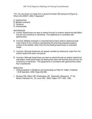
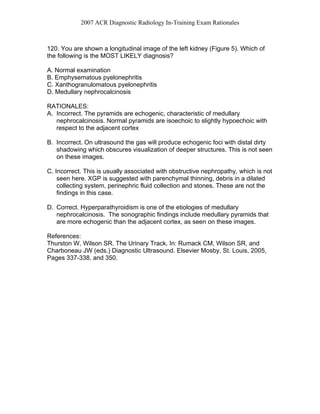
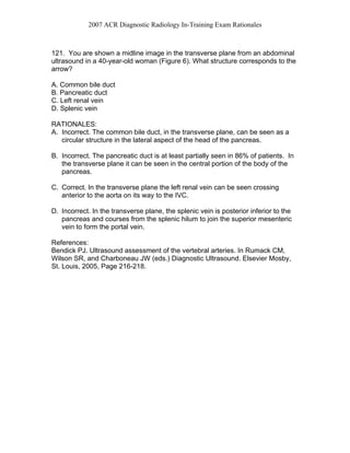
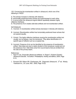
2007 ACR Diagnostic Radiology In-Training Exam Rationales
Section VI Ultrasound
116. You are shown longitudinal and transverse gray scale images of the uterus
(Figures 1A and 1B) of a 32 year-old woman with positive pregnancy test. These
findings are most consistent with which ONE of the following types of pregnancy?
A. Normal intrauterine
B. Heterotopic
C. Cornual (Interstitial)
D. Fimbrial
RATIONALES:
A. Incorrect. Normal intrauterine pregnancy implants eccentrically within the
endometrium and not at the interstitial portion of the fallopian tube. The
implantation in this case is at the interstitial portion of the fallopian tube as the
overlying myometrium is very thin (<5mm).
B. Incorrect. For heterotopic pregnancy, one has to demonstrate both intra and
extra uterine pregnancy present at the same time.
C. Correct. Cornual pregnancy (interstitial pregnancy) accounts for less than 2 %
of pregnancies and is important to recognize as these patients present very
late. A major complication is a ruptured uterus. Sonographically it is identified
by eccentric location and usually an echogenic band can be seen connecting
the ectopic pregnancy with the endometrial stripe, called interstitial line (sign).
D. Incorrect. Fimbrial pregnancy is an ectopic pregnancy outside the uterus in
the fimbrial portion of the fallopian tube.
References:
Paspulati RM and McElroy TM. Ectopic Pregnancy. In :Dogra V, Rubens
DJ(eds): Ultrasound Secrets. 1st Edition. Philadelphia; Hanley and Belfus; 2004,
Page 75-80.
2007 ACR Diagnostic Radiology In-Training Exam Rationales
117. You are shown longitudinal gray scale image of the testis in an
asymptomatic 57 year old man (Figure 2). A color flow Doppler ultrasound of the
abnormal area did not reveal any flow. What is the MOST LIKELY diagnosis?
A. Tubular ectasia of rete testis
B. Intratesticular varicocele
C. Testicular tumor
D. Testicular abscess
RATIONALES:
A. Correct. Tubular ectasia is a benign condition and occurs usually in men older
than 55 years of age. It is located in or adjacent to the mediastinum testis. It is
usually bilateral and is accompanied by spermatocele.
B. Incorrect. Larger serpiginous structures are present in intratesticular
varicoceles. The appearance is similar to extratesticular varicoceles, and
would have blood flow within.
C. Incorrect. Testicular cystic tumors can occur anywhere; however mediastinum
testis is not a characteristic location for them. The tubular ectasia occurs
classically adjacent to mediastinum testis and is postero-lateral in location.
Cystic immature teratoma is the main differential consideration and it usually
has rind of echogenic tissue around it.
D. Incorrect. Testicular abscesses have irregular, shaggy margins with
peripheral hyperemia. They usually occur as complication of epididymo-
orchitis or secondary to infection in testicular hematoma.
References:
1. Dogra VS, Gottlieb RH, Deborah RJ, Liao L. Benign Intratesticular Cystic Lesions:
US Features. Radiographics 2001;21:S273-S281.
2. Dogra VS, Oka M, Gottlieb RH, Deborah RJ. Sonography of the Scrotum.
Radiology 2003; 227: 18-36.
3. Rubenstein RA, Dogra VS, Seftel AD and Resnick MI. Benign Intrascrotal
Lesions. J Urol. 2004 May;171(5):1765-72.
2007 ACR Diagnostic Radiology In-Training Exam Rationales
118. You are shown a transverse image of the uterus from a 24-year-old woman
with a positive urine pregnancy test (Figure 3). What is the MOST LIKELY
diagnosis?
A. Ectopic pregnancy
B. Blighted ovum
C. Complete hydatidiform mole
D. Normal 8-week intrauterine pregnancy
RATIONALES:
A. Incorrect. Although ectopic pregnancy should be considered in any female patient
with positive pregnancy test and absence of intrauterine pregnancy, the large
echogenic mass occupying the central portion of uterus would make complete
hydatidiform mole the most likely diagnosis.
B. Incorrect. Blighted ovum is characterized by gestational sac without an embryonic
pole but of such size that an embryonic pole/yolk sac would be expected. The
submitted image demonstrated multiple small intrauterine cystic areas in the
context of a large central echogenic intrauterine mass, which would be more
consistent with complete hydatidiform mole.
C. Correct. The submitted image demonstrates an enlarged uterus with large
echogenic mass occupying the central portion of the uterus. Multiple small cystic
areas are present and there is no discernable endometrial stripe or fetal parts.
Considering this appearance and clinical presentation, complete hydatidiform mole
is the most likely diagnosis.
D. Incorrect. The submitted image demonstrates no evidence of a embryonic pole,
which would be expected at 8 weeks. In addition, the uterus is grossly abnormal in
appearance.
References:
1. Ultrasonography in Obstetrics and Gynecology by Peter W. Callen. Publisher – W B
Saunders. 2000, Pages 847-848.
2. Rumack CM, Wilson SR, Charboneau JW. Diagnostic Ultrasound. 3rd
ed.
Mosby Yearbook Inc., St. Louis, MO. 2005, Pages 1576-1578.
Complete hydatidiform mole
2007 ACR Diagnostic Radiology In-Training Exam Rationales
119. You are shown an image from a second trimester OB ultrasound (Figure 4).
What is the MOST LIKELY diagnosis?
A. Gastroschisis
B. Bladder exstrophy
C. Teratoma
D. Omphalocele
RATIONALES:
A. Correct. Bowel loops are seen to extend through an anterior abdominal wall defect
and are not covered by a membrane. This appearance is consistent with
gastroschisis.
B. Incorrect. Bladder exstrophy is characterized by lower anterior abdominal wall
mass inferior to the umbilicus representing the protruding exposed posterior
surface of the bladder rather than the free floating bowel loops on submitted
image.
C. Incorrect. Although teratomas can appear complex by ultrasound, origin from the
anterior abdominal wall is not typical.
D. Incorrect. Although bowel loops are seen to extend through an anterior abdominal
wall defect, these bowel loops are floating free within the amniotic fluid and are not
covered by a membrane. This appearance is consistent with gastroschisis rather
than omphalocele.
References:
1. Ultrasonography in Obstetrics and Gynecology by Peter W. Callen. Publisher
– W B Saunders. 2000, Page 492-494.
2. Rumack CM, Wilson SR, Charboneau JW. Diagnostic Ultrasound. 3rd
ed.
Mosby Yearbook Inc., St. Louis, MO. 2005, Pages 1377-1382, 1385.
2007 ACR Diagnostic Radiology In-Training Exam Rationales
120. You are shown a longitudinal image of the left kidney (Figure 5). Which of
the following is the MOST LIKELY diagnosis?
A. Normal examination
B. Emphysematous pyelonephritis
C. Xanthogranulomatous pyelonephritis
D. Medullary nephrocalcinosis
RATIONALES:
A. Incorrect. The pyramids are echogenic, characteristic of medullary
nephrocalcinosis. Normal pyramids are isoechoic to slightly hypoechoic with
respect to the adjacent cortex
B. Incorrect. On ultrasound the gas will produce echogenic foci with distal dirty
shadowing which obscures visualization of deeper structures. This is not seen
on these images.
C. Incorrect. This is usually associated with obstructive nephropathy, which is not
seen here. XGP is suggested with parenchymal thinning, debris in a dilated
collecting system, perinephric fluid collection and stones. These are not the
findings in this case.
D. Correct. Hyperparathyroidism is one of the etiologies of medullary
nephrocalcinosis. The sonographic findings include medullary pyramids that
are more echogenic than the adjacent cortex, as seen on these images.
References:
Thurston W, Wilson SR. The Urinary Track. In: Rumack CM, Wilson SR, and
Charboneau JW (eds.) Diagnostic Ultrasound. Elsevier Mosby, St. Louis, 2005,
Pages 337-338, and 350.
2007 ACR Diagnostic Radiology In-Training Exam Rationales
121. You are shown a midline image in the transverse plane from an abdominal
ultrasound in a 40-year-old woman (Figure 6). What structure corresponds to the
arrow?
A. Common bile duct
B. Pancreatic duct
C. Left renal vein
D. Splenic vein
RATIONALES:
A. Incorrect. The common bile duct, in the transverse plane, can be seen as a
circular structure in the lateral aspect of the head of the pancreas.
B. Incorrect. The pancreatic duct is at least partially seen in 86% of patients. In
the transverse plane it can be seen in the central portion of the body of the
pancreas.
C. Correct. In the transverse plane the left renal vein can be seen crossing
anterior to the aorta on its way to the IVC.
D. Incorrect. In the transverse plane, the splenic vein is posterior inferior to the
pancreas and courses from the splenic hilum to join the superior mesenteric
vein to form the portal vein.
References:
Bendick PJ. Ultrasound assessment of the vertebral arteries. In Rumack CM,
Wilson SR, and Charboneau JW (eds.) Diagnostic Ultrasound. Elsevier Mosby,
St. Louis, 2005, Page 216-218.
2007 ACR Diagnostic Radiology In-Training Exam Rationales
122. Concerning the reverberation artifact in ultrasound, which one of the
following is TRUE?
A. The echoes increase in intensity with distance.
B. Horizontally positioned linear echoes are superimposed on each other.
C. It occurs when the ultrasound signal reflects repeatedly between highly
reflective interfaces.
D. The presence of air creates side-lobe artifacts and not reverberation artifact.
RATIONALES:
A. Incorrect. In reverberation artifact echoes decrease in intensity with distance.
B. Incorrect. Reverberation artifact has horizontally positioned linear echoes that
are spaced equally.
C. Correct. The highly reflective interfaces causing the reverberation artifact are
usually, but not always, near the transducer. They allow identification of a
specific type of reflector such as a surgical clip.
D. Incorrect. Presence of air can be identified by the presence of reverberation
artifact. Side lobes are due to radial vibration of the transducer crystal and not
the presence of air. Side lobes arise from sound beams that are emitted from
the side of the primary beam.
References:
1. Ryan K Lee. Grayscale Ultrasound artifacts. In :Dogra V, Rubens DJ(eds):
Ultrasound Secrets. 1st Edition. Philadelphia; Hanley and Belfus; 2004, Page
8-14.
2. Rumack CM, Wilson SR, Charboneau JW. Diagnostic Ultrasound. 3rd
ed. Mosby
Yearbook Inc., St. Louis, MO. 2005, Page 19-22.
2007 ACR Diagnostic Radiology In-Training Exam Rationales
123. Theca lutein cysts are MOST commonly associated with:
A. Stein-Leventhal syndrome.
B. gestational trophoblastic disease.
C. elevated serum progesterone levels.
D. a normal singleton pregnancy.
RATIONALES:
A. Incorrect. Patients with Stein-Leventhal syndrome represent a subset of
women with polycystic ovary disease that have the clinical manifestations of
oligomenorrhea/amenorrhea, hirsutism, and obesity. The ovaries of polycystic
ovary disease can be normal in appearance or enlarged with increased
number of small subcapsular follicles. This is significantly different from the
large, septated theca lutein cysts, which can replace the ovaries with elevated
hCG levels.
B. Correct. Theca lutein cysts are associated with elevated hCG levels, which
can be seen with gestational trophoblastic disease and exogenous hCG used
for infertility treatment.
C. Incorrect. Theca lutein cysts are associated with elevated hCG levels rather
than elevated progesterone levels.
D. Incorrect. Not commonly seen in normal singleton pregnancy.
References:
1. Ultrasonography in Obstetrics and Gynecology by Peter W. Callen. Publisher
– W B Saunders. 2000, Page 863.
2. Rumack CM, Wilson SR, Charboneau JW. Diagnostic Ultrasound. 3rd
ed.
Mosby Yearbook Inc., St. Louis, MO. 2005, Page 557-558.
2007 ACR Diagnostic Radiology In-Training Exam Rationales
124. How is the amniotic fluid index calculated?
A. Greatest single vertical measurement of fluid pocket in any quadrant.
B. Greatest vertical measurement of fluid pocket in each of 4 quadrants summed.
C. Subjective assessment of amniotic fluid volume in each half of uterus graded
on scale of 1 to 5 and summed.
D. Greatest vertical measurement of fluid pocket in each of 4 quadrants summed
and divided by 4.
RATIONALES:
A. Incorrect. Measuring the greatest single vertical fluid pocket in any quadrant
is known as the maximum vertical pocket (MVP) measurement and can be
used as a method of assessing amniotic fluid.
B. Correct. This is the correct method for determining the amniotic fluid index.
C. Incorrect. Amniotic fluid index does not involve subjective assessment of
amniotic fluid or grading scale, and divides the uterus in quadrants rather than
halves.
D. Incorrect. Although the amniotic fluid index does use the deepest amniotic fluid
pocket measurement in 4 quadrants summed, dividing by 4 would merely give the
average of the fluid pocket measurements.
References:
1. Ultrasonography in Obstetrics and Gynecology by Peter W. Callen. Publisher
– W B Saunders. 2000, Page 641-644.
2. Rumack CM, Wilson SR, Charboneau JW. Diagnostic Ultrasound. 3rd
ed.
Mosby Yearbook Inc., St. Louis, MO. 2005, Page 1396-1397.
2007 ACR Diagnostic Radiology In-Training Exam Rationales
125. A didelphic uterus appears on ultrasound as:
A. two separate unfused uterine horns and a single uterine body.
B. two separate unfused uterine horns and two separate unfused uterine bodies.
C. a normal uterine contour of uterine horns and body but with two separated
endometrial canals.
D. a single normal uterine horn and body with secondary rudimentary uterine
horn present.
RATIONALES:
A. Incorrect. Two separate unfused uterine horns and singe uterine body
describes a bicornuate uterus. Bicornuate uterus can have fusion of the entire
uterine body or just the lower uterine segment.
B. Correct. Didelphic uterus has two separate unfused uterine horns and
bodies. Clinically, patient also can have two separate cervices and septated
vagina.
C. Incorrect. Normal uterine contour with regards to uterine horns and body but
with two separated endometrial canals describes a septated uterus. The
septation can be thick or thin.
D. Incorrect. Single normal uterine horn and body with second rudimentary
uterine horn present describes a unicornuate uterus.
References:
1. Ultrasonography in Obstetrics and Gynecology by Peter W. Callen. Publisher
– W B Saunders. 2000, Page 824-828.
2. Rumack CM, Wilson SR, Charboneau JW. Diagnostic Ultrasound. 3rd
ed.
Mosby Yearbook Inc., St. Louis, MO. 2005, Page 534-538.
2007 ACR Diagnostic Radiology In-Training Exam Rationales
126. Fetal head circumference during the second trimester of pregnancy on an
obstetrical ultrasound should be measured at the level of the:
A. frontal horns and cerebellum.
B. paired thalami and cerebellum.
C. paired thalami and cavum septi pellucidi.
D. cavum septi pellucidi and cerebellum.
RATIONALES:
A. Incorrect. Cerebellar hemispheres should not be visible in plane used to
measure head circumference.
B. Incorrect. Cerebellar hemispheres should not be visible in plane used to
measure head circumference.
C. Correct. Head circumference is measured at the level of the paired thalami and
third ventricle of mid brain, cavum septi pellucidi anteriorly, and includes the
tentorial hiatus posteriorly.
D. Incorrect. Cerebellar hemispheres should not be visible in plane used to measure
head circumference.
References:
1. Ultrasonography in Obstetrics and Gynecology by Peter W. Callen. Publisher – W B
Saunders. 2000, Page 155-157.
2. Middleton WD, Kurtz AB, Hertzberg BS: Ultrasound: The Requisites, 2nd
ed. St.
Louis, Mosby, 2004, Page 161-162.
Rumack CM, Wilson SR, Charboneau JW. Diagnostic Ultrasound. 3rd
ed.
Mosby Yearbook Inc., St. Louis, MO. 2005, Page 1495-1496
2007 ACR Diagnostic Radiology In-Training Exam Rationales
127. Concerning programmable ultrasound scanner settings, which of the
following is TRUE?
A. Time gain compensation (TGC) decreases the amplification (gain) applied to
deeper tissues to create a uniform signal intensity at all depths.
B. Dynamic range refers to the ratio of the highest to the lowest amplitude
displayed on the screen in decibels.
C. M-mode ultrasound uses a substantially increased amount of acoustic energy
to form an image as compared to grayscale imaging.
D. Pulse repetition frequency is a measure of the amplitude of the ultrasound
pulse.
RATIONALES:
A. Incorrect. Time gain compensation (TGC) increases the amplification (gain)
applied to deeper tissues to create a uniform signal intensity at all depths.
B. Correct. Dynamic range refers to the ratio of the highest to the lowest
amplitude displayed on the screen in decibels.
C. Incorrect. M-mode ultrasound uses an equivalent amount of acoustic energy
to form an image as compared to grayscale imaging.
D. Incorrect. Pulse repetition frequency is a measure of the time interval
between ultrasound pulses.
Literature Citation:
Rumack CM, Wilson SR, Charboneau JW. Diagnostic Ultrasound. 3rd
ed.
Mosby Yearbook Inc., St. Louis, MO. 2005, Page 9-12.
2007 ACR Diagnostic Radiology In-Training Exam Rationales
128. Concerning ultrasound interaction with matter, an ultrasound beam
reflected from small (2 mm) kidney stones would:
A. be specular reflection.
B. be non-specular reflection.
C. provide a strong echo.
D. produce reverberation artifacts.
RATIONALES:
A. Incorrect. A specular reflector is a smooth boundary between two media,
where the dimensions of the boundary are much larger than the wavelength of
the incident ultrasound energy. As the wavelength becomes smaller, the
boundary becomes rough, resulting in a nonspecular reflector surface. Since the
kidney stones are small, the resulting reflection would be non-specular.
B. Correct. A specular reflector is a smooth boundary between two media, where
the dimensions of the boundary are much larger than the wavelength of the
incident ultrasound energy. As the wavelength becomes smaller, the boundary
becomes rough, resulting in a nonspecular reflector surface. Since the kidney
stones are small, the resulting reflection would be non-specular.
C. Incorrect. Since the resulting reflection is non-specular, sound is reflected in
all directions and the amplitudes of the returning echoes are significantly weaker
then echoes from tissue boundaries.
D. Incorrect. Reverberation artifacts are generally produced by large objects
producing multiple reflections within the object.
References: The Essential Physics of Medical Imaging by Bushberg JT et. al.,
Second Edition, Chapter 16: Ultrasound
2007 ACR Diagnostic Radiology In-Training Exam Rationales
129. Concerning renal calculi, which of the following is TRUE?
A. Ultrasound should be the initial imaging test in all patients with acute renal
colic.
B. Transvaginal or transperineal scanning may be useful in detecting distal
ureteral calculi not demonstrated by the transabdominal approach.
C. Color and power Doppler twinkling artifacts are demonstrated in less than
50% of renal calculi.
D. Asymmetry of ureteral jets is seen only with high-grade ureteral obstruction.
RATIONALES:
A. Incorrect. Unenhanced helical CT is the accepted initial imaging test in
patients with renal colic. Ultrasound would be the initial test in pregnant
patients.
B. Correct. Better visualization of the distal ureters, especially in obese patients,
is possible with a transvaginal or transperineal approach.
C. Incorrect. It has been shown that most (83%) of renal calculi will demonstrate
this artifact.
D. Incorrect. Asymmetry of the ureteral jets can also be seen with low-grade
obstruction.
References:
Thurston W, Wilson SR. The Urinary Track. In: Rumack CM, Wilson SR, and
Charboneau JW (eds.) Diagnostic Ultrasound. Elsevier Mosby, St. Louis, 2005,
Page 345-350.
2007 ACR Diagnostic Radiology In-Training Exam Rationales
130. Regarding the anatomy of the lower extremity veins, which one of the
following statements is TRUE?
A. The popliteal vein is formed by the confluence of the anterior tibial and
posterior tibial veins.
B. The femoral and popliteal veins are duplicated in approximately 25% of
patients.
C. The first deep branches of the popliteal vein traveling into the calf are the
paired peroneal veins.
D. The gastrocnemius and soleal veins accompany an artery of the same name.
RATIONALES:
A. Incorrect. The popliteal vein is formed by the junction of the anterior tibial,
posterior tibial and peroneal veins.
B. Correct. Duplication can involve only a portion of the vein segment, or the
veins can be duplicated along their entire course.
C. Incorrect. The first deep branches are the anterior tibial veins.
D. Incorrect. These veins are muscular veins and do not have accompanying
arteries.
References:
1. Zwiebel WJ. Extremity Venous Anatomy, Terminology, and US features of
normal veins. In Zwiebel, Pellerito (eds.) Introduction to Vascular Ultrasound
5th
ed. Elsevier Saunders, Philadelphia, PA. 2005, Page 417-418.
2. Lewis BD. The Peripheral Veins. In Rumack CM, Wilson SR, and
Charboneau JW (eds.) Diagnostic Ultrasound. Elsevier Mosby, St. Louis,
2005, Page 1022.
2007 ACR Diagnostic Radiology In-Training Exam Rationales
131. Concerning portal hypertension, which of the following is TRUE?
A. It is possible for flow to be reversed in the right portal vein, and normal in
direction in the left portal vein.
B. A normal appearing liver by ultrasound excludes portal hypertension.
C. Spleen size correlates well with the portal pressure.
D. In portal hypertension secondary to cirrhosis the hepatic artery will be visibly
decreased in size on color Doppler.
RATIONALES:
A. Correct. If there is a large peri-umbilical vein collateral, the flow will be normal
in direction (antegrade) in the left portal vein as the peri-umbilical collateral
originates from the left portal system.
B. Incorrect. Portal hypertension may be present in patients with livers that have
a normal appearance by ultrasound.
C. Incorrect. The size of the spleen does not correlate well with the portal
pressure.
D. Incorrect. The hepatic artery is typically enlarged in cirrhosis. Increased
hepatic arterial blood flow is an attempt to compensate for the decreased
portal vein blood flow caused by elevated resistance in the hepatic sinusoids.
References:
Pellerito JS. Ultrasound Assessment of the Splanchnic (Mesenteric) Arteries. In
Zwiebel, Pellerito (eds.) Introduction to Vascular Ultrasound 5th
ed. Elsevier
Saunders, Philadelphia, PA. 2005, Page 585-590.
2007 ACR Diagnostic Radiology In-Training Exam Rationales
132. Which of the following is true regarding the upper extremity venous
ultrasound exam?
A. It typically includes evaluation of the veins to the wrist.
B. The subclavian, axillary, basilic, brachial, and cephalic veins are components
of the deep venous system.
C. Evaluation for compressibility of all upper-extremity veins is essential in
excluding a diagnosis of deep vein thrombosis.
D. It is important in evaluating normal subclavian flow to indirectly assess
patency of the innominate vein and the superior vena cava.
RATIONALES:
A. Incorrect. The forearm veins are only evaluated if there is clinical suspicion for
forearm venous thrombosis.
B. Incorrect. The basilic and cephalic veins are superficial veins.
C. Incorrect.. The subclavian vein often cannot be evaluated for compression, as
it lies deep to the clavicle.
D. Correct. Transmitted cardiac pulsatility and respiratory phasicity in the medial
subclavian vein and caudal internal jugular vein is used to assess for the
presence of a more proximal stenosis or occlusion in the innominate and/or
superior vena cava.
References:
Zwiebel WJ. Technique for Extremity Venous Ultrasound Examination. In
Zwiebel, Pellerito (eds.) Introduction to Vascular Ultrasound 5th
ed. Elsevier
Saunders, Philadelphia, PA. 2005, Page 432-435.
2007 ACR Diagnostic Radiology In-Training Exam Rationales
133. Concerning renal allograft complications, which of the following is TRUE?
A. Postoperative ultrasound is useful in differentiating rejection from acute tubular
necrosis.
B. Arterial stenosis is the most common vascular complication.
C. Urinomas occur 1-2 months or later after surgery.
D. Hydronephrosis 1-2 weeks post-op is typically due to obstructing debris, such
as blood clots in the ureter.
RATIONALES:
A. Incorrect. Sonography is neither sensitive nor specific for allograft rejection.
B. Correct. About 10% of transplant patients can develop renal arterial stenosis,
typically located at the renal artery anastomosis with the external iliac artery.
C. Incorrect. Urinomas occur in the immediate postoperative period.
D. Incorrect. Mild hydronephrosis of the allograft during the first or second week
post transplantation is typically due to postoperative edema at the insertion
site of the ureter into the bladder.
References:
Pellerito JS, Zwiebel WJ. Ultrasound assessment of native renal vessels and
renal allografts. In Zwiebel, Pellerito (eds.) Introduction to Vascular Ultrasound 5th
ed. Elsevier Saunders, Philadelphia, PA. 2005, Page 627-630.
2007 ACR Diagnostic Radiology In-Training Exam Rationales
134. Concerning axial resolution in ultrasound, which of the following is TRUE?
A. It is worse at greater depths.
B. It is generally better with lower frequency transducers.
C. It is requires a high Q factor.
D. It is generally better than lateral resolution.
RATIONALES:
A. Incorrect Axial resolution is the same at all depths.
B. Incorrect. The axial resolution is dependent on the pulse length with smaller
spatial pulse lengths improving the axial resolution. Smaller spatial pulse lengths
are achieved with higher frequency transducers because the wavelength is much
smaller and thus the spatial pulse length is much smaller.
C. Incorrect. A high Q transducer has a narrow bandwidth and a corresponding
long spatial pulse length which degrades the axial resolution.
D. Correct. Axial resolution is generally better than lateral resolution. Axial
resolution is of the order of 1mm or less while lateral resolution could be on the
order of 2 to 5 mm.
References: The Essential Physics of Medical Imaging by Bushberg JT et. al.,
Second Edition, Chapter 16: Ultrasound.
2007 ACR Diagnostic Radiology In-Training Exam Rationales
135. Concerning polyhydramnios, which of the following is CORRECT?
A. Is not diagnosed in the first trimester.
B. When associated with UPJ obstruction, it results in low urine output.
C. Diabetes mellitus is a frequent cause of increased amniotic fluid.
D. Polyhydramnios rarely spontaneously resolves.
RATIONALES:
A. Incorrect. Polyhydramnios, an excessive accumulation of amniotic fluid may
occur throughout the pregnancy.
B. Incorrect. Paradoxically, polyhydramnios associated with UPJ obstruction
results in high urine output.
C. Correct. Diabetes mellitus is a common cause of polyhydramnios and
frequently involves patients with poor diabetic control.
D. Incorrect. Polyhydramnios frequently resolves spontaneously. These
pregnancies are not associated with increase in either morbidity or mortality.
References:
Callen PW. Ultrasonography in Obstetrics and Gynecology, Fourth Edition, 2000,
Page 650-652.
136. Concerning omphalocele, which of the following is TRUE?
A. It is a defect in the anterior abdominal wall with extrusion of abdominal
contents adjacent to the umbilical cord.
B. The incidence of associated anomalies has been reported to be as high as
88%.
C. An increase in incidence is associated with increased paternal age.
D. Its prevalence is approximately twice that of gastroschisis.
RATIONALES:
A. Incorrect. An omphalocele is a defect in the anterior abdominal wall with
extrusion of abdominal contents into the base of the umbilical cord.
B. Correct. Associated anomalies have been reported to be as high as 88%, with
chromosomal anomalies in up to 60%
C. Incorrect. Omphaloceles increase in frequency with an increase in maternal
age.
2007 ACR Diagnostic Radiology In-Training Exam Rationales
D. Incorrect. The incidence of omphaloceles is similar to that of gastroschisis, 2.5
in 10,000 births.
References:
Callen PW. Ultrasonography in Obstetrics and Gynecology, Fourth Edition, 2000,
Page 498-499.
137. Concerning autosomal-recessive polycystic kidney disease (ARPKD), which
of the following is CORRECT?
A. Children with ARPKD have both kidney and liver disease.
B. Children with ARPKD do not survive beyond the first year of life.
C. During the last trimester of pregnancy, ARPKD and multicystic dysplastic
kidney disease cannot be differentiated.
D. When seen in late stages of fetal life the kidneys in ARPKD are normal to
small in size.
RATIONALES:
A. Correct. If the child survives the renal disease in infancy, the liver disease will
become clinically apparent.
B. Incorrect. ARPKD children may survive a number of years.
C. Incorrect. Multicystic dysplastic kidney size is always proportional to the size
and number of visible cysts as compared to ARPKD, where the size and
number of visible cysts will not account for the renal size.
D. Incorrect. With ARPKD the kidneys are 3 to 10 times normal size in the late
stages of fetal life.
Literature Citation:
Callen PW. Ultrasonography in Obstetrics and Gynecology, Fourth Edition, 2000,
Page 543.
2007 ACR Diagnostic Radiology In-Training Exam Rationales
138. Concerning nuchal translucency, which of the following is TRUE?
A. It is thickening of the cranial and nuchal soft tissues in the first trimester.
B. It decreases with increasing gestational age.
C. The most common aneuploidy seen with an abnormal nuchal translucency is
trisomy 21.
D. Nuchal translucency should be measured with the fetal neck in the transverse
orientation.
RATIONALES:
A. Incorrect. Nuchal translucency is a measurement of the thickness of only the
nuchal soft tissues.
B. Incorrect. Normal nuchal translucency increases with increasing gestational
age.
C. Correct. The most common aneuploidy seen with an abnormal nuchal
thickness is trisomy 21.
D. Incorrect. Nuchal translucency should be measured on a sagittal section of
the fetal neck.
References:
Callen PW. Ultrasonography in Obstetrics and Gynecology, Fourth Edition, 2000,
Page 38-39

23205065

  • 1.
    2007 ACR DiagnosticRadiology In-Training Exam Rationales Section VI Ultrasound 116. You are shown longitudinal and transverse gray scale images of the uterus (Figures 1A and 1B) of a 32 year-old woman with positive pregnancy test. These findings are most consistent with which ONE of the following types of pregnancy? A. Normal intrauterine B. Heterotopic C. Cornual (Interstitial) D. Fimbrial RATIONALES: A. Incorrect. Normal intrauterine pregnancy implants eccentrically within the endometrium and not at the interstitial portion of the fallopian tube. The implantation in this case is at the interstitial portion of the fallopian tube as the overlying myometrium is very thin (<5mm). B. Incorrect. For heterotopic pregnancy, one has to demonstrate both intra and extra uterine pregnancy present at the same time. C. Correct. Cornual pregnancy (interstitial pregnancy) accounts for less than 2 % of pregnancies and is important to recognize as these patients present very late. A major complication is a ruptured uterus. Sonographically it is identified by eccentric location and usually an echogenic band can be seen connecting the ectopic pregnancy with the endometrial stripe, called interstitial line (sign). D. Incorrect. Fimbrial pregnancy is an ectopic pregnancy outside the uterus in the fimbrial portion of the fallopian tube. References: Paspulati RM and McElroy TM. Ectopic Pregnancy. In :Dogra V, Rubens DJ(eds): Ultrasound Secrets. 1st Edition. Philadelphia; Hanley and Belfus; 2004, Page 75-80.
  • 2.
    2007 ACR DiagnosticRadiology In-Training Exam Rationales 117. You are shown longitudinal gray scale image of the testis in an asymptomatic 57 year old man (Figure 2). A color flow Doppler ultrasound of the abnormal area did not reveal any flow. What is the MOST LIKELY diagnosis? A. Tubular ectasia of rete testis B. Intratesticular varicocele C. Testicular tumor D. Testicular abscess RATIONALES: A. Correct. Tubular ectasia is a benign condition and occurs usually in men older than 55 years of age. It is located in or adjacent to the mediastinum testis. It is usually bilateral and is accompanied by spermatocele. B. Incorrect. Larger serpiginous structures are present in intratesticular varicoceles. The appearance is similar to extratesticular varicoceles, and would have blood flow within. C. Incorrect. Testicular cystic tumors can occur anywhere; however mediastinum testis is not a characteristic location for them. The tubular ectasia occurs classically adjacent to mediastinum testis and is postero-lateral in location. Cystic immature teratoma is the main differential consideration and it usually has rind of echogenic tissue around it. D. Incorrect. Testicular abscesses have irregular, shaggy margins with peripheral hyperemia. They usually occur as complication of epididymo- orchitis or secondary to infection in testicular hematoma. References: 1. Dogra VS, Gottlieb RH, Deborah RJ, Liao L. Benign Intratesticular Cystic Lesions: US Features. Radiographics 2001;21:S273-S281. 2. Dogra VS, Oka M, Gottlieb RH, Deborah RJ. Sonography of the Scrotum. Radiology 2003; 227: 18-36. 3. Rubenstein RA, Dogra VS, Seftel AD and Resnick MI. Benign Intrascrotal Lesions. J Urol. 2004 May;171(5):1765-72.
  • 3.
    2007 ACR DiagnosticRadiology In-Training Exam Rationales 118. You are shown a transverse image of the uterus from a 24-year-old woman with a positive urine pregnancy test (Figure 3). What is the MOST LIKELY diagnosis? A. Ectopic pregnancy B. Blighted ovum C. Complete hydatidiform mole D. Normal 8-week intrauterine pregnancy RATIONALES: A. Incorrect. Although ectopic pregnancy should be considered in any female patient with positive pregnancy test and absence of intrauterine pregnancy, the large echogenic mass occupying the central portion of uterus would make complete hydatidiform mole the most likely diagnosis. B. Incorrect. Blighted ovum is characterized by gestational sac without an embryonic pole but of such size that an embryonic pole/yolk sac would be expected. The submitted image demonstrated multiple small intrauterine cystic areas in the context of a large central echogenic intrauterine mass, which would be more consistent with complete hydatidiform mole. C. Correct. The submitted image demonstrates an enlarged uterus with large echogenic mass occupying the central portion of the uterus. Multiple small cystic areas are present and there is no discernable endometrial stripe or fetal parts. Considering this appearance and clinical presentation, complete hydatidiform mole is the most likely diagnosis. D. Incorrect. The submitted image demonstrates no evidence of a embryonic pole, which would be expected at 8 weeks. In addition, the uterus is grossly abnormal in appearance. References: 1. Ultrasonography in Obstetrics and Gynecology by Peter W. Callen. Publisher – W B Saunders. 2000, Pages 847-848. 2. Rumack CM, Wilson SR, Charboneau JW. Diagnostic Ultrasound. 3rd ed. Mosby Yearbook Inc., St. Louis, MO. 2005, Pages 1576-1578. Complete hydatidiform mole
  • 4.
    2007 ACR DiagnosticRadiology In-Training Exam Rationales 119. You are shown an image from a second trimester OB ultrasound (Figure 4). What is the MOST LIKELY diagnosis? A. Gastroschisis B. Bladder exstrophy C. Teratoma D. Omphalocele RATIONALES: A. Correct. Bowel loops are seen to extend through an anterior abdominal wall defect and are not covered by a membrane. This appearance is consistent with gastroschisis. B. Incorrect. Bladder exstrophy is characterized by lower anterior abdominal wall mass inferior to the umbilicus representing the protruding exposed posterior surface of the bladder rather than the free floating bowel loops on submitted image. C. Incorrect. Although teratomas can appear complex by ultrasound, origin from the anterior abdominal wall is not typical. D. Incorrect. Although bowel loops are seen to extend through an anterior abdominal wall defect, these bowel loops are floating free within the amniotic fluid and are not covered by a membrane. This appearance is consistent with gastroschisis rather than omphalocele. References: 1. Ultrasonography in Obstetrics and Gynecology by Peter W. Callen. Publisher – W B Saunders. 2000, Page 492-494. 2. Rumack CM, Wilson SR, Charboneau JW. Diagnostic Ultrasound. 3rd ed. Mosby Yearbook Inc., St. Louis, MO. 2005, Pages 1377-1382, 1385.
  • 5.
    2007 ACR DiagnosticRadiology In-Training Exam Rationales 120. You are shown a longitudinal image of the left kidney (Figure 5). Which of the following is the MOST LIKELY diagnosis? A. Normal examination B. Emphysematous pyelonephritis C. Xanthogranulomatous pyelonephritis D. Medullary nephrocalcinosis RATIONALES: A. Incorrect. The pyramids are echogenic, characteristic of medullary nephrocalcinosis. Normal pyramids are isoechoic to slightly hypoechoic with respect to the adjacent cortex B. Incorrect. On ultrasound the gas will produce echogenic foci with distal dirty shadowing which obscures visualization of deeper structures. This is not seen on these images. C. Incorrect. This is usually associated with obstructive nephropathy, which is not seen here. XGP is suggested with parenchymal thinning, debris in a dilated collecting system, perinephric fluid collection and stones. These are not the findings in this case. D. Correct. Hyperparathyroidism is one of the etiologies of medullary nephrocalcinosis. The sonographic findings include medullary pyramids that are more echogenic than the adjacent cortex, as seen on these images. References: Thurston W, Wilson SR. The Urinary Track. In: Rumack CM, Wilson SR, and Charboneau JW (eds.) Diagnostic Ultrasound. Elsevier Mosby, St. Louis, 2005, Pages 337-338, and 350.
  • 6.
    2007 ACR DiagnosticRadiology In-Training Exam Rationales 121. You are shown a midline image in the transverse plane from an abdominal ultrasound in a 40-year-old woman (Figure 6). What structure corresponds to the arrow? A. Common bile duct B. Pancreatic duct C. Left renal vein D. Splenic vein RATIONALES: A. Incorrect. The common bile duct, in the transverse plane, can be seen as a circular structure in the lateral aspect of the head of the pancreas. B. Incorrect. The pancreatic duct is at least partially seen in 86% of patients. In the transverse plane it can be seen in the central portion of the body of the pancreas. C. Correct. In the transverse plane the left renal vein can be seen crossing anterior to the aorta on its way to the IVC. D. Incorrect. In the transverse plane, the splenic vein is posterior inferior to the pancreas and courses from the splenic hilum to join the superior mesenteric vein to form the portal vein. References: Bendick PJ. Ultrasound assessment of the vertebral arteries. In Rumack CM, Wilson SR, and Charboneau JW (eds.) Diagnostic Ultrasound. Elsevier Mosby, St. Louis, 2005, Page 216-218.
  • 7.
    2007 ACR DiagnosticRadiology In-Training Exam Rationales 122. Concerning the reverberation artifact in ultrasound, which one of the following is TRUE? A. The echoes increase in intensity with distance. B. Horizontally positioned linear echoes are superimposed on each other. C. It occurs when the ultrasound signal reflects repeatedly between highly reflective interfaces. D. The presence of air creates side-lobe artifacts and not reverberation artifact. RATIONALES: A. Incorrect. In reverberation artifact echoes decrease in intensity with distance. B. Incorrect. Reverberation artifact has horizontally positioned linear echoes that are spaced equally. C. Correct. The highly reflective interfaces causing the reverberation artifact are usually, but not always, near the transducer. They allow identification of a specific type of reflector such as a surgical clip. D. Incorrect. Presence of air can be identified by the presence of reverberation artifact. Side lobes are due to radial vibration of the transducer crystal and not the presence of air. Side lobes arise from sound beams that are emitted from the side of the primary beam. References: 1. Ryan K Lee. Grayscale Ultrasound artifacts. In :Dogra V, Rubens DJ(eds): Ultrasound Secrets. 1st Edition. Philadelphia; Hanley and Belfus; 2004, Page 8-14. 2. Rumack CM, Wilson SR, Charboneau JW. Diagnostic Ultrasound. 3rd ed. Mosby Yearbook Inc., St. Louis, MO. 2005, Page 19-22.
  • 8.
    2007 ACR DiagnosticRadiology In-Training Exam Rationales 123. Theca lutein cysts are MOST commonly associated with: A. Stein-Leventhal syndrome. B. gestational trophoblastic disease. C. elevated serum progesterone levels. D. a normal singleton pregnancy. RATIONALES: A. Incorrect. Patients with Stein-Leventhal syndrome represent a subset of women with polycystic ovary disease that have the clinical manifestations of oligomenorrhea/amenorrhea, hirsutism, and obesity. The ovaries of polycystic ovary disease can be normal in appearance or enlarged with increased number of small subcapsular follicles. This is significantly different from the large, septated theca lutein cysts, which can replace the ovaries with elevated hCG levels. B. Correct. Theca lutein cysts are associated with elevated hCG levels, which can be seen with gestational trophoblastic disease and exogenous hCG used for infertility treatment. C. Incorrect. Theca lutein cysts are associated with elevated hCG levels rather than elevated progesterone levels. D. Incorrect. Not commonly seen in normal singleton pregnancy. References: 1. Ultrasonography in Obstetrics and Gynecology by Peter W. Callen. Publisher – W B Saunders. 2000, Page 863. 2. Rumack CM, Wilson SR, Charboneau JW. Diagnostic Ultrasound. 3rd ed. Mosby Yearbook Inc., St. Louis, MO. 2005, Page 557-558.
  • 9.
    2007 ACR DiagnosticRadiology In-Training Exam Rationales 124. How is the amniotic fluid index calculated? A. Greatest single vertical measurement of fluid pocket in any quadrant. B. Greatest vertical measurement of fluid pocket in each of 4 quadrants summed. C. Subjective assessment of amniotic fluid volume in each half of uterus graded on scale of 1 to 5 and summed. D. Greatest vertical measurement of fluid pocket in each of 4 quadrants summed and divided by 4. RATIONALES: A. Incorrect. Measuring the greatest single vertical fluid pocket in any quadrant is known as the maximum vertical pocket (MVP) measurement and can be used as a method of assessing amniotic fluid. B. Correct. This is the correct method for determining the amniotic fluid index. C. Incorrect. Amniotic fluid index does not involve subjective assessment of amniotic fluid or grading scale, and divides the uterus in quadrants rather than halves. D. Incorrect. Although the amniotic fluid index does use the deepest amniotic fluid pocket measurement in 4 quadrants summed, dividing by 4 would merely give the average of the fluid pocket measurements. References: 1. Ultrasonography in Obstetrics and Gynecology by Peter W. Callen. Publisher – W B Saunders. 2000, Page 641-644. 2. Rumack CM, Wilson SR, Charboneau JW. Diagnostic Ultrasound. 3rd ed. Mosby Yearbook Inc., St. Louis, MO. 2005, Page 1396-1397.
  • 10.
    2007 ACR DiagnosticRadiology In-Training Exam Rationales 125. A didelphic uterus appears on ultrasound as: A. two separate unfused uterine horns and a single uterine body. B. two separate unfused uterine horns and two separate unfused uterine bodies. C. a normal uterine contour of uterine horns and body but with two separated endometrial canals. D. a single normal uterine horn and body with secondary rudimentary uterine horn present. RATIONALES: A. Incorrect. Two separate unfused uterine horns and singe uterine body describes a bicornuate uterus. Bicornuate uterus can have fusion of the entire uterine body or just the lower uterine segment. B. Correct. Didelphic uterus has two separate unfused uterine horns and bodies. Clinically, patient also can have two separate cervices and septated vagina. C. Incorrect. Normal uterine contour with regards to uterine horns and body but with two separated endometrial canals describes a septated uterus. The septation can be thick or thin. D. Incorrect. Single normal uterine horn and body with second rudimentary uterine horn present describes a unicornuate uterus. References: 1. Ultrasonography in Obstetrics and Gynecology by Peter W. Callen. Publisher – W B Saunders. 2000, Page 824-828. 2. Rumack CM, Wilson SR, Charboneau JW. Diagnostic Ultrasound. 3rd ed. Mosby Yearbook Inc., St. Louis, MO. 2005, Page 534-538.
  • 11.
    2007 ACR DiagnosticRadiology In-Training Exam Rationales 126. Fetal head circumference during the second trimester of pregnancy on an obstetrical ultrasound should be measured at the level of the: A. frontal horns and cerebellum. B. paired thalami and cerebellum. C. paired thalami and cavum septi pellucidi. D. cavum septi pellucidi and cerebellum. RATIONALES: A. Incorrect. Cerebellar hemispheres should not be visible in plane used to measure head circumference. B. Incorrect. Cerebellar hemispheres should not be visible in plane used to measure head circumference. C. Correct. Head circumference is measured at the level of the paired thalami and third ventricle of mid brain, cavum septi pellucidi anteriorly, and includes the tentorial hiatus posteriorly. D. Incorrect. Cerebellar hemispheres should not be visible in plane used to measure head circumference. References: 1. Ultrasonography in Obstetrics and Gynecology by Peter W. Callen. Publisher – W B Saunders. 2000, Page 155-157. 2. Middleton WD, Kurtz AB, Hertzberg BS: Ultrasound: The Requisites, 2nd ed. St. Louis, Mosby, 2004, Page 161-162. Rumack CM, Wilson SR, Charboneau JW. Diagnostic Ultrasound. 3rd ed. Mosby Yearbook Inc., St. Louis, MO. 2005, Page 1495-1496
  • 12.
    2007 ACR DiagnosticRadiology In-Training Exam Rationales 127. Concerning programmable ultrasound scanner settings, which of the following is TRUE? A. Time gain compensation (TGC) decreases the amplification (gain) applied to deeper tissues to create a uniform signal intensity at all depths. B. Dynamic range refers to the ratio of the highest to the lowest amplitude displayed on the screen in decibels. C. M-mode ultrasound uses a substantially increased amount of acoustic energy to form an image as compared to grayscale imaging. D. Pulse repetition frequency is a measure of the amplitude of the ultrasound pulse. RATIONALES: A. Incorrect. Time gain compensation (TGC) increases the amplification (gain) applied to deeper tissues to create a uniform signal intensity at all depths. B. Correct. Dynamic range refers to the ratio of the highest to the lowest amplitude displayed on the screen in decibels. C. Incorrect. M-mode ultrasound uses an equivalent amount of acoustic energy to form an image as compared to grayscale imaging. D. Incorrect. Pulse repetition frequency is a measure of the time interval between ultrasound pulses. Literature Citation: Rumack CM, Wilson SR, Charboneau JW. Diagnostic Ultrasound. 3rd ed. Mosby Yearbook Inc., St. Louis, MO. 2005, Page 9-12.
  • 13.
    2007 ACR DiagnosticRadiology In-Training Exam Rationales 128. Concerning ultrasound interaction with matter, an ultrasound beam reflected from small (2 mm) kidney stones would: A. be specular reflection. B. be non-specular reflection. C. provide a strong echo. D. produce reverberation artifacts. RATIONALES: A. Incorrect. A specular reflector is a smooth boundary between two media, where the dimensions of the boundary are much larger than the wavelength of the incident ultrasound energy. As the wavelength becomes smaller, the boundary becomes rough, resulting in a nonspecular reflector surface. Since the kidney stones are small, the resulting reflection would be non-specular. B. Correct. A specular reflector is a smooth boundary between two media, where the dimensions of the boundary are much larger than the wavelength of the incident ultrasound energy. As the wavelength becomes smaller, the boundary becomes rough, resulting in a nonspecular reflector surface. Since the kidney stones are small, the resulting reflection would be non-specular. C. Incorrect. Since the resulting reflection is non-specular, sound is reflected in all directions and the amplitudes of the returning echoes are significantly weaker then echoes from tissue boundaries. D. Incorrect. Reverberation artifacts are generally produced by large objects producing multiple reflections within the object. References: The Essential Physics of Medical Imaging by Bushberg JT et. al., Second Edition, Chapter 16: Ultrasound
  • 14.
    2007 ACR DiagnosticRadiology In-Training Exam Rationales 129. Concerning renal calculi, which of the following is TRUE? A. Ultrasound should be the initial imaging test in all patients with acute renal colic. B. Transvaginal or transperineal scanning may be useful in detecting distal ureteral calculi not demonstrated by the transabdominal approach. C. Color and power Doppler twinkling artifacts are demonstrated in less than 50% of renal calculi. D. Asymmetry of ureteral jets is seen only with high-grade ureteral obstruction. RATIONALES: A. Incorrect. Unenhanced helical CT is the accepted initial imaging test in patients with renal colic. Ultrasound would be the initial test in pregnant patients. B. Correct. Better visualization of the distal ureters, especially in obese patients, is possible with a transvaginal or transperineal approach. C. Incorrect. It has been shown that most (83%) of renal calculi will demonstrate this artifact. D. Incorrect. Asymmetry of the ureteral jets can also be seen with low-grade obstruction. References: Thurston W, Wilson SR. The Urinary Track. In: Rumack CM, Wilson SR, and Charboneau JW (eds.) Diagnostic Ultrasound. Elsevier Mosby, St. Louis, 2005, Page 345-350.
  • 15.
    2007 ACR DiagnosticRadiology In-Training Exam Rationales 130. Regarding the anatomy of the lower extremity veins, which one of the following statements is TRUE? A. The popliteal vein is formed by the confluence of the anterior tibial and posterior tibial veins. B. The femoral and popliteal veins are duplicated in approximately 25% of patients. C. The first deep branches of the popliteal vein traveling into the calf are the paired peroneal veins. D. The gastrocnemius and soleal veins accompany an artery of the same name. RATIONALES: A. Incorrect. The popliteal vein is formed by the junction of the anterior tibial, posterior tibial and peroneal veins. B. Correct. Duplication can involve only a portion of the vein segment, or the veins can be duplicated along their entire course. C. Incorrect. The first deep branches are the anterior tibial veins. D. Incorrect. These veins are muscular veins and do not have accompanying arteries. References: 1. Zwiebel WJ. Extremity Venous Anatomy, Terminology, and US features of normal veins. In Zwiebel, Pellerito (eds.) Introduction to Vascular Ultrasound 5th ed. Elsevier Saunders, Philadelphia, PA. 2005, Page 417-418. 2. Lewis BD. The Peripheral Veins. In Rumack CM, Wilson SR, and Charboneau JW (eds.) Diagnostic Ultrasound. Elsevier Mosby, St. Louis, 2005, Page 1022.
  • 16.
    2007 ACR DiagnosticRadiology In-Training Exam Rationales 131. Concerning portal hypertension, which of the following is TRUE? A. It is possible for flow to be reversed in the right portal vein, and normal in direction in the left portal vein. B. A normal appearing liver by ultrasound excludes portal hypertension. C. Spleen size correlates well with the portal pressure. D. In portal hypertension secondary to cirrhosis the hepatic artery will be visibly decreased in size on color Doppler. RATIONALES: A. Correct. If there is a large peri-umbilical vein collateral, the flow will be normal in direction (antegrade) in the left portal vein as the peri-umbilical collateral originates from the left portal system. B. Incorrect. Portal hypertension may be present in patients with livers that have a normal appearance by ultrasound. C. Incorrect. The size of the spleen does not correlate well with the portal pressure. D. Incorrect. The hepatic artery is typically enlarged in cirrhosis. Increased hepatic arterial blood flow is an attempt to compensate for the decreased portal vein blood flow caused by elevated resistance in the hepatic sinusoids. References: Pellerito JS. Ultrasound Assessment of the Splanchnic (Mesenteric) Arteries. In Zwiebel, Pellerito (eds.) Introduction to Vascular Ultrasound 5th ed. Elsevier Saunders, Philadelphia, PA. 2005, Page 585-590.
  • 17.
    2007 ACR DiagnosticRadiology In-Training Exam Rationales 132. Which of the following is true regarding the upper extremity venous ultrasound exam? A. It typically includes evaluation of the veins to the wrist. B. The subclavian, axillary, basilic, brachial, and cephalic veins are components of the deep venous system. C. Evaluation for compressibility of all upper-extremity veins is essential in excluding a diagnosis of deep vein thrombosis. D. It is important in evaluating normal subclavian flow to indirectly assess patency of the innominate vein and the superior vena cava. RATIONALES: A. Incorrect. The forearm veins are only evaluated if there is clinical suspicion for forearm venous thrombosis. B. Incorrect. The basilic and cephalic veins are superficial veins. C. Incorrect.. The subclavian vein often cannot be evaluated for compression, as it lies deep to the clavicle. D. Correct. Transmitted cardiac pulsatility and respiratory phasicity in the medial subclavian vein and caudal internal jugular vein is used to assess for the presence of a more proximal stenosis or occlusion in the innominate and/or superior vena cava. References: Zwiebel WJ. Technique for Extremity Venous Ultrasound Examination. In Zwiebel, Pellerito (eds.) Introduction to Vascular Ultrasound 5th ed. Elsevier Saunders, Philadelphia, PA. 2005, Page 432-435.
  • 18.
    2007 ACR DiagnosticRadiology In-Training Exam Rationales 133. Concerning renal allograft complications, which of the following is TRUE? A. Postoperative ultrasound is useful in differentiating rejection from acute tubular necrosis. B. Arterial stenosis is the most common vascular complication. C. Urinomas occur 1-2 months or later after surgery. D. Hydronephrosis 1-2 weeks post-op is typically due to obstructing debris, such as blood clots in the ureter. RATIONALES: A. Incorrect. Sonography is neither sensitive nor specific for allograft rejection. B. Correct. About 10% of transplant patients can develop renal arterial stenosis, typically located at the renal artery anastomosis with the external iliac artery. C. Incorrect. Urinomas occur in the immediate postoperative period. D. Incorrect. Mild hydronephrosis of the allograft during the first or second week post transplantation is typically due to postoperative edema at the insertion site of the ureter into the bladder. References: Pellerito JS, Zwiebel WJ. Ultrasound assessment of native renal vessels and renal allografts. In Zwiebel, Pellerito (eds.) Introduction to Vascular Ultrasound 5th ed. Elsevier Saunders, Philadelphia, PA. 2005, Page 627-630.
  • 19.
    2007 ACR DiagnosticRadiology In-Training Exam Rationales 134. Concerning axial resolution in ultrasound, which of the following is TRUE? A. It is worse at greater depths. B. It is generally better with lower frequency transducers. C. It is requires a high Q factor. D. It is generally better than lateral resolution. RATIONALES: A. Incorrect Axial resolution is the same at all depths. B. Incorrect. The axial resolution is dependent on the pulse length with smaller spatial pulse lengths improving the axial resolution. Smaller spatial pulse lengths are achieved with higher frequency transducers because the wavelength is much smaller and thus the spatial pulse length is much smaller. C. Incorrect. A high Q transducer has a narrow bandwidth and a corresponding long spatial pulse length which degrades the axial resolution. D. Correct. Axial resolution is generally better than lateral resolution. Axial resolution is of the order of 1mm or less while lateral resolution could be on the order of 2 to 5 mm. References: The Essential Physics of Medical Imaging by Bushberg JT et. al., Second Edition, Chapter 16: Ultrasound.
  • 20.
    2007 ACR DiagnosticRadiology In-Training Exam Rationales 135. Concerning polyhydramnios, which of the following is CORRECT? A. Is not diagnosed in the first trimester. B. When associated with UPJ obstruction, it results in low urine output. C. Diabetes mellitus is a frequent cause of increased amniotic fluid. D. Polyhydramnios rarely spontaneously resolves. RATIONALES: A. Incorrect. Polyhydramnios, an excessive accumulation of amniotic fluid may occur throughout the pregnancy. B. Incorrect. Paradoxically, polyhydramnios associated with UPJ obstruction results in high urine output. C. Correct. Diabetes mellitus is a common cause of polyhydramnios and frequently involves patients with poor diabetic control. D. Incorrect. Polyhydramnios frequently resolves spontaneously. These pregnancies are not associated with increase in either morbidity or mortality. References: Callen PW. Ultrasonography in Obstetrics and Gynecology, Fourth Edition, 2000, Page 650-652. 136. Concerning omphalocele, which of the following is TRUE? A. It is a defect in the anterior abdominal wall with extrusion of abdominal contents adjacent to the umbilical cord. B. The incidence of associated anomalies has been reported to be as high as 88%. C. An increase in incidence is associated with increased paternal age. D. Its prevalence is approximately twice that of gastroschisis. RATIONALES: A. Incorrect. An omphalocele is a defect in the anterior abdominal wall with extrusion of abdominal contents into the base of the umbilical cord. B. Correct. Associated anomalies have been reported to be as high as 88%, with chromosomal anomalies in up to 60% C. Incorrect. Omphaloceles increase in frequency with an increase in maternal age.
  • 21.
    2007 ACR DiagnosticRadiology In-Training Exam Rationales D. Incorrect. The incidence of omphaloceles is similar to that of gastroschisis, 2.5 in 10,000 births. References: Callen PW. Ultrasonography in Obstetrics and Gynecology, Fourth Edition, 2000, Page 498-499. 137. Concerning autosomal-recessive polycystic kidney disease (ARPKD), which of the following is CORRECT? A. Children with ARPKD have both kidney and liver disease. B. Children with ARPKD do not survive beyond the first year of life. C. During the last trimester of pregnancy, ARPKD and multicystic dysplastic kidney disease cannot be differentiated. D. When seen in late stages of fetal life the kidneys in ARPKD are normal to small in size. RATIONALES: A. Correct. If the child survives the renal disease in infancy, the liver disease will become clinically apparent. B. Incorrect. ARPKD children may survive a number of years. C. Incorrect. Multicystic dysplastic kidney size is always proportional to the size and number of visible cysts as compared to ARPKD, where the size and number of visible cysts will not account for the renal size. D. Incorrect. With ARPKD the kidneys are 3 to 10 times normal size in the late stages of fetal life. Literature Citation: Callen PW. Ultrasonography in Obstetrics and Gynecology, Fourth Edition, 2000, Page 543.
  • 22.
    2007 ACR DiagnosticRadiology In-Training Exam Rationales 138. Concerning nuchal translucency, which of the following is TRUE? A. It is thickening of the cranial and nuchal soft tissues in the first trimester. B. It decreases with increasing gestational age. C. The most common aneuploidy seen with an abnormal nuchal translucency is trisomy 21. D. Nuchal translucency should be measured with the fetal neck in the transverse orientation. RATIONALES: A. Incorrect. Nuchal translucency is a measurement of the thickness of only the nuchal soft tissues. B. Incorrect. Normal nuchal translucency increases with increasing gestational age. C. Correct. The most common aneuploidy seen with an abnormal nuchal thickness is trisomy 21. D. Incorrect. Nuchal translucency should be measured on a sagittal section of the fetal neck. References: Callen PW. Ultrasonography in Obstetrics and Gynecology, Fourth Edition, 2000, Page 38-39