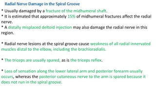
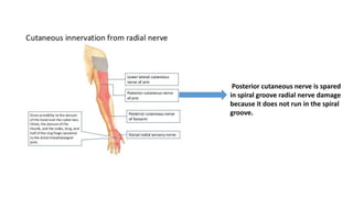
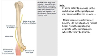
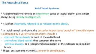
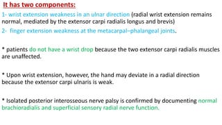
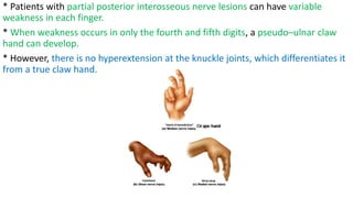
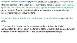
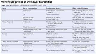
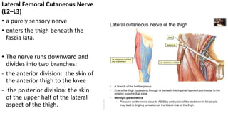
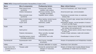
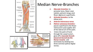
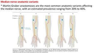
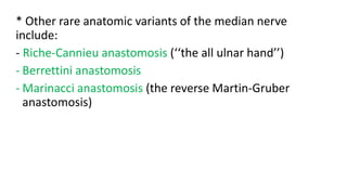
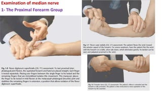
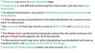
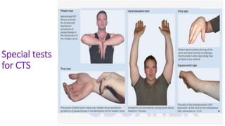
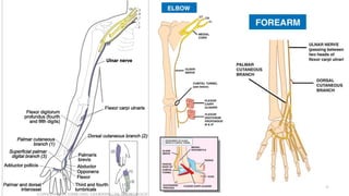
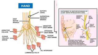
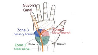
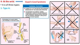
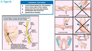
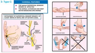
This document discusses peripheral nerve diseases, focusing on individual nerve lesions. It describes the anatomy and clinical presentations of lesions of the dorsal scapular, long thoracic, suprascapular, subscapular, thoracodorsal, anterior thoracic, axillary, musculocutaneous, and median nerves. It also discusses median nerve variants, carpal tunnel syndrome, and ulnar nerve anatomy.
6. Mononeuropathy Multiplex
* involvement of several isolated nerves.
* The nerves involved are often widely separated (e.g., right median and left
femoral nerve).
* It is usually due to a disseminated vasculitis that affects individual nerves
27. Lesions of Individual Nerves
1- Dorsal Scapular Nerve (C4–C5)
* May occur in body builders
* the lateral displacement of the vertebral
border of the scapula, which is rotated,
with the inferior angle displaced laterally.
* Rhomboid paresis is evident if the elbow
can be pressed back only weakly against
resistance (keeping the hand on the hip).
* Weakness is also evident when the
patient attempts to push the palm
backward against resistance with the arm
folded behind the back.
28. 2- Long Thoracic Nerve (C5–C7)
Causes:
- Sport injury
- The overhead arm positioning during
surgery with general anesthesia .
- iatrogenic : first rib resections,
mastectomies with axillary node
dissection,thoracotomy,scalenectomies,
infraclavicular plexus anesthesia, and
chest tube insertion
- neuralgic amyotrophy (Parsonage–
Turner syndrome)
- radiation therapy for breast cancer
- familial long thoracic nerve
- Lyme disease (painful)
30. NERVE LESIONS
Etiologies :
1- proximal upper brachial
plexopathies
2- trauma to shoulder
3- after repair of rotator cuff
tears
4- reported after driving for 2
hours speaking on a mobile
telephone with the phone
cradled between the ear and
shoulder,
5- ganglion cysts
Clinical :
• shoulder pain radiating into thearm worsens
by movements that adduct the scapula or
rotate the head away from the involved
shoulder.
• weakness, and eventual atrophy
• Supraspinatus paresis results in weakness of
arm abduction
• infraspinatus paresis results in impaired
external rotations at the shoulder joint.
• Suprascapular involvement may also occur
with neuralgic amyotrophy
• Isolated infraspinatus paresis may also result
from a nerve injury in body builders
31. 4- Subscapular Nerves (C5–C7)
• Purely motor nerves
• The upper subscapular nerves supply the subscapularis
• the lower subscapular nerves supply the teres major.
• Subscapular nerve palsies usually occur with posterior cord brachial plexus
lesions.
• The arm is externally rotated, with some paresis of internal rotation
• The patient may complain of difficulty in scratching the lower back.
5- Thoracodorsal Nerve (pure motor) (C6–C8)
• Supply latissimus dorsi
• having the patient adduct the horizontally raised upper arm against resistance or
by palpating the muscle bellies when the patient coughs
• Its lesion causes weak proximal arm adduction
• usually in in body builders and posterior cord brachial plexus lesion
32. 5- Anterior Thoracic Nerves (C5–T1)
• the lateral nerve supplies upper portions of the pectoralis major,
• the medial division supplies the lower portion of this muscle and the pectoralis
minor.
• It is tested by having the patient hold the arm in front of the body. The two
portions can be seen and palpated when the patient resists attempts by the
examiner to force the arm laterally
• Their Lesion can cause weak Adduction and medial rotation of the upper arm
• Isolated injury can occur from a seatbelt injury
• Bilateral medial anterior thoracic neuropathies in a weight lifter were thought to
be due to concomitant pectoralis minor hypertrophy causing intramuscular
entrapment of the nerves
35. MONONEUROPATHIES
• Mononeuropathy is defined as a disorder of a single peripheral nerve.
• may result from compression, traction, laceration, thermal, or chemical injury.
• May include axon loss, demyelination, or a combination of both.
segmental demyelination but
leaving the axons intact
Axonal loss + intact endoneurial
tubes , perineurium and
epineurium
The axons and endoneurium
are damaged
The axons, endoneurium, and
perineurium are disrupted, but
the epineurium is intact
complete disruption of the
nerve and all supporting
structures
36.
37.
38.
39. Introduction
>> Carpal tunnel syndrome is the most common ( 42% of workers
and with annual incidence of 193 per 100,000 in all women) followed
by:
>> Morton neuroma
>> Ulnar neuropathy
>> Meralgia paresthetica
>> Radial neuropathy
40. MEDIAN NERVE
Basic Anatomy
* It forms from the terminal divisions of the medial and lateral cords of the brachial
plexus, receiving contributions from the C5 to T1 nerve roots
* It courses medial to the brachial artery throughout the upper arm.
* In the distal arm, the nerve may pass beneath the ligament of Struthers, which is
present in some individuals (1% to 13%) and represents a rare cause of median
nerve entrapment.
* The nerve then passes under the bicipital aponeurosis at the elbow.
* Moving distally, the median nerve then travels between the two heads of the
pronator teres, deep to the humeral head and superficial to the ulnar head.
* The nerve then continues distally between the flexor digitorum superficialis and
flexor digitorum profundus muscles.
41. 41
* Approximately 4 cm distal to the medial
epicondyle, the anterior interosseus nerve
branches from the main trunk of the median
nerve.
* The anterior interosseus nerve innervates the
flexor pollicis longus, flexor digitorum profundus
to the second and third digits, and pronator
quadratus.
* Just proximal to the distal wrist crease, the
palmar cutaneous branch of the median nerve
leaves the main nerve trunk.
* It travels between the palmaris longus and
flexor carpi radialis tendons and proceeds
outside the carpal tunnel to provide sensation
for the thenar eminence and proximal lateral
palm.
42.
43.
44.
45. Median nerve anatomic variants
* Martin-Gruber anastomoses are the most common anatomic variants affecting
the median nerve, with an estimated prevalence ranging from 20% to 40%.
46. * Other rare anatomic variants of the median nerve
include:
- Riche-Cannieu anastomosis (‘‘the all ulnar hand’’)
- Berrettini anastomosis
- Marinacci anastomosis (the reverse Martin-Gruber
anastomosis)
53. Clinical Findings and Syndromes
A - In the Upper Arm:
1- usually secondary to trauma in which radial and ulnar injured also because of
proximity to each other in the upper arm (triad neuropathy).
* Or due to Pressure palsies, like the Saturday night palsy and the honeymooner’s
palsy.
54. 2- Supracondylar Spur/Ligament of Struthers
3- Supracondylar Fractures usually occur
in children and may cause median nerve
damage.
This canal which is
formed by this accessory
ligament can make
compression on median
nerve
55. B- In The Forearm
1- Musculotendinous Median Neuropathies
* The bicipital aponeurosis, which crosses from lateral to medial over the antecubital
fossa and serves to attach the biceps brachii tendon indirectly to the ulna, may irritate
the median nerve.
* It is similar as Ligament of Struthers but Occasionally, resisted forearm flexion in the
supinated position for 30 seconds may precipitate symptoms.
2- Pronator teres syndrome:
* The only median-innervated muscle that is not affected by this syndrome is the
pronator teres itself.
* Dull aching pain in the proximal forearm that is worsened by repetitive or forceful
pronation.
* tenderness of the pronator teres to palpation.
* Median-innervated hand sensation is often normal,
* A Tinel sign can often be elicited at the antecubital fossa.
56.
57.
58. C- The Anterior Interosseous syndrome
* may occur secondary to trauma, fractures, Parsonage-Turner syndrome,
anomalous muscles and/ or tendons, or without a known cause.
* Patients usually complain of weakness or clumsiness in grasping objects with
their first two digits (e.g., grasping the handle of a coffee cup).
* There are usually no complaints of pain, and because this nerve does not carry
cutaneous sensation, no numbness occurs.
* There is weakness of the flexor digitorum profundus (to the second and third
digits), flexor pollicis longus, and pronator quadratus.
59. * Patients with rheumatoid arthritis can have spontaneous and painless rupture of
the flexor digitorum profundus and flexor pollicis longus tendons, mimicking an
anterior interosseous palsy.
* To exclude this possibility:
- have the patient open and relax the hand.
- If the tendons are intact, pressing your thumb firmly across the ventral aspect of
the forearm about 2 to 3 inches proximal to the wrist should cause passive finger
flexion.
* Unlike carpal tunnel syndrome, anterior interosseous neuropathies are generally
due to noncompressive causes such as idiopathic brachial neuritis/neuralgic
amyotrophy.
60.
61. Carpal tunnel syndrome
Clinical Presentation
* compression at the wrist, causing carpal tunnel syndrome classically present with
numbness, tingling, and other paresthesia affecting the first through third fingers
and the lateral aspect of the fourth finger.
* Some reports sensory symptoms affecting the entire hand or radiation from the
hand to the proximal upper extremity.
* These may be exacerbated by wrist flexion,including driving a car, and often wake
the patient from sleep at night.
* Shaking or flicking the affected hand may alleviate the sensation, which can be
quite painful.
62. * Weakness may occur in more advanced stage
* weakened grip and difficulty performing fine motor tasks, and they may drop
items easily.
* On physical examination: may reveal mild flattening of the thenar eminence or
frank atrophy.
* A Tinel sign consists of paresthesia in the distal distribution of a sensory nerve
when it is percussed.
* the sensitivity of Tinel sign may be as low as 30% to 43% with specificity up to
65%.
* The Phalen test is performed by having the patient flex the wrists and press the
dorsum of both hands together for 30 to 60 seconds.
* A false-positive result may occur when interpreting musculoskeletal wrist pain as
a positive result, and specificity is only 15% to 17% range.
* Sensitivity of Phalen testing is better, but only around 50% to 67%
64. * Patients with positive Tinel and Phalen testing have lower nerve conduction
velocities than those with negative testing.
* A negative Phalen test is a strong predictor of normal nerve conduction studies.
* Strength testing for suspected carpal tunnel syndrome should include upper limb
and especially forearm and intrinsic hand muscles with special focus on the
abductor pollicis brevis, opponens pollicis, and flexor pollicis longus muscles.
* loss of sensation in the palmar first through third digits and lateral fourth digit.
* Sensation of the proximal lateral palm may be spared in carpal tunnel syndrome
because of its innervation by the palmar cutaneous branch of the median nerve.
* More proximal lesions of the median nerve may shows negative a Tinel sign at
the wrist and positive Phalen testing.
65. Electrodiagnostic Testing in Carpal Tunnel Syndrome
* It is recommended to perform an initial median sensory nerve conduction study
across the wrist at a distance of 13 cm or 14 cm.
* If normal, a shorter segment study (7 cm to 8 cm) of the median mixed nerve is
recommended.
* These studies should be performed with a radial or ulnar sensory or mixed
response across the same distance in the same limb for comparison of interlatency
differences.
* A median motor response recording over the abductor pollicis brevis is also
recommended, along with measurement of another motor response in the same
limb.
* Optional testing includes needle EMG of C5 to T1 muscles (to exclude cervical
radiculopathy as a contributing factor) and supplementary nerve conduction
studies
66. Electrodiagnostic testing in MartinGruber anastomosis
* A Martin-Gruber anastomosis may result in low distal amplitude of the median
nerve compound muscle action potential (CMAP) when recording over the
abductor pollicis brevis and stimulating at the wrist.
* With proximal nerve stimulation, the median CMAP amplitude will be higher.
* This can create confusion and concern for overstimulation of the median nerve at
the elbow, with costimulation of the ulnar nerve.
* The presence of a MartinGruber anastomosis can be confirmed by stimulating
the median nerve at the elbow and recording a CMAP in an ulnar-innervated hand
muscle, typically the first dorsal interosseus or abductor digiti minimi.
67. Ultrasonographic Testing in Carpal Tunnel Syndrome
* It should be high-frequency ultrasound probes
* It has 89% sensitivity and 90% specificity
* Nerves are enlarged at sites of compression.
* a cross-sectional area of the median nerve of greater than 12 mm is considered
abnormal.
* Routine ultrasound studies also help us in revealing :
- contributing factors
- anatomic variants
- intraneural tumors
- bifid median nerve
- the persistent median artery which is an accessory artery that arises from the
ulnar artery in the proximal forearm
- Other non-nerve findings may include coexistent tenosynovitis or accessory
musculature
68. Treatment of Carpal Tunnel Syndrome
Generally :
1- Nonsurgical treatments (splinting + local corticosteroid injections) for those
early in the course of symptoms without evidence of median nerve denervation.
* A trial period of 2 to 7 weeks is recommended to observe for improvement.
2- Carpal tunnel release if nonsurgical therapies have failed.
((Heat therapy, electrical stimulation, iontophoresis, and laser therapies were
among the treatments with no evidence to support their use))
69. * For patients with mild carpal tunnel syndrome involving abnormal findings
isolated to the median mixed or sensory responses, the authors begin a trial of:
Splinting + NSAIDS
* If failed consider a trial of local corticosteroid injections.
* Decompressive surgery to transect the transverse carpal ligament may
subsequently be considered in patients with significant residual symptoms.
* For patients with severe carpal tunnel syndrome with prolonged, low amplitude
CMAPs and denervated median-innervated thenar muscles by needle EMG
suddenly consider surgery
74. Basic Anatomy
* It arises from the C8 to T1 nerve roots and the medial cord of the brachial plexus
and descends through the posteromedial arm, anterior to the medial head of the
triceps brachii
* The first branch off the ulnar nerve is typically a sensory branch to the elbow,
followed by a motor branch to the flexor carpi ulnaris.
* Compression in the arm is rare but may occur at the arcade of Struthers.
* The ulnar nerve then passes through the epicondylar (ulnar) groove, located
between the medial epicondyle and the olecranon.
* An anomalous anconeus epitrochlearis muscle overlying the ulnar nerve and
extending between the olecranon and medial epicondyle may be a source of
compression in some individuals.
75. * The cubital tunnel is located just distal to the elbow.
* The medial collateral ligament of the elbow defines the floor of the cubital tunnel,
while the roof consists of the thickened fascia known as the Osborne (arcuate) ligament.
* Within the proximal forearm, the ulnar nerve is located medial and superior to the
flexor digitorum profundus and deep to the flexor carpi ulnaris and gives branches to
innervate these muscles along its course.
* The ulnar nerve lies lateral to the flexor carpi ulnaris and medial to the ulnar artery in
the distal forearm.
* Compression of the ulnar nerve in the forearm is rare.
* The Guyon canal, or ulnar tunnel, is a site of potential ulnar nerve entrapment at the
wrist.
* The ulnar nerve passes through the Guyon canal with the ulnar artery.
83. Clinical Findings and Syndromes
At the arm:
Causes :
- trauma
- Compression by a crutch or by
the arm hanging over a chair
(Saturday night palsy).
- Arcade of struther
- Clinical findings as its compression
At elbow but here flexor carpi ulnaris
Also affected.
84. 2- At elbow:
Causes:
- trauma
- Ulnar nerve entrapment at the elbow include between the two heads of the
flexor carpi ulnaris (cubital tunnel syndrome) and by the Osborne fascia, when
present.
85.
86. Other possible findings:
* Provocative maneuvers, like flexing the forearm for a minute, may precipitate
symptoms.
* The patient should also repetitively flex the elbow while the ulnar nerve is
palpated in the postcondylar groove to rule out “snapping” or dislocation of either
the ulnar nerve or medial triceps tendon, both of which can cause ulnar nerve
irritation at the elbow.
87.
88. 3- At The Forearm
A) Flexor–Pronator Fascia
* It may be irritated by a thickened flexor–pronator fascia which is 5cm distal to the
medial epicondyle
B) The Dorsal Ulnar Cutaneous Nerve palsy
* Occasionally, the dorsal ulnar cutaneous branch may be compressed or
transected as it leaves the ulnar nerve and passes to the dorsum of the wrist
between the flexor carpi ulnaris tendon and the ulna.
* Ulnar fractures or repairs, lacerations, or blunt trauma may involve this nerve.
* patients present with numbness or hypesthesia in the dorsomedial third of the
hand.
* With some lesions, a Tinel sign can be elicited when tapping on the medial
border of the ulna a few centimeters proximal to the wrist.
89.
90. 4- At the wrist
• It is of three types:
1- Type A:
Guyon’s Canal Syndrome
94. Electrodiagnostic Testing
* Electrodiagnostic testing is considered the gold standard for the diagnosis of
ulnar neuropathy
* AANEM guidelines for the diagnosis of ulnar neuropathy at the elbow
recommend performing ulnar sensory nerve conduction studies and motor nerve
conduction studies to the abductor digiti minimi.
* Elbow position during testing (with flexion between 70 degrees and 90 degrees
recommended) should be recorded and adequate warming maintained.
* A minimum of 10cm should be present between the above-elbow and below
elbow sites of stimulation.
95.
96. * The diagnosis of ulnar neuropathy at the elbow can be made when the
conduction velocity across the elbow is greater than or equal to 10 m/s slower
than the wrist to below-elbow segment.
* Other supportive findings include a drop in CMAP amplitude of greater than 20%
across the elbow.
* Sensitivity has ranged from 37% to 86%, while specificity has been greater than
95%.
97. * The 10-cm segment between the above elbow and below-elbow sites is divided
into 2-cm segments.
* Stimulation is applied at each point, with attention to the latency differences and
CMAP waveforms.
* Using this approach, the sensitivity of nerve conduction studies is improved,
detecting up to 90% of patients with clinical evidence of ulnar neuropathy at the
elbow.
* Needle EMG is essential in assessing ulnar neuropathy at the elbow.
* Examination of ulnar-innervated muscles is helpful for both localization and
assessment of severity.
* Other muscles with C7 and C8 innervation should also be sampled to assess for
the presence of coexistent cervical radiculopathy or brachial plexopathy.
98. Ultrasonographic Testing
* The normal ulnar nerve cross sectional area is 8 mm to 11 mm at the elbow, with
larger values suggesting focal compression.
* It is helpful in:
- pinpointing the area of pathology
- identify alternative etiologies for ulnar neuropathy at the elbow like anatomic
variants (eg, anomalous anconeus epitrochlearis muscle), ganglion cysts, and
other mass lesions
* Testing of the dorsal ulnar cutaneous sensory nerve response can be added when
assessing lesions distal to the elbow, assisting in localization of ulnar nerve lesions
at or proximal to the wrist.
* The dorsal ulnar cutaneous sensory nerve response will be normal with the
lesions at the wrist, but abnormal with lesions in the region of the proximal
forearm or elbow.
99. Treatment of Ulnar Neuropathy at the Elbow
* For mild symptoms just conservative treatment with elbow pads and avoid
flexion of the elbow for 3months.
* If is failed or who have evidence of significant axonal loss on initial
electrodiagnostic examination, surgical management is indicated.
* Three general approaches are used:
1- Simple decompression through excision of the arcuate ligament
2- Medial epicondylectomy
3- Ulnar nerve transposition.
* Unlike in CTS , corticosteroid injections have not shown efficacy in ulnar
neuropathy at the elbow.
* Ultrasound and low-level laser therapy have shown some promise but are
neither widely accepted nor available.
102. * The radial nerve receives contributions from the C5– C8 spinal nerves.
* These contributions pass through the upper, middle, and lower trunks and
posterior cord of the brachial plexus.
* The radial nerve supplies the extensor muscles of the arm and forearm as well
as the skin covering them.
Radial Nerve in the Upper Arm
* As it winds around the humerus, or just proximal to this section, the radial nerve
supplies the triceps muscle.
* After its course in the spiral groove, it then supplies the brachioradialis and the
extensor carpi radialis longus and brevis muscles.
* The nerve then bifurcates into a superficial (sensory) branch and a deep (motor)
branch.
103. Radial Nerve in the Forearm
* The superficial branch passes distally into the hand, where it supplies the skin of
the radial aspect of the dorsum of the hand and the dorsum of the first four
fingers.
* The sensory autonomous zone of the radial nerve is the skin over the first
interosseous space.
* The deep branch of the radial nerve passes deep through the fibrous arch of the
supinator muscle (the arcade of Frohse) to enter the posterior compartment of
the forearm.
* The nerve continues in this compartment as the purely motor posterior
interosseous nerve, which innervates the remaining wrist and finger extensors
104. * These include the following:
(1) Supinator : a forearm supinator;
(2) extensor digitorum: an extensor of the second through the fifth MCP joints
(3) extensor digiti minimi: an extensor of the fifth MCP joint
(4) extensor carpi ulnaris: an ulnar extensor of the wrist
(5) abductor pollicis longus: an abductor of the carpometacarpal joint of the thumb
(6) extensor pollicis longus: an extensor of the interphalangeal joint of the thumb;
(7) extensor pollicis brevis: an extensor of the metacarpophalangeal joint of the thumb
(8) extensor indicis: an extensor of the second finger.
111. * A high radial palsy (rare) in the axilla can cause both triceps weakness and
posterior arm sensory loss, two deficits that distinguish it from more common
radial nerve injuries at the spiral groove.
* Injuries affecting the proximal radial nerve may be differentiated from posterior
cord involvement by confirming normal deltoid (axillary branch off the posterior
cord) and latissimus dorsi (thoracodorsal branch off the posterior cord) strength.
112. * Patients with C7 palsies can be distinguished from posterior cord or radial nerve injuries
because they usually have numbness in both the volar and dorsal aspects of the third
digit (middle finger), with sensory loss not extending proximal to the wrist.
* Additionally, C7 muscles innervated by the median nerve, including the pronator teres
and flexor carpi radialis longus, would also be weak.
* A complete radial palsy presents with sensory loss along the posterior arm and forearm,
the lower anterolateral arm, as well as the dorsolateral hand.
* All extensors of the arm are weak.
* The triceps are weak, and the triceps reflex is absent.
* Secondary to brachioradialis paralysis, elbow flexion may be somewhat weak compared
with the normal side.
113. * There is a wrist drop from extensor carpi radialis (longus and brevis) and extensor
carpi ulnaris weakness.
* The fingers cannot extend at the knuckle joint.
* Supination is partially weak, with residual supination performed by the biceps
brachii.
* The wrist and hand appear limp, with the fingers semiflexed and the metacarpal
bone of the thumb volar to the palm
* Distal finger extension is possible secondary to lumbrical function.
Because the lesion is in
the arm the triceps
spared but if the lesion
is in axilla triceps is also
affected
114.
115. * Usually damaged by a fracture of the midhumeral shaft.
* It is estimated that approximately 15% of midhumeral fractures affect the radial
nerve.
* A distally misplaced deltoid injection may also damage the radial nerve in this
region.
* Radial nerve lesions at the spiral groove cause weakness of all radial innervated
muscles distal to the elbow, including the brachioradialis.
* The triceps are usually spared, as is the triceps reflex.
* Loss of sensation along the lower lateral arm and posterior forearm usually
occurs, whereas the posterior cutaneous nerve to the arm is spared because it
does not run in the spiral groove.
116. Posterior cutaneous nerve is spared
in spiral groove radial nerve damage
because it does not run in the spiral
groove.
117. Note:
• In some patients, damage to the
radial nerve at the spiral groove
may cause mild triceps weakness.
• This is because supplementary
branches to the lateral and medial
heads from the radial nerve
originate in the spiral groove,
where they may be injured.
118. * Radial tunnel syndrome is an uncommon cause of lateral elbow pain almost
always being initially misdiagnosed.
* it is often incorrectly referred to as resistant tennis elbow .
* In radial tunnel syndrome, the posterior interosseous branch of the radial nerve
is entrapped by a variety of mechanisms include :
- aberrant fibrous bands in front of the radial head
- anomalous blood vessels that compress the nerve
- extrinsic masses, or a sharp tendinous margin of the extensor carpi radialis
brevis.
* These entrapments may exist alone or in combination.
119.
120. * pain just below the lateral epicondyle of the humerus That may develop after
an acute twisting injury or direct trauma to the soft tissues overlying the
posterior interosseous branch of the radial nerve, or the onset may be more
insidious, without an obvious inciting factor.
* The pain is constant and worsens with active supination of the wrist.
* Patients often note the inability to hold a coffee cup or hammer.
* Sleep disturbance is common.
* On physical examination, elbow range of motion is normal.
* Grip strength on the affected side may be diminished.
121. * three important signs that allow the clinician to distinguish radial tunnel
syndrome from tennis elbow:
(1) tenderness to palpation distal to the radial head in the muscle mass of the
extensors, rather than over the more proximal lateral epicondyle, as in tennis
elbow
(2) increasing pain on active resisted supination of the forearm owing to
compression of the radial nerve by the arcade of Frohse as a result of contraction
of the muscle mass
(3) a positive result on the middle finger test.
* The middle finger test is performed by having the patient extend the forearm,
wrist, and middle finger and sustain this action against resistance.
* Patients with radial tunnel syndrome exhibit increased lateral elbow pain
secondary to fixation and compression of the radial nerve by the extensor carpi
radialis brevis muscle.
122. Clinical pearl:
* Radial tunnel syndrome
should have no objective
motor or sensory deficits on
examination or
electrodiagnostic studies.
* If these are present,
especially finger extension
weakness, one must
consider other diagnoses,
specifically posterior
interosseous nerve
compression at the
supinator.
123. Testing
* Electromyography helps to distinguish cervical radiculopathy and radial
tunnel syndrome from tennis elbow.
* Plain radiographs are indicated in all patients who present with radial tunnel
syndrome to rule out occult bony pathology.
* MRI of the elbow to role out ganglion cysts or lipomas
* Acute gout affecting the elbow may be difficult to distinguish
Treatment
* NSAIDs + physical therapy.
* The local application of heat and cold also may be beneficial.
* injection of the radial nerve at the elbow with a local anesthetic and steroid may
be a reasonable next step.
* Finally surgical exploration and decompression of the radial nerve are indicated.
124. * Supinator syndrome is a posterior interosseous nerve palsy secondary to
compression where this nerve enters the supinator m..
* The posterior interosseous nerve passes between the superficial and deep heads
of this muscle,
* This pain is most often the result of repetitive microtrauma to the muscle caused
by such activities as turning a screwdriver, prolonged ironing, handshaking, or
digging with a trowel
* pain localized to the supinator muscle, which worsens after a few minutes of
forced supination.
* sudden or progressive finger extension weakness.
* Denervation of the supinator is classically spared
* Sensation is normal in the territory of the superficial sensory radial nerve
* the brachioradialis muscle has normal strength.
125.
126. * The trigger point is pathognomonic of myofascial pain syndrome
* Mechanical stimulation of the trigger point by palpation or stretching produces
not only intense local pain but also referred pain.
* In addition, an involuntary withdrawal of the stimulated muscle, called a jump
sign, is often seen and is characteristic of myofascial pain syndrome.
* Increased plasma myoglobin has been reported in some patients with supinator
syndrome.
Treatment
* prolonged relaxation of the affected muscle and local anesthetic or saline
solution is the starting point.
* Antidepressants + Pregabalin and gabapentin
* Botulinum toxin type A directly into trigger points has been used with success in
patients who have not responded to traditional treatment modalities.
127. Causes : trauma, diabetic mononeuropathy, compression at the supinator muscle
(supinator syndrome), or a space-occupying mass, or as a manifestation of
Parsonage-Turner syndrome.
* The most common soft tissue mass compressing the posterior interosseous
nerve is a lipoma, followed by nerve sheath tumors, ganglion cysts, and
hypertrophic synovium (rheumatoid arthritis).
* Considering it carries no cutaneous sensory fibers, sensation remains normal.
* dull ache in the proximal forearm extensor muscles near the radial head.
128. It has two components:
1- wrist extension weakness in an ulnar direction (radial wrist extension remains
normal, mediated by the extensor carpi radialis longus and brevis)
2- finger extension weakness at the metacarpal–phalangeal joints.
* patients do not have a wrist drop because the two extensor carpi radialis muscles
are unaffected.
* Upon wrist extension, however, the hand may deviate in a radial direction
because the extensor carpi ulnaris is weak.
* Isolated posterior interosseous nerve palsy is confirmed by documenting normal
brachioradialis and superficial sensory radial nerve function.
129. * Patients with partial posterior interosseous nerve lesions can have variable
weakness in each finger.
* When weakness occurs in only the fourth and fifth digits, a pseudo–ulnar claw
hand can develop.
* However, there is no hyperextension at the knuckle joints, which differentiates it
from a true claw hand.
130. * Isolated damage to the superficial sensory radial nerve can occur from trauma,
tight handcuffs or watches, venipuncture, surgery for de Quervain tenosynovitis,
and occasionally from scissor-like pinching between the brachioradialis and
extensor carpi radialis longus tendons.
* numbness, hyperesthesia, and burning pain on the dorsolateral aspect of the
hand.
* The superficial sensory radial nerve pierces the antebrachial fascia
approximately two thirds down the forearm at the lateral (radial) border, between
the tendons of the brachioradialis and extensor carpi radialis longus
131. * During pronation, the brachioradialis tendon closes the space between these two
tendons in a scissor-like fashion, potentially irritating the nerve where it pierces
the fascia.
* In these patients, pain and numbness can be exacerbated by forced pronation
and ulnar wrist deviation,
* A Tinel sign can often be elicited.
* This diagnosis may be confirmed with electrodiagnostic testing.
132.
133.
134.
135. * Cheiralgia paresthetica also should be differentiated from cervical
radiculopathy involving the C6 or C7 roots, although patients with cervical
radiculopathy generally present not only with pain and numbness but also
with reflex and motor changes.
* Cervical radiculopathy and radial nerve entrapment may coexist as the “double
crush” syndrome.
* The double crush syndrome is seen most commonly with median nerve
entrapment at the wrist or carpal tunnel syndrome.
* Workup: palin X-ray, ESR ,CBC, ANA, MRI elbow, EMG
Treatment:
- NSAIDS
- Injection local anesthetic + steroid
- decompression surgery
136.
137. Electrodiagnostic Testing in suspected radial nerve entrapment
* requires nerve conduction studies and needle EMG.
* Typically, the radial motor response is recorded over the extensor indicis proprius
following stimulation at the forearm, lateral elbow, and spiral groove.
* This permits assessment of potential areas of entrapment, and special attention
to CMAP amplitudes and dispersion is necessary.
* Stimulation at Erb point is helpful in localizing proximal humeral lesions, although
costimulation of the brachial plexus may contaminate the recordings.
138. * A superficial radial sensory response is valuable for differentiating between
common radial and posterior interosseous nerve pathology.
* As with any mononeuropathy, nerve conduction studies of other upper extremity
nerves may be needed to confirm isolated involvement of the radial nerve.
* Needle EMG can assist in localization and prognosis.
* Additional examination of nonradial muscles is helpful and often necessary in
excluding brachial plexopathy or cervical radiculopathy as a cause of symptoms.
139. Ultrasonographic Testing
* Focal enlargement has been documented at sites of compression and can be
helpful in localization.
* Radial nerve cross-sectional area is typically less than 10 mm in the upper arm
and antecubital fossa, with the superficial radial sensory nerve measuring only 1
mm to 3 mm
* The posterior interosseous nerve cross-sectional area measures approximately 2
mm
* Interestingly, distal enlargement of the posterior interosseous nerve has been
documented in cases of proximal radial nerve lesions, suggesting a double crush
syndrome.
140.
141. Medial Cutaneous Nerves of the Arm and
Forearm (C8–T1)
• may be injured in the axilla, affecting the
medial cord of the brachial plexus.
• may be affected in isolation by a stretch
injury or by injury due to the placement of
an arterial graft
• The nerve may also be damaged as a
complication of an ulnar nerve surgery at the
elbow
Intercostobrachial Nerve (T2)
• Nerve injury results in pain and dysesthesia
in the axilla and medial arm.
• largest sensory nerve in the body and
damage to the nerve may occur often
following surgeries for breast cancer and
axillary lymphadenectomy
142.
143.
144.
145. It may be injured in
a modified radical
mastectomy and
other surgical
procedures
involving the
axilla and lateral
pectoral region
146.
147. Iliohypogastric (T12–L1)
• mixed nerve
• Supply :
- the internal oblique and transversus abdominis muscles
- the skin over the outer buttock and hip, and the anterior the anterior abdominal
wall above the pubis.
• Site of injury:
- The lumbar plexus
- At the posterior or anterior abdominal wall, or distally near the inguinal ring.
• Lesions :
- little motor deficit
- pain or sensory loss
- Painful iliohypogastric neuropathies are not uncommon after lower abdominal
surgery
148. ilioinguinal nerve (L1):
supplies :
- Motor: the internal oblique and transversus abdominis muscles.
- Sensory: the medial thigh below the inguinal ligament , the skin of the symphysis
pubis and the external genitalia.
Sites Of injury:
- the lumbar plexus
- posterior abdominal wall and at the anterior abdominal wall
- within the inguinal canal.
• Neuropathy may occur after pregnancy, likely due to the stretching of the nerve .
• Lower abdominal and inguinal pain and paresthesias in professional ice hockey
players
149. The diagnostic triad for ilioinguinal/iliohypogastric nerve postsurgical entrapment
syndrome consists of the following :
1. Sharp, burning pain emanating from the incision site and radiating to the
suprapubic, labial, or thigh areas
2. Paresthesia over the appropriate nerve distribution
3. Pain relief after infiltration with a local anesthetic
• The onset of symptoms may be immediate or may occur months to years after
the offending surgical procedure.
• The symptoms are exacerbated as a result of stretching, coughing, sneezing, and
Valsalva and can be relieved with hip flexion or by adopting a stooped posture
when ambulating.
150. Genitofemoral nerve (L1-L2):
• a predominantly sensory nerve
• divides into two branches:
- External spermatic (genital branch L1): skin of the scrotum and adjacent medial thigh.
- Lumboinguinal (femoral branch L2): supplies the skin of the upper thigh over the femoral
triangle.
Sites of injury:
1- the lumbar plexus
2- within the abdomen
3- in the femoral or inguinal region (e.g., after inguinal herniorrhaphy, appendectomy, cesarean
section, hysterectomy, vasectomy, blunt abdominal trauma, and even due to wearing tight jeans).
• Injury causes pain (genitofemoral neuralgia) and sensory loss in the area of the cutaneous
supply of the nerve.
• The cremasteric reflex may be lost on the side of the nerve lesion.
151. Lower Extremity
•The major nerves of the thigh are:
1- The femoral nerve (ventrally)
2- The sciatic nerve (dorsally).
•The leg and foot are mainly supplied by the terminal
branches of the sciatic nerve:
-The peroneal nerve (ventrally)
-The tibial nerve (dorsally).
153. Femoral Nerve (L2–L4)
• a mixed nerve
• Beneath the inguinal ligament (lateral to
the femoral artery) enter the thigh.
• Within the femoral triangle, it separates
into the anterior and posterior divisions.
• hanging leg syndrome : Severe,
combined, bilateral femoral and sciatic
neuropathies in the context of alcohol
intoxication .
154.
155. MOTOR MANIFESTATION:
Wasting of quadriceps femoris.
Loss of extension of knee.
Weak flexion of hip (psoas major is
intact).
SENSORY EFFECT:
loss of sensation over areas
supplied (antero-medial) aspect of
thigh & medial side of knee, leg &
foot.
156. Etiology:
• Compression
Iliopsoas, pelvic, or retroperitoneal
hematoma
Anticoagulation
Hemophilia
• Pelvic mass (tumor, abscess, cyst)
• Aortic or iliac aneurysm
• Inguinal lymph node
• Hyperextension stretch injury
• Dancing “Hanging leg syndrome”
• Direct injury
• Pelvic fracture
• Iatrogenic
• Radiation injury
• Ischemia
-Diabetes
-IV drug abuse
-Common iliac artery occlusion
- Intraoperative hypotension
- Aortic clamping during vascular
surgery
• Surgical operations or procedures
• Vaginal delivery
157.
158. • A proximal lesion of the femoral nerve (e.g., at the lumbar plexus or within the
pelvis) results in the following signs:
1. Atrophy: There is wasting of the musculature of the anterior part of the thigh.
2. Motor signs: There is weakness or paralysis of hip flexion (iliacus, psoas, and
rectus femoris muscles) and an inability to extend the leg (quadriceps femoris).
With paralysis of the Sartorius muscle, lateral thigh rotation may be impaired.
3. Sensory symptoms and signs: Sensory loss, paresthesias, and, occasionally, pain
occur on the anteromedial thigh and inner leg as far as the ankle.
4. Reflex signs: The patellar reflex is depressed or absent
159. • Lesions at the inguinal ligament result in similar findings, but thigh flexion is
spared because of the more proximal origin of the femoral nerve branches to the
iliacus and psoas muscles.
• A purely motor syndrome (quadriceps atrophy and paresis) ….lesions within the
femoral triangle (the posterior division of the femoral nerve distal to the origin of
the saphenous branch) .
• Unilateral or bilateral anterior femoral cutaneous nerve injury, clinically sparing
femoral branches to the saphenous nerve and quadriceps muscles, This purely
sensory syndrome
• Isolated medial femoral cutaneous neuropathy, resulting in paresthesias and
dysesthesias over the anteromedial thigh, after penetrating injury.
160. • A purely sensory syndrome (pain, paresthesias, and sensory loss) may result
when the saphenous nerve alone is damaged.
- The nerve may be entrapped proximally in the thigh by fibrous bands or branches
of femoral vessels.
- Isolated saphenous involvement may occur with femoral thrombectomy and
femoral-popliteal bypass surgery and after popliteal vein aneurysm resection
with saphenous vein interposition .
- Schwannomas of the nerve may develop in the thigh
- Spontaneous saphenous neuralgia may result from nerve compromise in the
subsartorial canal
>>> Medial knee and leg pain result, and tenderness over the subsartorial canal is
present.
- The saphenous nerve may be damaged at the knee after medial arthrotomy or
arthroscopy, in association with coronary artery bypass graft surgery, and by
lacerations .
161. • Damage to the infrapatellar branch of the
saphenous nerve results in numbness and
paresthesias in the skin over the patella
(gonyalgia paresthetica)
>> When bending the knee, occasional pins-and-
needles sensations are produced. >> Gonyalgia
paresthetica usually develops insidiously without
acute trauma and is often accompanied by sharp
pain below and lateral to the knee.
>> The infrapatellar branch may be injured during
arthroscopy or other knee operations, by
accidental trauma, or by nerve compression
162.
163. Obturator Nerve (L2–L4)
• a mixed nerve
• Within the obturator canal, it
supplies the obturator externus
muscle and then divides into two
branches, the anterior and
posterior branches, which descend
into the medial thigh.
• The anterior division is mixed
supplies those muscles in the
picture and the skin over the
medial thigh.
• The posterior division is motor
• The adductor magnus may also be
innervated by the sciatic nerve.
• Motor function : having the patient
adduct the extended leg against
resistance
164. NERVE LESIONS
Site of lesions:
- lumbar plexus
- near the sacroiliac joint
- the lateral pelvic wall
- within the obturator canal.
Etiology :
- orthopedic, gynecological, or urological
procedures or injuries
- obturator hernia
- after pelvic surgery, after femoral artery
procedures
- after bilateral total knee arthroplasty with
prolonged tourniquet use
- Bilateral obturator nerve injuries may occur
during urologic surgery owing to prolonged hip
flexion, resulting in the stretching of each
nerve at the bony obturator foramen
- cancer
Symptoms:
• leg weakness and cannot stabilize the
hip joint.
• wasting of the inner aspect of the
thigh
• paresis of adduction of the thigh
• a sensory : the medial aspect of the
thigh.
Obturator neuralgia:
• pain radiating from the obturator
nerve territory to the inner thigh, may
occur from compression of the
obturator nerve in the obturator canal
• It is worse when standing or in a
monopodal stance and walking may
cause the pain and a limp.
165. Lateral Femoral Cutaneous Nerve
(L2–L3)
• a purely sensory nerve
• enters the thigh beneath the
fascia lata.
• The nerve runs downward and
divides into two branches:
- the anterior division: the skin of
the anterior thigh to the knee
- the posterior division: the skin
of the upper half of the lateral
aspect of the thigh.
166. NERVE LESIONS : sites
- within the abdomen (e.g., by iliopsoas hemorrhage)
- in the inguinal region .
- Prolonged sitting in the lotus position
• Tight-fitting pants (e.g., hip-huggers) may be a precipitating factor for meralgia
paresthetica, or after long-distance walking or cycling, after abdominal surgery
167. Gluteal Nerves (L4–S2)
• purely motor nerves include : the superior gluteal (from rami L4–S1) and inferior gluteal
(from rami L5–S2) nerves, which supply the musculature of the buttocks.
• The superior gluteal nerve supply muscles which are abductors and internal rotators of
the thigh.
• Lesions of the superior gluteal nerve:
- May occur within the lumbosacral plexus, pelvis, greater sciatic foramen, or buttock.
Etiologies :
- hip surgery, pelvic or hip trauma, hip fracture or dislocation
- iliac artery aneurysm, fall on the buttock, injection
- entrapment of the nerve between the tendinous edge of the piriformis muscle and the
ilium
- On walking, the pelvis tilts toward the side of the unaffected raised leg (Trendelenburg’s
sign).
- There is paresis or paralysis of thigh abduction and medial rotation.
- Isolated complete paralysis of the tensor fasciae latae may develop secondary to
intramuscular injection
168. • The inferior gluteal nerve:
- Supply the gluteus maximus muscle
(L5–S2), the main hip extensor
- It may be injured within the
lumbosacral plexus, pelvis, greater
sciatic foramen, or buttock.
- paresis or paralysis of hip extension,
which is most noticeable when the
patient attempts to climb stairs.
- Mostlt in a basketball player
169. Posterior Femoral Cutaneous Nerve (S1–S3)
• a purely sensory
• It leaves the pelvis through the greater sciatic
notch and descends into the buttock deep into
the gluteus muscle.
• the skin of the posterior thigh and popliteal
fossa.
• Sites of the lesion:
- The sacral plexus
- greater sciatic foramen
- buttock
• injections, lacerations, falls onto the buttocks,
presacral tumors, and prolonged bicycle riding
• Posterior femoral cutaneous neuralgia due to a
venous malformation consists of attacks of pain
in the lateral scrotum, posterolateral perineum,
and posterior thigh to the popliteal fossa
170. Pudendal Nerve (S1–S4)
• It leaves the pelvis through the greater
sciatic notch below the piriformis muscle
and reaches the perineum.
• This mixed nerve:
- motor : the perineal muscles and external
anal sphincter
- sensory : the skin of the perineum, penis (or
clitoris), scrotum (or labia majus), and anus
• Buttock injections, pelvic fractures, hip
surgery, and prolonged bicycle riding
(pedaller’s penis)
• erectile impotence, and difficulty with
bladder and bowel control.
• gluteal pain and associated perineal
anesthesia or paresthesia
172. Course & Distribution
It leaves the pelvis through
greater sciatic foramen, below
the piriformis & passes in the
gluteal region (between
ischial tuberosity & greater
trochanter) then to posterior
compartment of thigh.
Termination:
In the middle of the back of the
thigh It divides into 2 terminal
branches:
Tibial &
Common Peroneal (Fibular).
173. Branches of Sciatic Nerve
1. Cutaneous:
To all leg & foot
EXCEPT:
Areas supplied by
the saphenous
nerve (branch of
femoral nerve).
174. 2. Muscular:
• To Hamstrings:
(flexors of knee & extensors of the
hip).
(through tibial part) to:
1. Hamstring part of Adductor
Magnus.
2. Long head of Biceps Femoris.
3. Semitendinosus.
4. Semimembranosus.
NB. The short head of biceps
receives its branch from the
lateral popliteal (common
peroneal) nerve.
175. • The sciatic nerve is most
frequently injured by…?
I- Badly placed intramuscular
injections in the gluteal
region.
• To avoid this,
injections should be done into
the gluteus maximus or medius
(into the upper outer quadrant
of buttock
• Most nerve lesions are
incomplete, and in 90% of
injuries, the common peroneal
nerve is the mostly affected.
Why?
- The common peroneal nerve
fibers lie superficial in the
sciatic nerve.
CAUSES OF SCIATIC
NERVE INJURY
II-Posterior
dislocation of
the hip joint
176. SCIATIC NERVE INJURY
MOTOR EFFECT:
• Marked wasting of the
muscles below the knee.
• Weak flexion of the knee
(sartorius & gracilis are
intact).
• Weak extension of hip
(gluteus maximus is intact).
• All the muscles below the
knee are paralyzed, and the
weight of the foot causes it
to assume the plantar-flexed
position, or Foot Drop.
• (Stamping gait).
177. Sensory Loss
• Sensation is lost below the
knee, except for a narrow
area down the medial side
of the lower part of the leg
(blue) and along the medial
border of the foot as far as
the ball of the big toe, which
is supplied by the saphenous
nerve (femoral nerve).
178. EFFECT OF SCIATIC NERVE INJURY
MOTOR EFFECT Paralysis of Movements affected
Hamstrings Flexion of knee &
Extension of hip
All muscles of
Leg & Foot
All movements of the
leg & Foot
SENSORY EFFECT Loss of sensation of
the areas supplied
by sciatic nerve
(below knee).
EXCEPT area supplied
by the (Saphenous
nerve).
179. SCIATICA
• Sciatica describes the
condition in which
patients have pain
along the sensory
distribution of the
sciatic nerve.
• Thus the pain is
experienced in the
posterior aspect of the
thigh, the posterior
and lateral sides of the
leg, and the lateral
part of the foot.
180. Causes of Sciatica :
Prolapse of an intervertebral disc, with pressure on
one or roots of the lower lumbar and sacral spinal
nerves,
Pressure on the sacral plexus or sciatic nerve by an
intrpelvic tumor,
Inflammation of the sciatic nerve or its terminal
branches.
181. LESIONS OF THE SCIATIC NERVE PROPER
• Sites of injury: the sacral plexus, the pelvis, the gluteal region, or at the sciatic
notch.
• Etiology : too many
>>> severe, combined, bilateral femoral and sciatic neuropathies have been
described in the context of alcohol intoxication (hanging leg syndrome)
• Sciatic lesions tend to affect the peroneal division more than the tibial division in
about 75% of cases
• Cyclic sciatic pain and sensorimotor changes may occur with sciatic
endometriosis (catamenial sciatica)
182. High sciatic lesions result in the following signs :
1- Deformity:
- A flail foot is present because of paralysis of the dorsiflexors and plantar flexors
of the foot.
(foot drop) .
2- Atrophy: hamstrings and all the muscles below the knee.
3- Motor signs:
paresis or paralysis of knee flexion (hamstrings), foot eversion (peronei), foot
inversion (tibialis anterior), foot dorsiflexion (tibialis anterior and anterior leg
musculature), foot plantar flexion (gastrocnemius and soleus), toe dorsiflexion
(extensors of the toes), and toe plantar flexion (plantar flexors of the toes).
4- Reflex signs: a decrease or absence of the Achilles reflex (S1–S2), which is
served by the tibial nerve.
5- Trophic changes: Loss of hair and changes in the toenails and skin texture may
occur in the distal leg below the knee.
183. 6- Sensory signs:
- outer aspect of the leg and the dorsum
of the foot (common peroneal N.)
- on the sole and the inner aspect of the
foot (tibial nerve).
- The skin of the medial leg as far as the
medial malleolus is spared because it is
innervated by the saphenous nerve (a
branch of the femoral nerve).
- pain in the sensory distribution (Tests
that stretch the sciatic nerve (e.g.,
Lasegue test, Gower test) accentuate
this pain)
184.
185.
186. * A bedside test maneuver in which the hip is placed passively in adduction, internal rotation,
and flexion of the hip (AIF maneuver) may reproduce the pain and is considered diagnostic.
* Imaging shows hypertrophy of the piriformis muscle or abnormal vessels or bands in the region
of the piriformis muscle.
• Other etiologies :
- pressure by a wallet (credit-card–wallet sciatica) or by coins in a back pocket (car toll
neuropathy)
- as a consequence of yoga (lotus foot drop)
- toilet seat entrapment (toilet seat sciatic neuropathy)
- a complication of hip surgery
• Treatment :
- prolonged stretching of the piriformis muscle by flexion, adduction, and internal rotation of
the hip.
- CT- or MRI-guided corticosteroid injection
into the piriformis muscle ( used as a confirmatory test.
- Surgical sectioning of the piriformis muscle
187. Common Peroneal (Fibular) Nerve
Course:
Leaves the lateral angle of
the popliteal fossa & turns
around the lateral aspect of
neck of fibula, (Dangerous
Position).
Then divides into:
Superficial peroneal or
(Musculocutaneous):
which supply the Lateral
compartment of the leg.
Deep peroneal or
(Anterior Tibial):
which supply the Anterior
compartment of the leg.
188. Muscular Branches
To muscles of anterior & lateral
compartments of leg:
1. Dorsi flexors of ankle,
2. Extensors of toes,
3. Evertors of foot).
189. Common Peroneal Nerve
Injury
The common peroneal nerve is
in an exposed position as it
leaves the popliteal fossa it
winds around neck of the fibula
to enter peroneus longus
muscle, (Dangerous
Position)!!!!!!!!!!!!!
The common peroneal nerve is commonly injured
In Fractures of the neck of the fibula and
By pressure from casts or splints.
190. • The following clinical features are
present:
Motor:
• The muscles of the anterior and
lateral compartments of the leg
are paralyzed,
• As a result, the opposing
muscles, the plantar flexors of
the ankle joint and the invertors
of the subtalar joints, cause the
foot to be Plantar Flexed (Foot
Drop) and Inverted, an attitude
referred to as Talipes
Equinovarus.
Manifestations of
Common Peroneal Nerve
Injury
191. Common Peroneal Nerve
Injury
Sensory
Sensation is lost between the
first and second toes.
Dorsum of the foot and toes.
Medial side of the big toe.
Lateral side of the leg.
Superficial
peroneal
192. LESIONS OF THE COMMON PERONEAL NERVE
1. Lesions at the fibular head:
• Most cases ( the nerve is quite superficial and susceptible to injury )
• Stretch-induced peroneal neuropathy at the fibular head may be due to forceful
inversion and plantar flexion of the ankle while kicking a football (punter’s palsy)
• The nerve may also be stretched by flexion of the hip and knee while the patient is in
the lithotomy position (e.g., postpartum foot drop)
• Compression may also occur with chronic squatting and with protracted sitting in the
cross-legged position (e.g., strawberry pickers’ foot drop) or during yoga (yoga foot
drop)
• Bilateral peroneal palsy may occur during natural childbirth (pushing palsy)
• Drop foot, caused by peroneal neuropathy, may also occur during weight reduction
(slimmer’s paralysis)
• Entrapment of the peroneal nerve in the fibular tunnel (fibular tunnel syndrome)
may occur with a band at the origin of the peroneus longus muscle
• Other causes: nerve infarct, casts, ganglion, Baker cyst, cysts of the tibiofibular joint,
hematoma, tumor, or leprosy, most lesions are traumatic
193. >> Intraneural ganglia is another cause
>> the intraneural ganglia group had a greater body mass index, more
pain at the knee or in the peroneal distribution.
• With lesions at the fibular head:
>> the deep branch of the nerve is affected
>> When both branches (deep and superficial) are affected, there is
paresis or paralysis of toe and foot dorsiflexion and of foot eversion.
>> A variable sensory disturbance affects the entire dorsum of the foot
and toes and the lateral distal portion of the lower leg.
>> In some patients there is big toe drop rather than foot drop
• In Rupture of the tibialis anterior tendon , The absence of toe extensor,
foot eversion, and hip abduction weakness helps to distinguish TAT
rupture from other neuromuscular causes of foot drop, such as
peroneal neuropathy, lumbosacral plexopathy, or L5 radiculopathy.
194. 2. The anterior tibial (deep peroneal) nerve syndrome:
• Causes : compressive masses (e.g., ganglia, osteochondromas, aneurysms),
direct trauma (knee surgery) ,thrombosis of crural veins , and occlusion of
the anterior tibial artery.
• The deep peroneal nerve may also be compressed at the ankle (anterior
tarsal tunnel syndrome)
>> The anterior tarsal tunnel syndrome is caused by ankle fractures,
dislocations, sprains, ill-fitting shoes, or extreme ankle inversion and is due to
distal deep peroneal compression beneath the crural cruciate ligament
>> This compression results in paresis and atrophy of the extensor digitorum
brevis muscle alone.
>> The terminal sensory branch to the skin web between the first and second
toes may be affected by lesions at this location.
>> A distal peroneal neuropathy may occur because of nerve injury during
needle aspiration of the ankle joint.
>> The deep peroneal sensory branch can be tested by a Tinel sign which can
be elicited by percussion of the tendon, and numbness and tingling occur
over the first web space of the foot .
195.
196. 3. The superficial peroneal nerve
syndrome:
• affected in isolation by lesions at the
fibular head or by lesions more distally
in the leg.
• Paresis and atrophy of the peronei
(foot eversion)
• sensory :
>> the lateral distal portion of the lower
leg and dorsum of the foot
>> The web of skin between the first and
second toes is spared (this is the area of
supply of the deep peroneal nerve).
>> purely sensory syndrome may occur
if the sensory portion affected
The superficial
peroneal nerve
syndrome
197. 4. Lesions of the lateral
cutaneous nerve of the
calf:
• numbness, pain, and
sensory loss over the
posterolateral aspect
of the leg, extending
from the knee to the
lower third of the leg.
• Mononeuritis of this
nerve is rare.
198. LESIONS OF THE SURAL NERVE
• pain or paresthesias over the lateral ankle and border of the foot.
• The nerve may be damaged in the popliteal fossa, the calf, the ankle, or the foot.
• Trauma is the most common etiology for sural mononeuropathy
• The nerve may be entrapped by tendon sheath cysts, ganglia, Baker cysts, and scar tissue
• sural mononeuropathy was detected in conditions of generalized inflammation or vasculitis
199. Tibial Nerve
Course:
Descends through
popliteal fossa to
posterior compartment
of leg, accompanied
with posterior tibial
vessels.
Passes deep to flexor
retinaculum (through
the tarsal tunnel, behind
medial malleolus) to
reach the sole of foot
where it divides into 2
terminal branches
(Medial & Lateral
planter nerves.
200. Muscular Branches
1. Muscles of posterior
compartment of leg
(Planter flexors of
ankle, Flexors of toes
2. Intrinsic muscles of
sole.
3. ONE Invertor of foot
(tibialis posterior).
201. Tibial Nerve Injury
• Because of its deep and protected
position, the tibial nerve is rarely
injured.
• Complete division results in the
following clinical features:
• Motor:
• All the muscles in the back of the leg
and the sole of the foot are
paralyzed.
• The opposing muscles Dorsiflex the
foot at the ankle joint and Evert the
foot at the subtalar joint, an attitude
referred to as Taleps
Calcaneovalgus.
203. LESIONS OF THE TIBIAL NERVE
Sites : the popliteal space, the tarsal tunnel, the foot (abductor hallucis syndrome).
1- Lesions at the popliteal fossa:
• Motor: paresis or paralysis of plantar flexion and inversion of the foot, plantar
flexion of the toes, and movements of the intrinsic muscles of the foot.
• Sensory: the sole and the lateral border of the foot.
Etiologies: Baker cysts, trauma ,nerve tumors, and entrapment by the tendinous
arch .
>> Tibial nerve entrapment by the arch of origin of the soleus muscle can be
distinguished from tibial nerve compression at the ankle and S1 radiculopathy by
the presence of severe pain and tenderness and a positive Tinel sign in the
popliteal fossa
204. 2- Lesions within the tarsal tunnel:
• burning paresthesias in the sole of the foot and sensory loss affecting the skin of
the sole and medial heel.
• Sensory symptoms are usually precipitated by standing or walking or by pressure
applied at the ankle behind and below the medial malleolus (Tinel sign).
• Nocturnal pain is also quite common , attain relief by hanging the involved leg
out of bed.
• Motor deficits are minimal, but atrophy and paresis are present on examination
of the intrinsic muscles of the foot.
Etiologies: trauma to the ankle , ill-fitting footwear, casts, posttraumatic fibrosis,
ganglia or cysts, nerve tumors, abnormal muscles
205. 3- Lesions within the foot:
• pain, paresthesias, and sensory loss in the distribution area of the individual
nerve
• localized tenderness over the individual nerve and some intrinsic muscle atrophy
and paresis.
• The medial plantar nerve may be compressed by the calcaneonavicular ligament,
where the nerve pierces the abductor hallucis muscle, by a hypertrophic or
fibrous abductor hallucis muscle, or by tendon sheath cysts . The medial plantar
nerve may also be injured by trauma (e.g., foot fractures) or schwannomas.
• Distal medial plantar neuropathy has been noted to occur frequently in infantry
soldiers probably due to repeated mechanical injury causing nerve compression.
• Lateral plantar neuropathy may be caused by ankle injury, surgical scarring, or
schwannoma .
>> The most common site of lateral plantar nerve injury is at the passage of the
nerve through the abductor tunnel at the instep of the foot .Rarely, isolated medial
calcaneal nerve compression (e.g., by ganglia or fascial entrapment) may occur,
resulting in heel pain