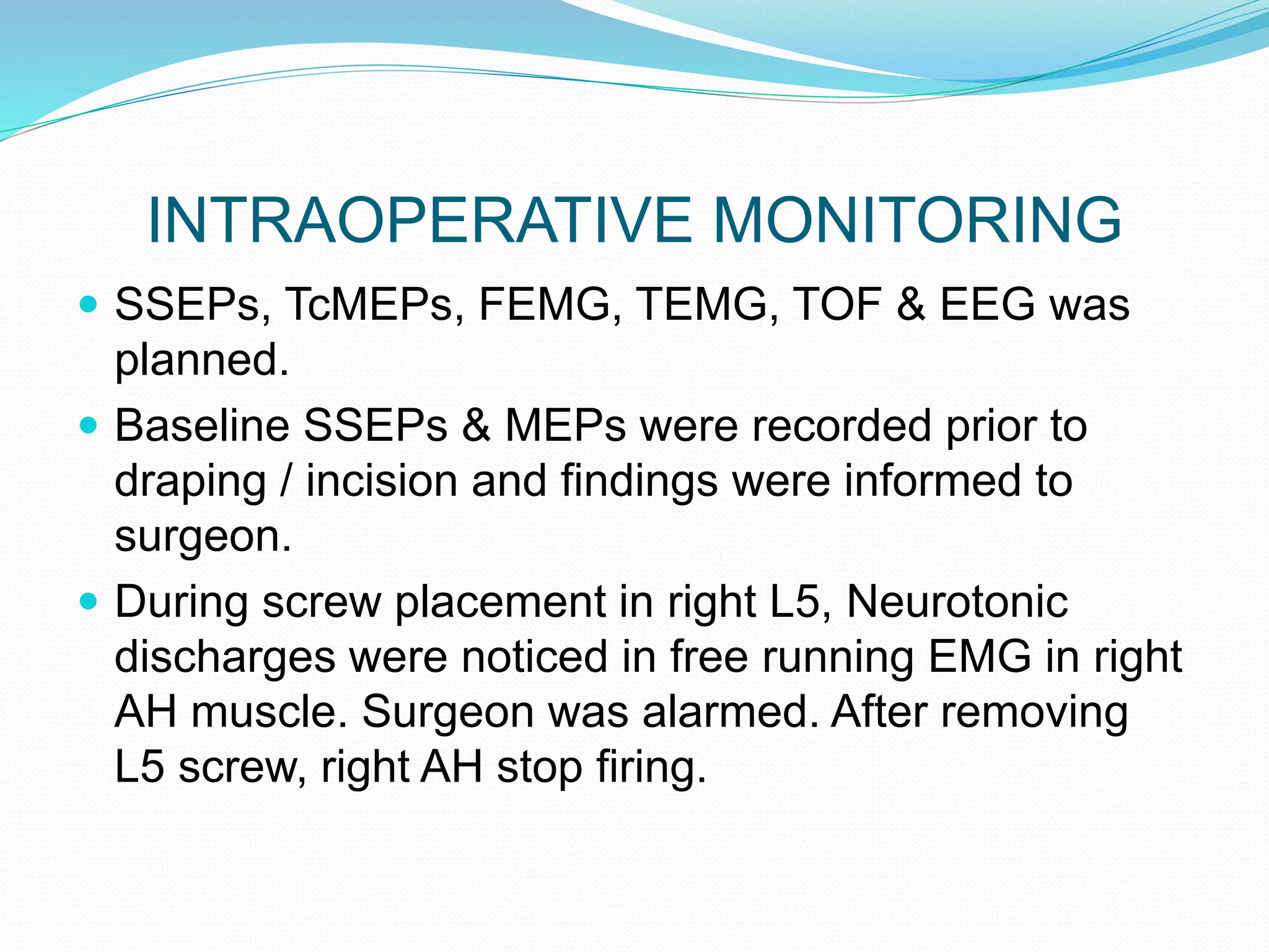
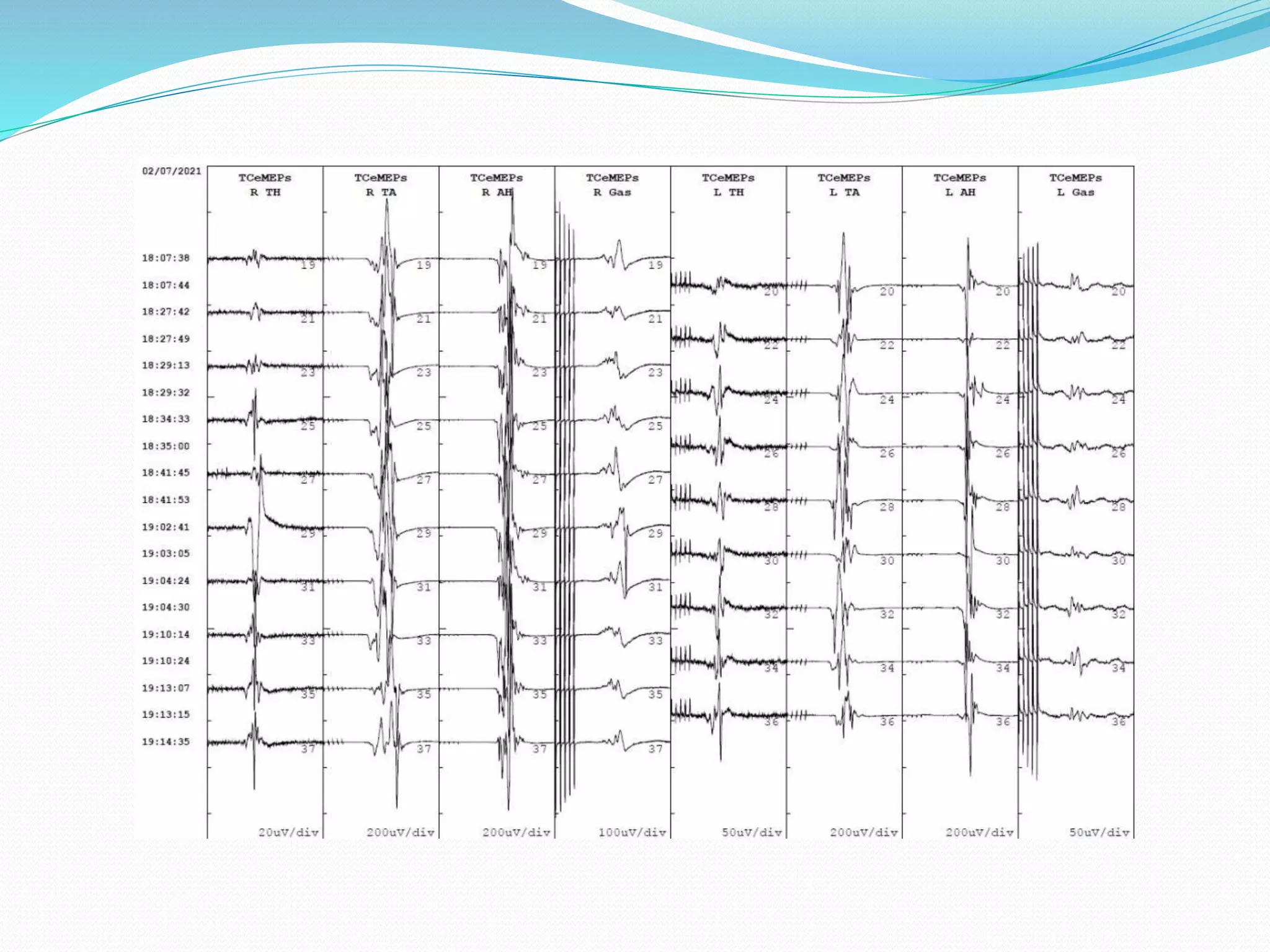
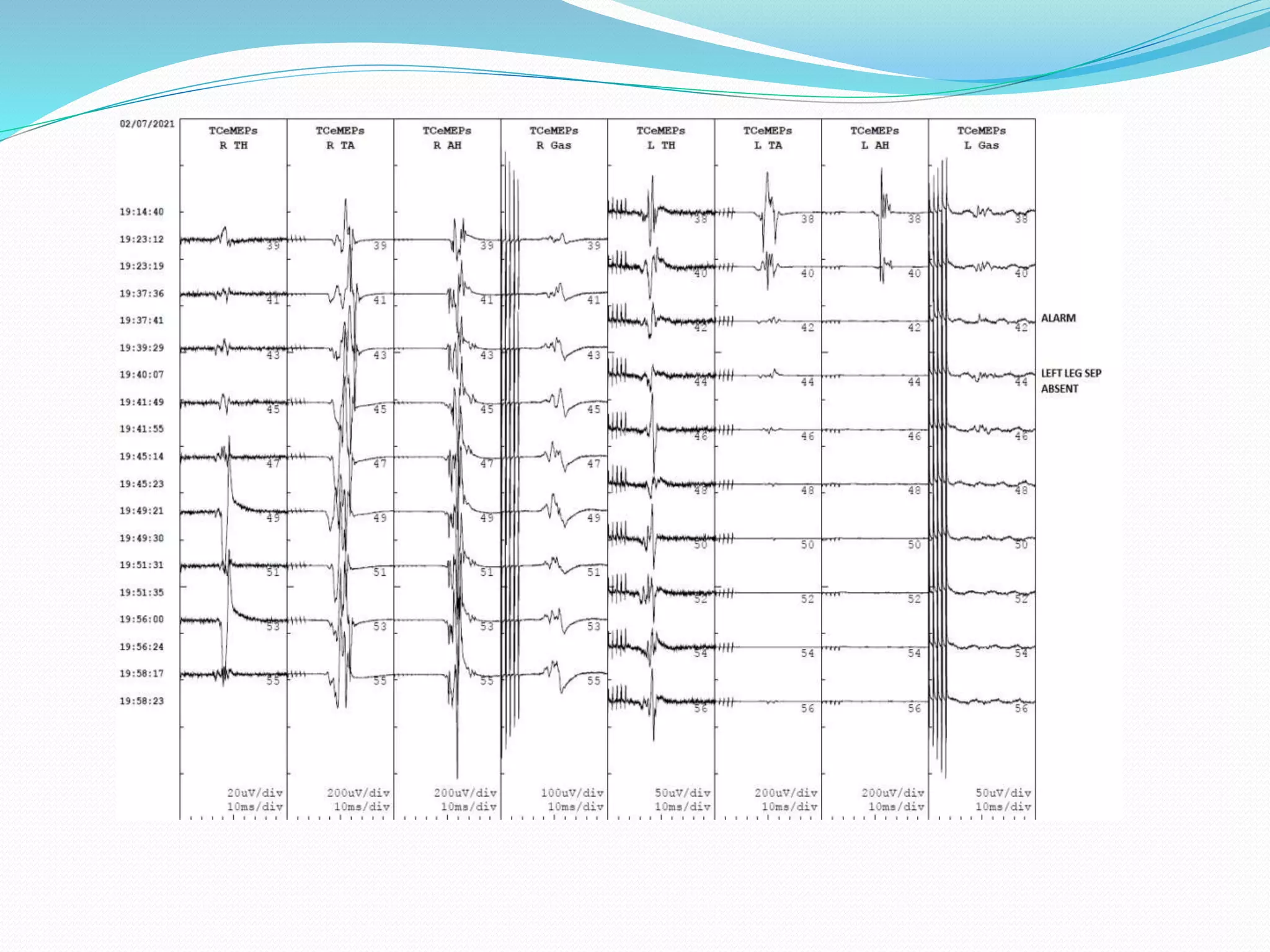
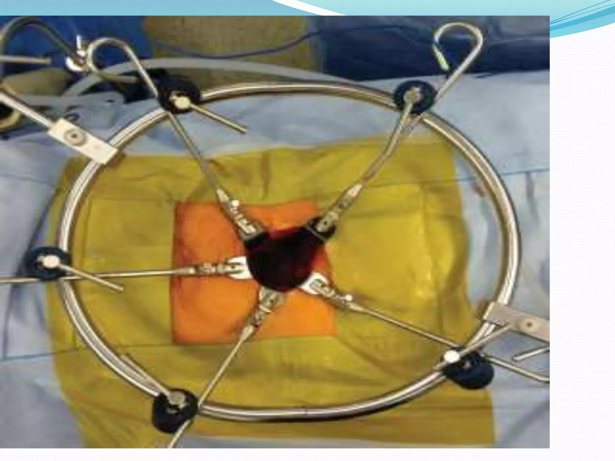
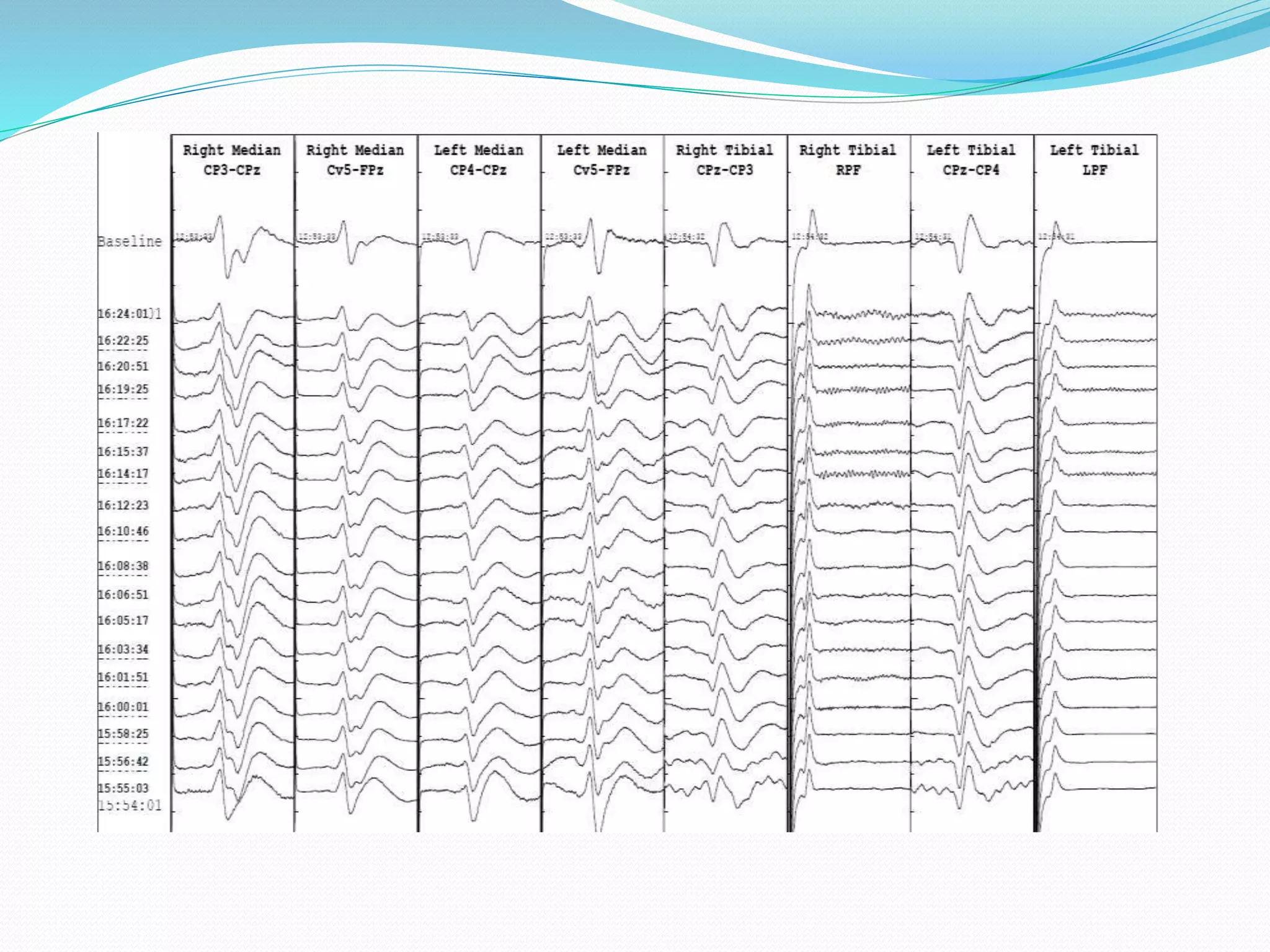
Syed Imran (CNIM, REEGT)
Senior Physiologist
Neurophysiology Department
The Wellington Hospital, London, UK
MULTIMODALITY??
 The combination of motor evoked potential (MEP),
somatosensory evoked potential (SSEP), free-run
and trigger electromyography (EMG).
THINK AHEAD
 What the case is?
 What neurological structures are at risk?
 What monitoring modalities are indicated or
planned?
 Does the patient have any neurological deficit?
ANAESTHSIA CONSIDERATION
 Total intravenous anaesthesia technique was used
 Induction was done with Propofol TCi and bolus of
Rocuronium (0.5 mg/kg) to facilitate intubation.
 Anaesthesia was maintained with infusion of
Propofol and remifentanil during the monitoring.
 Following the last IOM check and during wound
closure, Propofol was discontinued and sevoflurane
was commenced to achieve rapid patient wake up at
the conclusion of surgery.
IONM TECHNIQUE
 IONM was performed using a 16 channel Nihon
Kohden Neuromaster MEE-1000 with constant
current stimulator.
 All patients underwent multimodal monitoring,
recording of cortical SSEPs, cervical SSEPs, and
MEPs.
IONM TECHNIQUE
Somatosensory Evoked Potentials
 Sensory evoked potentials were recorded from the brain
(cortical SSEP) and spinal cord (cervical SSEP).
 SSEPs recorded from bilateral median nerve as a control
monitoring and bilateral Tibial nerve for surgical real time
monitoring. Peripheral recording was obtained from
popliteal fossa & cubital fossa bilaterally to stimulate the
nerve supramaximal or to rule out limb ischemia due to
position.
 Recorded from CP4/CP3-CPz derivations for Median
sseps & CPz-CP4/CP3 for Tibial sseps.
 2 channel EEG was also recorded to monitor depth of
anaesthesia.
IONM TECHNIQUE
Motor Evoked Potentials
 Transcranial Motor Evoked Potentials(TcMEPs) were
obtained from M1/M2 & M3/M4 interhemispheric
derivations which is 1cm above somatosensory strip.
 Muscles used in these cases were according to the surgical
levels but mainly TA & AH.
 Bilateral thenar muscle was used as control monitoring.
 FEMG was also recorded from same muscle group.
 TOF was recorded from thenar muscle to evaluate
neuromuscular block ratio.
ALERT CRITERIA
 SSEPs: 50% amplitude from baseline or 10%
increase in latency.
 MEPs: 70-80% drop in amplitude OR absent
response.
 FEMG: Neurotonic discharges or irritation from
muscles
CASE REPORT
1ST CASE;REPORT
 67 years old female with complain of back pain
radiating to both legs. She was Unable to sit due to
back pain. Had previous spine fusion L1-L4 few
years ago which did not improve her symptoms.
 Redo surgery for Transforaminal Lumbar Interbody
Fusion
(L4-S1)
INTRAOPERATIVE MONITORING
 SSEPs, TcMEPs, FEMG, TEMG, TOF & EEG was
planned.
 Baseline SSEPs & MEPs were recorded prior to
draping / incision and findings were informed to
surgeon.
 During screw placement in right L5, Neurotonic
discharges were noticed in free running EMG in right
AH muscle. Surgeon was alarmed. After removing
L5 screw, right AH stop firing.
Alarm Surgeon
Remove L5
Screw
Firing Stop
INTRAOPERATIVE MONITORING
 After few minutes, sudden 80% amplitude drop in
right AH MEP response was noticed and alarmed.
Screw was replaced carefully but right foot MEP
remain low until the end of procedure.
 MAP was raised to 80mmHg
 No change in SSEPs were noticed throughout.
POST OPERATIVE ASSESSMENT
 Motor assessment was done by the surgeon and
noticed mild weakness in right toe flexion and
abduction which was predicted by Neurophysiology
staff intraoperatively.
 This indicate importance of multi modality
neurophysiology monitoring during spine cases.
However, these deficit takes few weeks or months to
recover.
2nd CASE REPORT
 35 years old male with lower back pain; diagnosed
with spondylolisthesis. Surgery was planned for L4-
S1 anterior lumbar interbody fusion.
 During monitoring, a drop in left tibial SSEP & left leg
MEP was alarmed, soon after placement of frame
retractor. Signals were recovered post reposition of
retractor.
STEPS TAKEN
 Troubleshoot for any technical error
 Alarm Surgeon
 Ask anaesthetist to raise MAP >80-90mm Hg
 Check SpO2 saturation in the lost limb if it’s ischemic
changes.
 Ask surgeon to release retractor or reposition.
3RD CASE; REPORT
 History: Pain in lower back, walking difficulty,
spondylolisthesis.
 Surgical Plan: ALIF L5-S1
 Monitoring Plan: SSEPs, TcMEPs, FEMG, TOF, EEG
INTRAOPERATIVE MONITORING
 Baseline SEPs & MEPs were taken and informed to
surgeon prior opening.
 During cage placement changes in MEPs were
alarmed and no change in SSEPs were noticed.
 BP was raises; Steroid was given; Cage was
removed.
 Surgery aborted
POST OPERATIVE
ASSESSMENT
 Rt leg weakness
 Redo surgery after 48 hours
CONCLUSION
 Multimodality IONM is recommended in procedures
where there is a risk of neurological injury. The ability of
IONM to detect such complications has improved,
predominantly as multimodal monitoring techniques have
been introduced.
 SSEP record specifically the ascending dorsal tract of the
cord and provide no information on the integrity of the
descending anterior tracts (Motor pathway).
 However, Combined IONM techniques SSEPs, FEMG
and MEPs have good diagnostic characteristics to detect
neurological injury.
THANK YOU

Multimodality IONM in spine surgery

  • 1.
    Syed Imran (CNIM,REEGT) Senior Physiologist Neurophysiology Department The Wellington Hospital, London, UK
  • 2.
    MULTIMODALITY??  The combinationof motor evoked potential (MEP), somatosensory evoked potential (SSEP), free-run and trigger electromyography (EMG).
  • 3.
    THINK AHEAD  Whatthe case is?  What neurological structures are at risk?  What monitoring modalities are indicated or planned?  Does the patient have any neurological deficit?
  • 4.
    ANAESTHSIA CONSIDERATION  Totalintravenous anaesthesia technique was used  Induction was done with Propofol TCi and bolus of Rocuronium (0.5 mg/kg) to facilitate intubation.  Anaesthesia was maintained with infusion of Propofol and remifentanil during the monitoring.  Following the last IOM check and during wound closure, Propofol was discontinued and sevoflurane was commenced to achieve rapid patient wake up at the conclusion of surgery.
  • 5.
    IONM TECHNIQUE  IONMwas performed using a 16 channel Nihon Kohden Neuromaster MEE-1000 with constant current stimulator.  All patients underwent multimodal monitoring, recording of cortical SSEPs, cervical SSEPs, and MEPs.
  • 6.
    IONM TECHNIQUE Somatosensory EvokedPotentials  Sensory evoked potentials were recorded from the brain (cortical SSEP) and spinal cord (cervical SSEP).  SSEPs recorded from bilateral median nerve as a control monitoring and bilateral Tibial nerve for surgical real time monitoring. Peripheral recording was obtained from popliteal fossa & cubital fossa bilaterally to stimulate the nerve supramaximal or to rule out limb ischemia due to position.  Recorded from CP4/CP3-CPz derivations for Median sseps & CPz-CP4/CP3 for Tibial sseps.  2 channel EEG was also recorded to monitor depth of anaesthesia.
  • 7.
    IONM TECHNIQUE Motor EvokedPotentials  Transcranial Motor Evoked Potentials(TcMEPs) were obtained from M1/M2 & M3/M4 interhemispheric derivations which is 1cm above somatosensory strip.  Muscles used in these cases were according to the surgical levels but mainly TA & AH.  Bilateral thenar muscle was used as control monitoring.  FEMG was also recorded from same muscle group.  TOF was recorded from thenar muscle to evaluate neuromuscular block ratio.
  • 9.
    ALERT CRITERIA  SSEPs:50% amplitude from baseline or 10% increase in latency.  MEPs: 70-80% drop in amplitude OR absent response.  FEMG: Neurotonic discharges or irritation from muscles
  • 10.
  • 11.
    1ST CASE;REPORT  67years old female with complain of back pain radiating to both legs. She was Unable to sit due to back pain. Had previous spine fusion L1-L4 few years ago which did not improve her symptoms.  Redo surgery for Transforaminal Lumbar Interbody Fusion (L4-S1)
  • 12.
    INTRAOPERATIVE MONITORING  SSEPs,TcMEPs, FEMG, TEMG, TOF & EEG was planned.  Baseline SSEPs & MEPs were recorded prior to draping / incision and findings were informed to surgeon.  During screw placement in right L5, Neurotonic discharges were noticed in free running EMG in right AH muscle. Surgeon was alarmed. After removing L5 screw, right AH stop firing.
  • 13.
  • 14.
    INTRAOPERATIVE MONITORING  Afterfew minutes, sudden 80% amplitude drop in right AH MEP response was noticed and alarmed. Screw was replaced carefully but right foot MEP remain low until the end of procedure.  MAP was raised to 80mmHg  No change in SSEPs were noticed throughout.
  • 17.
    POST OPERATIVE ASSESSMENT Motor assessment was done by the surgeon and noticed mild weakness in right toe flexion and abduction which was predicted by Neurophysiology staff intraoperatively.  This indicate importance of multi modality neurophysiology monitoring during spine cases. However, these deficit takes few weeks or months to recover.
  • 18.
    2nd CASE REPORT 35 years old male with lower back pain; diagnosed with spondylolisthesis. Surgery was planned for L4- S1 anterior lumbar interbody fusion.  During monitoring, a drop in left tibial SSEP & left leg MEP was alarmed, soon after placement of frame retractor. Signals were recovered post reposition of retractor.
  • 26.
    STEPS TAKEN  Troubleshootfor any technical error  Alarm Surgeon  Ask anaesthetist to raise MAP >80-90mm Hg  Check SpO2 saturation in the lost limb if it’s ischemic changes.  Ask surgeon to release retractor or reposition.
  • 27.
    3RD CASE; REPORT History: Pain in lower back, walking difficulty, spondylolisthesis.  Surgical Plan: ALIF L5-S1  Monitoring Plan: SSEPs, TcMEPs, FEMG, TOF, EEG
  • 28.
    INTRAOPERATIVE MONITORING  BaselineSEPs & MEPs were taken and informed to surgeon prior opening.  During cage placement changes in MEPs were alarmed and no change in SSEPs were noticed.  BP was raises; Steroid was given; Cage was removed.  Surgery aborted
  • 33.
    POST OPERATIVE ASSESSMENT  Rtleg weakness  Redo surgery after 48 hours
  • 34.
    CONCLUSION  Multimodality IONMis recommended in procedures where there is a risk of neurological injury. The ability of IONM to detect such complications has improved, predominantly as multimodal monitoring techniques have been introduced.  SSEP record specifically the ascending dorsal tract of the cord and provide no information on the integrity of the descending anterior tracts (Motor pathway).  However, Combined IONM techniques SSEPs, FEMG and MEPs have good diagnostic characteristics to detect neurological injury.
  • 35.