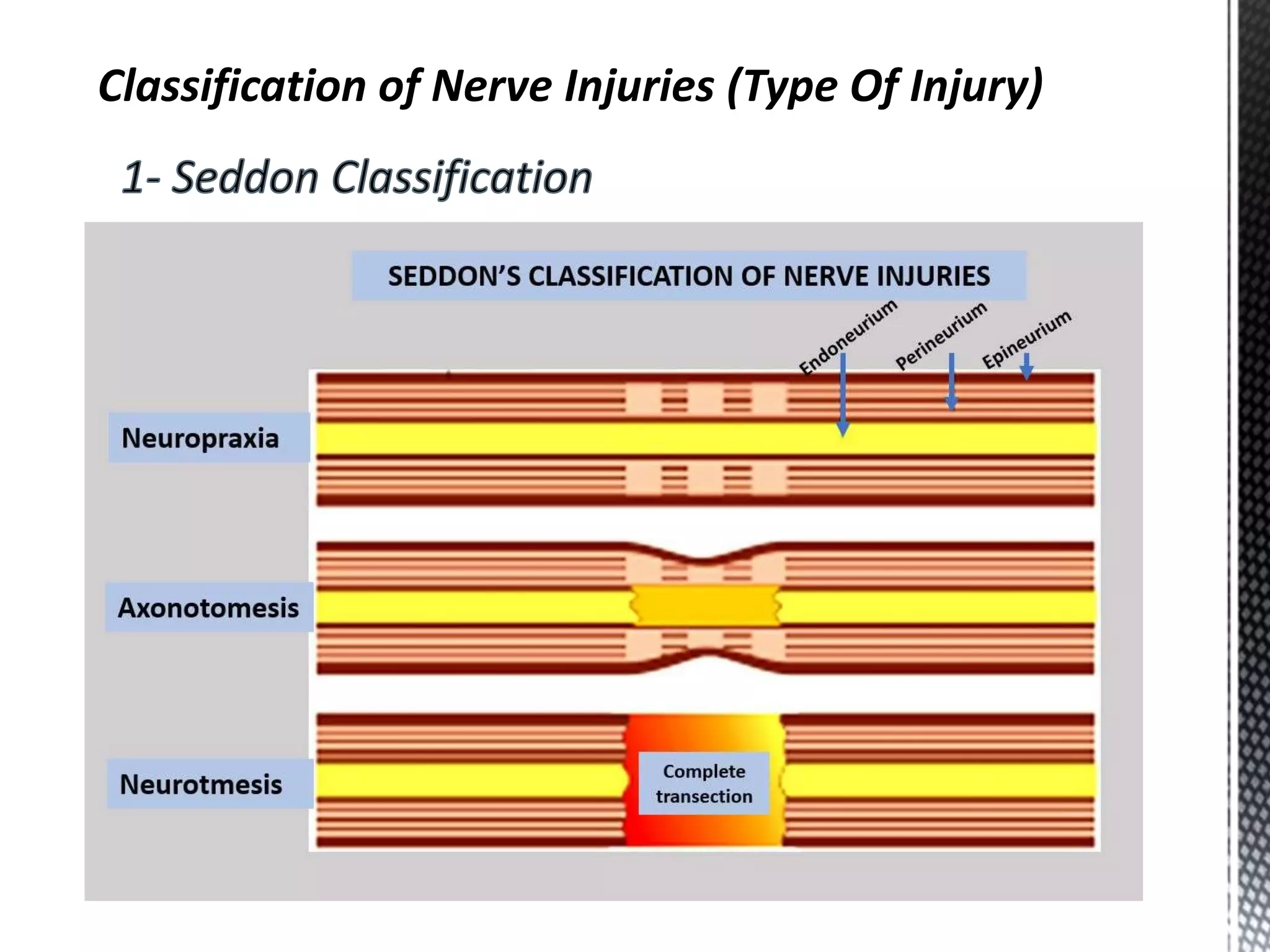
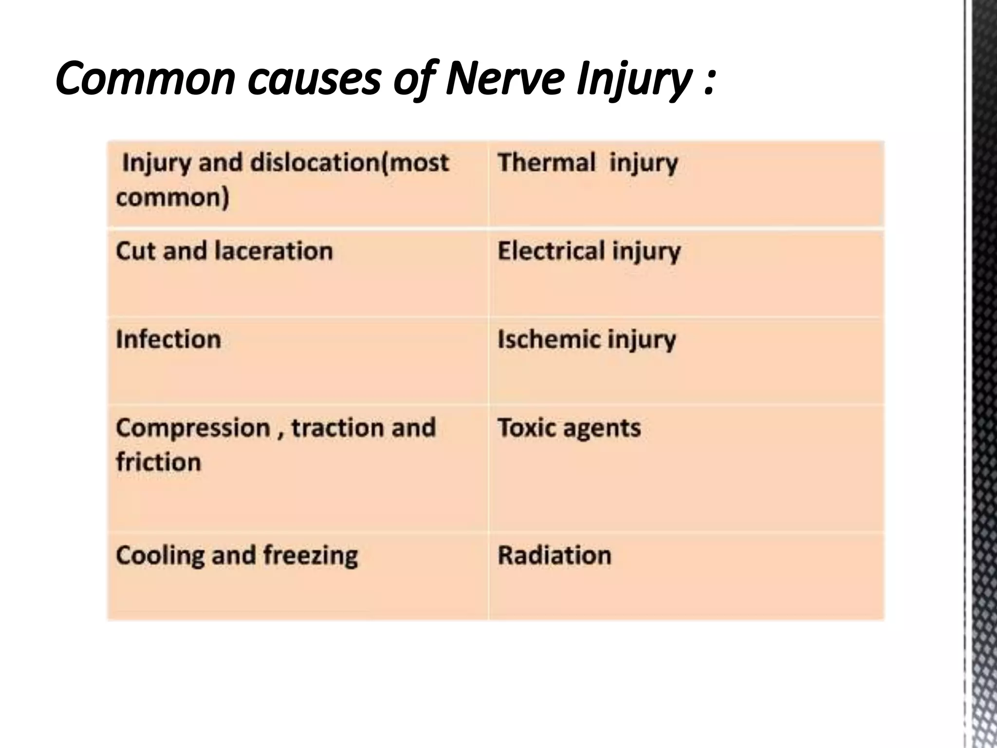
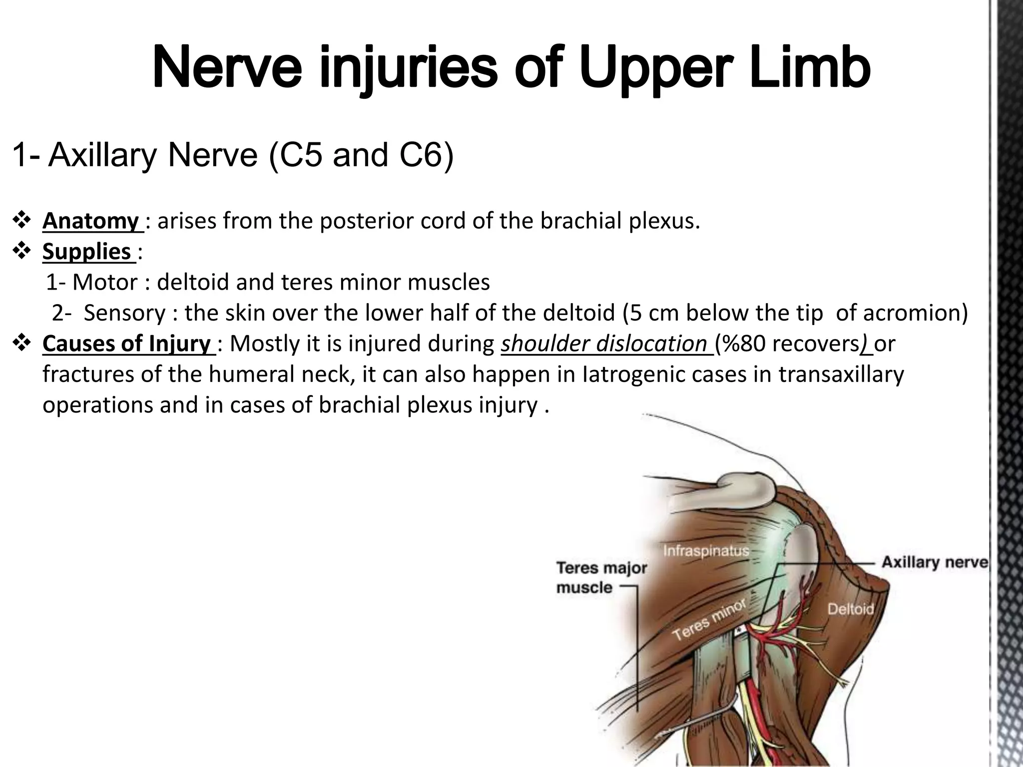
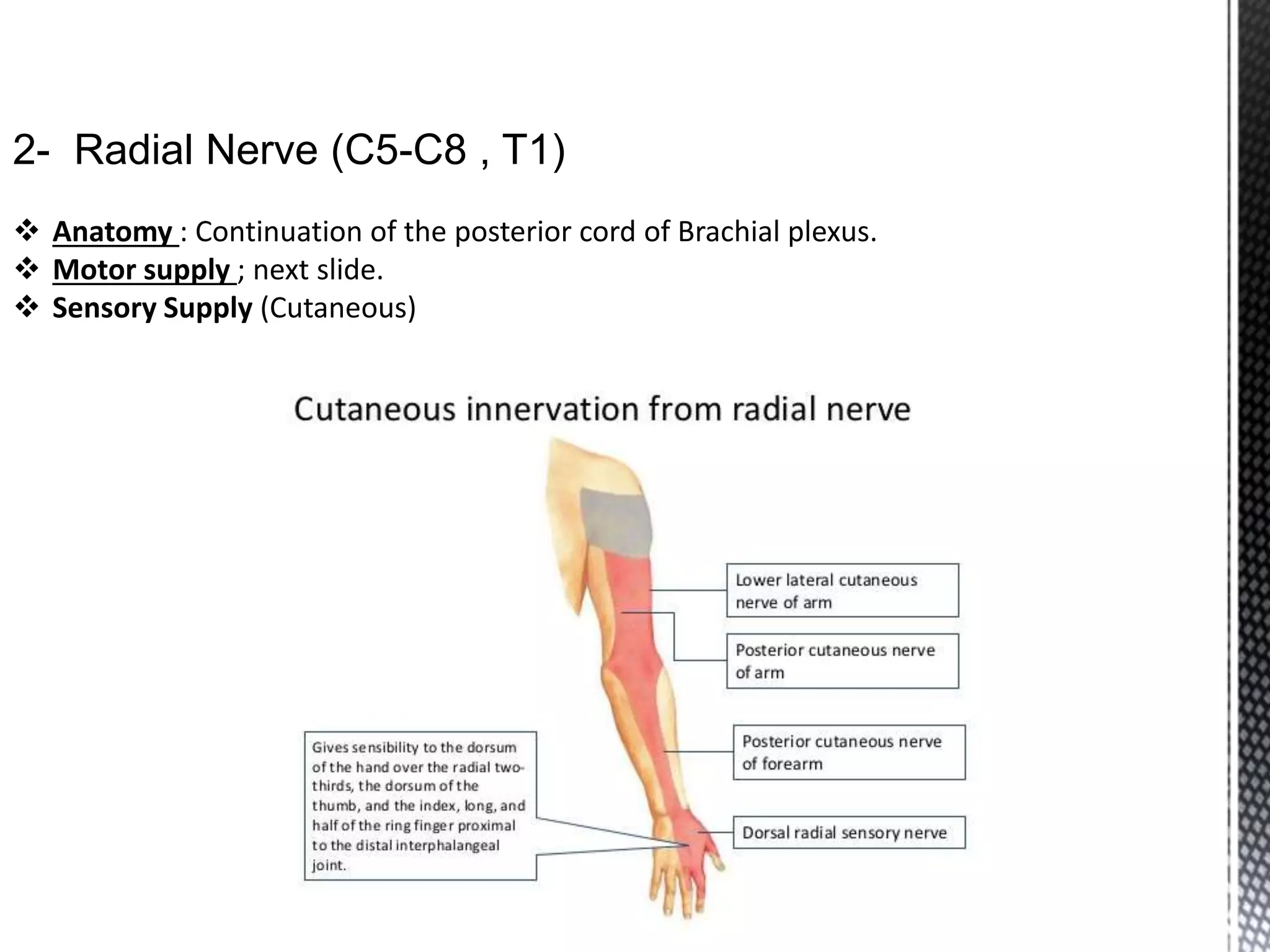
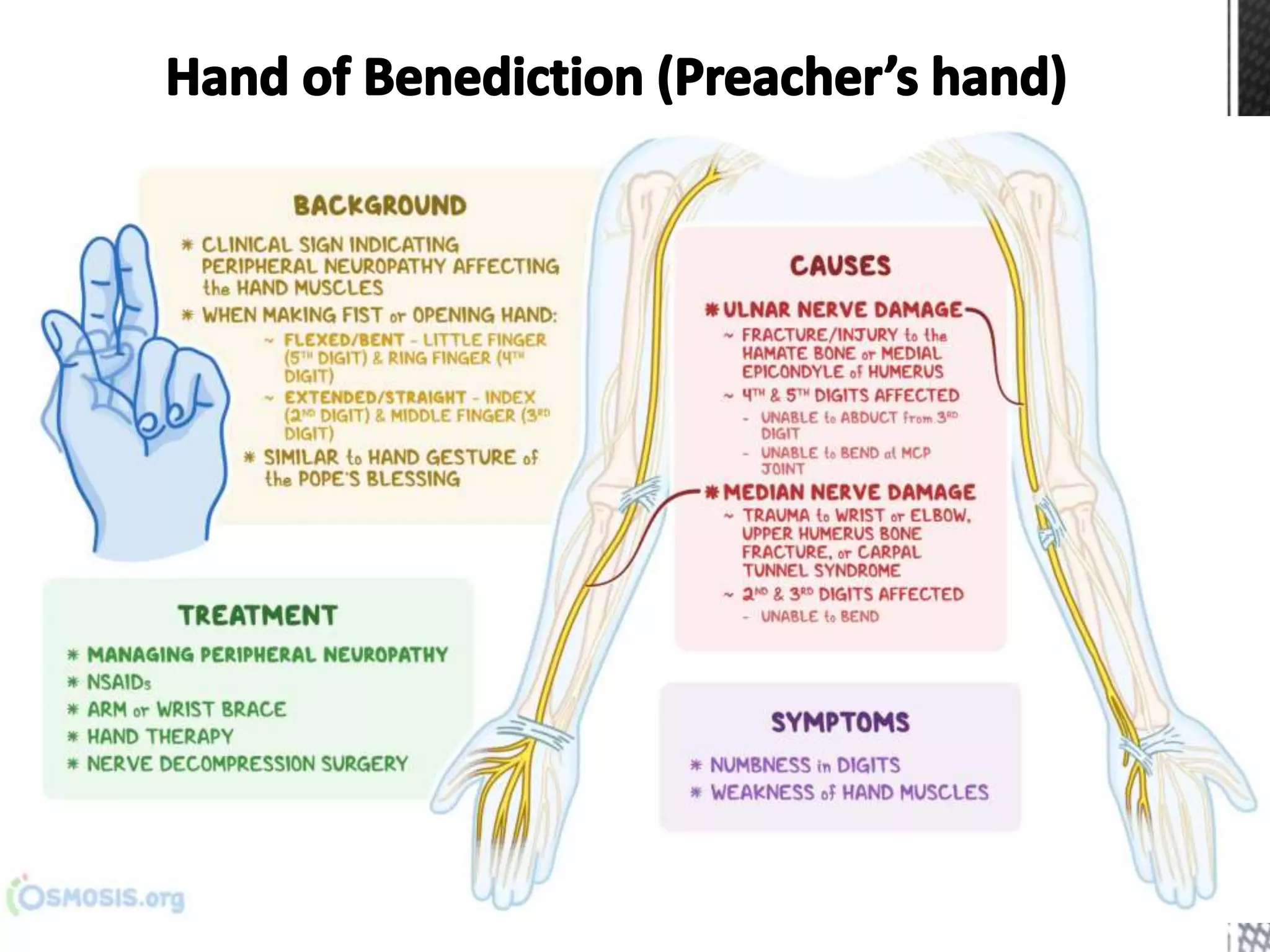
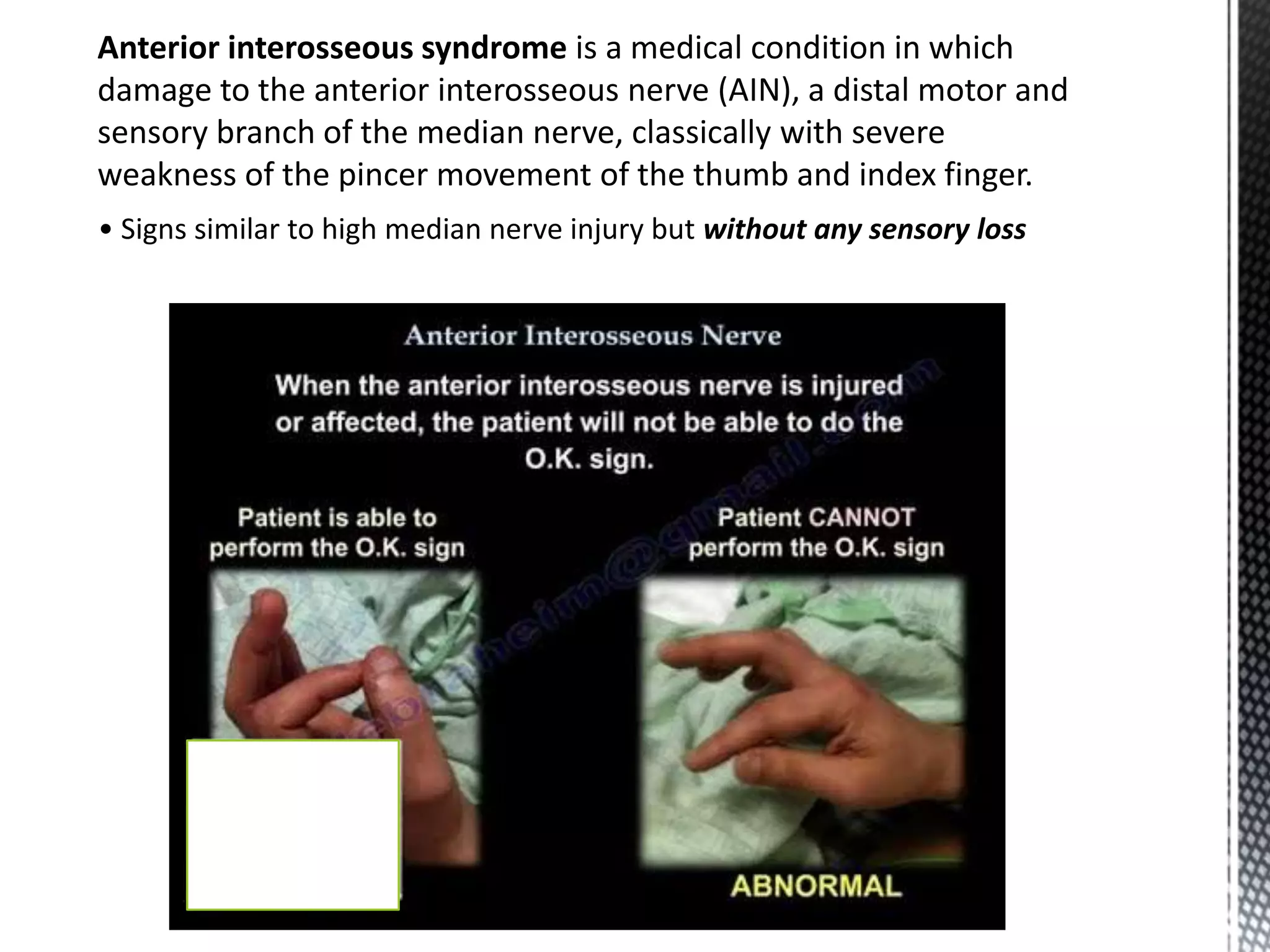
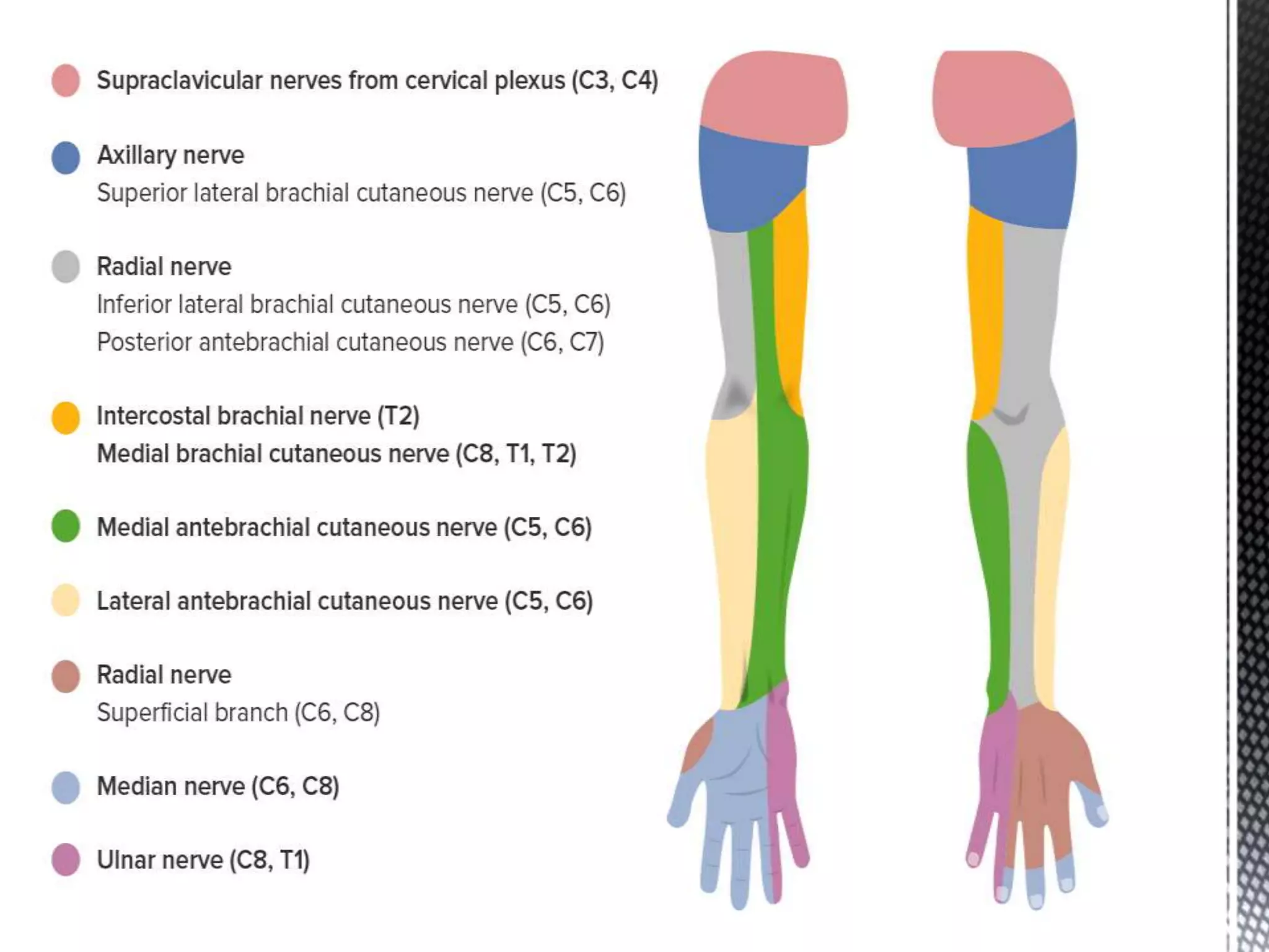
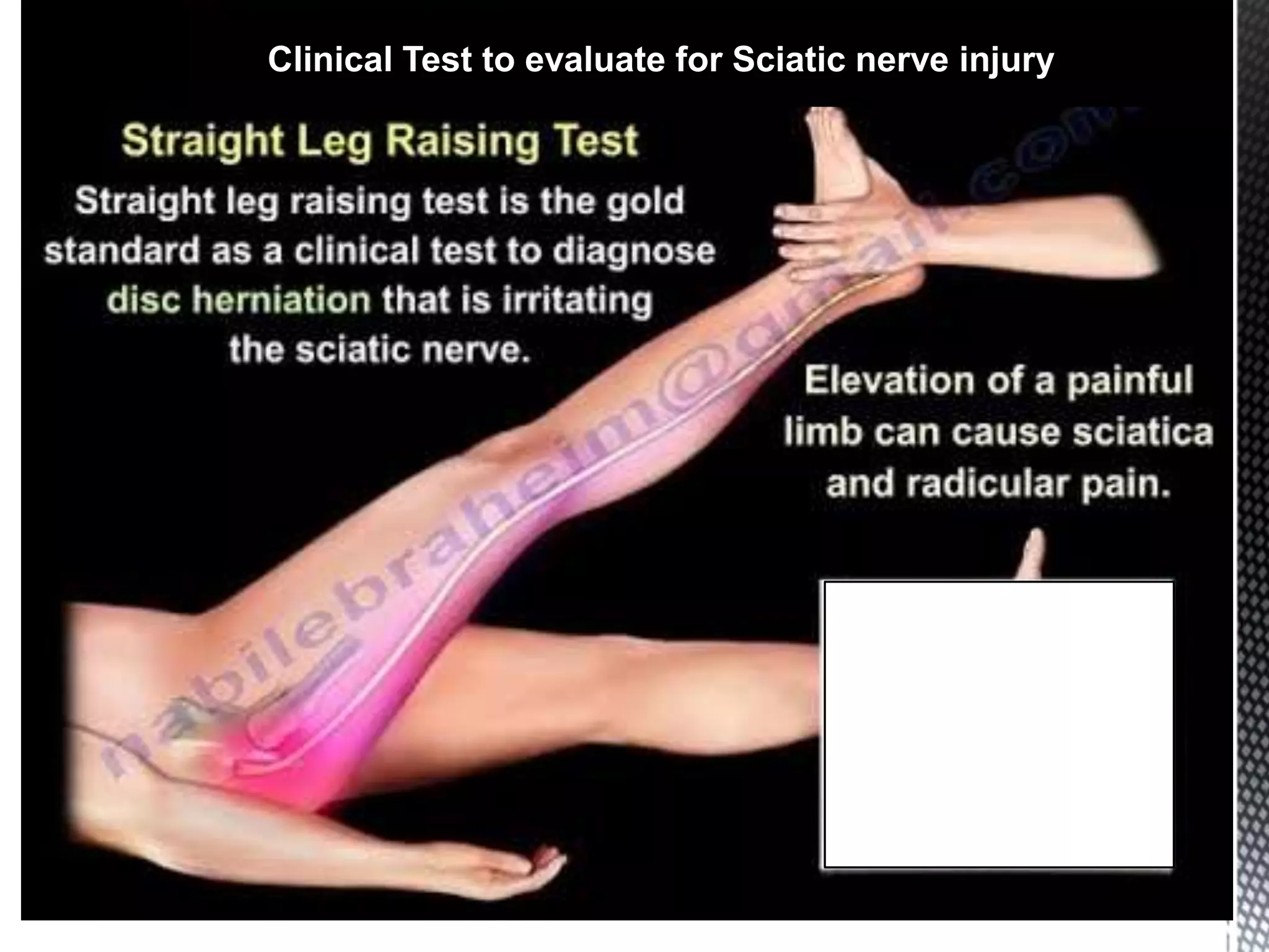
Prepared by Dr Ozhin Araz
Supervised by Dr Rawezh Majeed
Classification of Nerve Injuries (Type Of Injury)
Diagnosis of Nerve Injuries:
1- History
2- Examination
3- Investigations
 Blood tests: such as FBC, ESR, CRP, urea and electrolytes and liver
function tests.
 Nerve conduction
 Electromyography (EMG)
 Nerve biopsy.
 Cerebrospinal fluid (CSF) examination.
1- Axillary Nerve (C5 and C6)
 Anatomy : arises from the posterior cord of the brachial plexus.
 Supplies :
1- Motor : deltoid and teres minor muscles
2- Sensory : the skin over the lower half of the deltoid (5 cm below the tip of acromion)
 Causes of Injury : Mostly it is injured during shoulder dislocation (%80 recovers) or
fractures of the humeral neck, it can also happen in Iatrogenic cases in transaxillary
operations and in cases of brachial plexus injury .
• Careful testing will reveal a small area of numbness over the deltoid (the
‘sergeant’s patch’).
• Note : Although abduction can be mediated (by supraspinatus), it cannot be
maintained.
2- Radial Nerve (C5-C8 , T1)
 Anatomy : Continuation of the posterior cord of Brachial plexus.
 Motor supply ; next slide.
 Sensory Supply (Cutaneous)
Motor supply :
• Before the radial groove: long and medial heads of triceps
• After the radial groove: -
• - before crossing the elbow: lateral head of triceps, anconeous,
brachioradialis, extensor carpi radialis longus
• - After crossing the elbow: extensor carpi radialis brevis, the supinator
• After piercing the supinator: other extensor muscles of the forearm and
hand and the deep branch continues as Post. Interosseus nerve.
Low lesions (Below elbow) and Post. Interosseous Nerve)
• Due to fracture or dislocation at the elbow or to a local wound
• Complain of weakness , not being able to extend the MCP joints of the hand(Finger drop)
• In thumb, weakness of extension and retroposition
• Wrist extension is preserved and extends into radial deviation.
High lesions (Mid-Arm)
• Due to fracture of the humerus or after prolonged tourniquet pressure
• Wrist drop due to weakness of the radial extensors of the wrist
• Inability to extend MCP joints or elevate the thumb(Finger drop).
• Sensory loss to a small patch on the dorsum around the anatomical snuff box.
Very high lesions (Axilla or upper Arm)
• Due to trauma or operations around the shoulder
• Also common in Saturday night palsy or crutch palsy (Chronic compression of the nerve)
• In addition to high lesions, the triceps is paralysed and the triceps reflex is absent
 Anatomy : arises from the medial cord of Brachial plexus.
 Motor supply ; Sensory Supply (Cutaneous) :
As in cyclist
• Metacarpophalangeal joints of fourth and fifth finger are hyper extended
• Interphlangeal joint of fourth and fifth fingers are flexed
• Flattening of hypothenar eminence
• Hollowing between metacarpals on dorsum of hand due to paralysis of dorsal
interossei .
• More prominent if the injury at wrist level because the FDP is not affected.
Claw Hand (Ulnad Paradox)
'the closer to the Paw, the worse the Claw'
Froment’s Sign or Book test
• testing the action of Adductor Pollicis.
• In case of Paralysis , uses Flexor Pollicis Longus (Median N.)
4- Median Nerve
 Anatomy : formed by joining braches of medial and lateral cord of brachial plexus.
 Motor supply ; Sensory Supply (Cutaneous)
Causes :
1. Tenosynovitis of flexor tendons
2. Myxedema
3. Retention of fluid in pregnancy
4. Fracture dislocation of lunate
5. osteoarthritis
Features :
Sensory : feeling of burning pain in
lateral three and half digits especially at night
Motor
1. weakness of thenar muscles
2. ape thumb deformity
3. Positive Tinel’s sign and Phalen;s test
Anterior interosseous syndrome is a medical condition in which
damage to the anterior interosseous nerve (AIN), a distal motor and
sensory branch of the median nerve, classically with severe
weakness of the pincer movement of the thumb and index finger.
• Signs similar to high median nerve injury but without any sensory loss
• It is formed from the L4 to S3 segments of the sacral
plexus.
• It is the longest and thickest nerve in the body
• Pain caused by a compression or irritation of the sciatic
nerve by a problem in the lower back is called sciatica.
• High-stepping gait is characteristic.
• Commonest Cause of injury ;
1- Hip dislocation
2- Wrong placement of IM injection into the gluteal region.
3- Hip replacement surgeries.
Course & Distribution
It leaves the pelvis through greater sciatic foramen, below
the piriformis and passes in the gluteal region (between
ischial tuberosity & greater trochanter) then to posterior
compartment of thigh.
Termination: In the middle of the back of the thigh, It divides
into 2 branches :Tibial &Common Peroneal (Fibular).
Clinical Test to evaluate for Sciatic nerve injury
Foot drop is characterized by inability or impaired ability to raise the toes or
raise the foot from the ankle (dorsiflexion) it is mainly due to weakness,
irritation or damage to the deep fibular nerve (deep peroneal), including
the sciatic nerve, or paralysis of the muscles in the anterior portion of the lower
leg. (Tibialis Anterior)
Test : Ask the patient to dorsiflex the foot against resistance.
Conservative :
1- Splintage by Ankle foot orthosis
2- Physiotherapy
3- Electical Functional Stimulations.
Surgery : done if conservative management fails
• Repairs or decompresses a damaged nerve that fuses the foot
and ankle joint or transfers tendons from stronger leg muscles
 Formed by: Ventral rami of posterior division of
L2-4
 Root Value: L2-4
 Motor Supply: Hip Flexor (iliacus, Sartorius,
Pectineus), Knee Extensor(Quadriceps)
 Sensory Supply: Antero-medial thigh and antero-
medial leg and foot (Saphenous Nerve)
 Course:
• The nerve descends in the abdomen from
Lumbar Plexus through Psoas Major muscle.
• The nerve further travels downs into the thigh
behind the mid-inguinal point.
• It divides into anterior and posterior branches
which supply hip flexor and knee extensor
respectively.
SENSORY EFFECT: loss of sensation over areas supplied (antero-medial)
aspect of thigh & medial side of knee, leg & foot loss of sensation over areas
supplied (antero-medial) aspect of thigh & medial side of knee, leg & foot
Conservative management
1. Splintage of the paralysed limb
Preserve mobility of the joint
1. Physiotherapy
Operative management
1.Neurolysis
2.Nerve repair
3.Nerve grafting
4.Nerve Conduit Repair
 Neurolysis
• Application of physical or chemical agents to a nerve in order to cause a
temporary or sometimes perminant degeneration of targeted nerve fibres .
 Nerve repair
• Types:
• Primary repair: Indicated in clean sharp nerve injuries;
- done in the first 6 to 8 hours of injury
• Delayed primary repair:
- Done in the first 7 to 18 days of injury when
the wound is clean and there are no other
major complicating injuries
• Secondary repair: Done in crushed, avulsed injuries;
- done at a delay of 3-6 weeks
Techniques of nerve repair
1. Nerve suture : Indicated when the nerve ends
can be brought close to each other
• Techniques:
a. Epineural suture (Best technique)
b. Perineural suture (Trauma to nerve is a setback)
c. Group fascicular repair
1. Adequate exposure
2. Proper anesthesia
3. The nerve ends are then sharply transected perpendicular to the long axis.
4. Minimum of two epineural sutures with 8-0/ 9-0 nylon 180° to each other.
5. Careful alignment is the critical factor in this first step
2. Nerve grafting
• Indicated when the gap is more than 10 cm or end to end suture is likely to result
in tension at the suture line.
• Most common nerve used is sural nerve
• Nerve autografts are the gold standard of repair
 Nerve Conduit Repair
• Their ease of application and lack of donor site morbidity make them an attractive
option for nerve repair in many situations.
• Their use is currently limited to small-diameter peripheral nerves with small defects.
Peripheral Nerve Injury

Peripheral Nerve Injury

  • 1.
    Prepared by DrOzhin Araz Supervised by Dr Rawezh Majeed
  • 3.
    Classification of NerveInjuries (Type Of Injury)
  • 6.
    Diagnosis of NerveInjuries: 1- History 2- Examination 3- Investigations  Blood tests: such as FBC, ESR, CRP, urea and electrolytes and liver function tests.  Nerve conduction  Electromyography (EMG)  Nerve biopsy.  Cerebrospinal fluid (CSF) examination.
  • 7.
    1- Axillary Nerve(C5 and C6)  Anatomy : arises from the posterior cord of the brachial plexus.  Supplies : 1- Motor : deltoid and teres minor muscles 2- Sensory : the skin over the lower half of the deltoid (5 cm below the tip of acromion)  Causes of Injury : Mostly it is injured during shoulder dislocation (%80 recovers) or fractures of the humeral neck, it can also happen in Iatrogenic cases in transaxillary operations and in cases of brachial plexus injury .
  • 9.
    • Careful testingwill reveal a small area of numbness over the deltoid (the ‘sergeant’s patch’). • Note : Although abduction can be mediated (by supraspinatus), it cannot be maintained.
  • 10.
    2- Radial Nerve(C5-C8 , T1)  Anatomy : Continuation of the posterior cord of Brachial plexus.  Motor supply ; next slide.  Sensory Supply (Cutaneous)
  • 11.
    Motor supply : •Before the radial groove: long and medial heads of triceps • After the radial groove: - • - before crossing the elbow: lateral head of triceps, anconeous, brachioradialis, extensor carpi radialis longus • - After crossing the elbow: extensor carpi radialis brevis, the supinator • After piercing the supinator: other extensor muscles of the forearm and hand and the deep branch continues as Post. Interosseus nerve.
  • 12.
    Low lesions (Belowelbow) and Post. Interosseous Nerve) • Due to fracture or dislocation at the elbow or to a local wound • Complain of weakness , not being able to extend the MCP joints of the hand(Finger drop) • In thumb, weakness of extension and retroposition • Wrist extension is preserved and extends into radial deviation. High lesions (Mid-Arm) • Due to fracture of the humerus or after prolonged tourniquet pressure • Wrist drop due to weakness of the radial extensors of the wrist • Inability to extend MCP joints or elevate the thumb(Finger drop). • Sensory loss to a small patch on the dorsum around the anatomical snuff box. Very high lesions (Axilla or upper Arm) • Due to trauma or operations around the shoulder • Also common in Saturday night palsy or crutch palsy (Chronic compression of the nerve) • In addition to high lesions, the triceps is paralysed and the triceps reflex is absent
  • 15.
     Anatomy :arises from the medial cord of Brachial plexus.  Motor supply ; Sensory Supply (Cutaneous) :
  • 16.
  • 17.
    • Metacarpophalangeal jointsof fourth and fifth finger are hyper extended • Interphlangeal joint of fourth and fifth fingers are flexed • Flattening of hypothenar eminence • Hollowing between metacarpals on dorsum of hand due to paralysis of dorsal interossei . • More prominent if the injury at wrist level because the FDP is not affected.
  • 18.
    Claw Hand (UlnadParadox) 'the closer to the Paw, the worse the Claw'
  • 19.
    Froment’s Sign orBook test • testing the action of Adductor Pollicis. • In case of Paralysis , uses Flexor Pollicis Longus (Median N.)
  • 21.
    4- Median Nerve Anatomy : formed by joining braches of medial and lateral cord of brachial plexus.  Motor supply ; Sensory Supply (Cutaneous)
  • 25.
    Causes : 1. Tenosynovitisof flexor tendons 2. Myxedema 3. Retention of fluid in pregnancy 4. Fracture dislocation of lunate 5. osteoarthritis Features : Sensory : feeling of burning pain in lateral three and half digits especially at night Motor 1. weakness of thenar muscles 2. ape thumb deformity 3. Positive Tinel’s sign and Phalen;s test
  • 27.
    Anterior interosseous syndromeis a medical condition in which damage to the anterior interosseous nerve (AIN), a distal motor and sensory branch of the median nerve, classically with severe weakness of the pincer movement of the thumb and index finger. • Signs similar to high median nerve injury but without any sensory loss
  • 30.
    • It isformed from the L4 to S3 segments of the sacral plexus. • It is the longest and thickest nerve in the body • Pain caused by a compression or irritation of the sciatic nerve by a problem in the lower back is called sciatica. • High-stepping gait is characteristic. • Commonest Cause of injury ; 1- Hip dislocation 2- Wrong placement of IM injection into the gluteal region. 3- Hip replacement surgeries. Course & Distribution It leaves the pelvis through greater sciatic foramen, below the piriformis and passes in the gluteal region (between ischial tuberosity & greater trochanter) then to posterior compartment of thigh. Termination: In the middle of the back of the thigh, It divides into 2 branches :Tibial &Common Peroneal (Fibular).
  • 32.
    Clinical Test toevaluate for Sciatic nerve injury
  • 33.
    Foot drop ischaracterized by inability or impaired ability to raise the toes or raise the foot from the ankle (dorsiflexion) it is mainly due to weakness, irritation or damage to the deep fibular nerve (deep peroneal), including the sciatic nerve, or paralysis of the muscles in the anterior portion of the lower leg. (Tibialis Anterior) Test : Ask the patient to dorsiflex the foot against resistance.
  • 34.
    Conservative : 1- Splintageby Ankle foot orthosis 2- Physiotherapy 3- Electical Functional Stimulations. Surgery : done if conservative management fails • Repairs or decompresses a damaged nerve that fuses the foot and ankle joint or transfers tendons from stronger leg muscles
  • 35.
     Formed by:Ventral rami of posterior division of L2-4  Root Value: L2-4  Motor Supply: Hip Flexor (iliacus, Sartorius, Pectineus), Knee Extensor(Quadriceps)  Sensory Supply: Antero-medial thigh and antero- medial leg and foot (Saphenous Nerve)  Course: • The nerve descends in the abdomen from Lumbar Plexus through Psoas Major muscle. • The nerve further travels downs into the thigh behind the mid-inguinal point. • It divides into anterior and posterior branches which supply hip flexor and knee extensor respectively.
  • 36.
    SENSORY EFFECT: lossof sensation over areas supplied (antero-medial) aspect of thigh & medial side of knee, leg & foot loss of sensation over areas supplied (antero-medial) aspect of thigh & medial side of knee, leg & foot
  • 38.
    Conservative management 1. Splintageof the paralysed limb Preserve mobility of the joint 1. Physiotherapy Operative management 1.Neurolysis 2.Nerve repair 3.Nerve grafting 4.Nerve Conduit Repair
  • 39.
     Neurolysis • Applicationof physical or chemical agents to a nerve in order to cause a temporary or sometimes perminant degeneration of targeted nerve fibres .
  • 40.
     Nerve repair •Types: • Primary repair: Indicated in clean sharp nerve injuries; - done in the first 6 to 8 hours of injury • Delayed primary repair: - Done in the first 7 to 18 days of injury when the wound is clean and there are no other major complicating injuries • Secondary repair: Done in crushed, avulsed injuries; - done at a delay of 3-6 weeks Techniques of nerve repair 1. Nerve suture : Indicated when the nerve ends can be brought close to each other • Techniques: a. Epineural suture (Best technique) b. Perineural suture (Trauma to nerve is a setback) c. Group fascicular repair
  • 41.
    1. Adequate exposure 2.Proper anesthesia 3. The nerve ends are then sharply transected perpendicular to the long axis. 4. Minimum of two epineural sutures with 8-0/ 9-0 nylon 180° to each other. 5. Careful alignment is the critical factor in this first step
  • 42.
    2. Nerve grafting •Indicated when the gap is more than 10 cm or end to end suture is likely to result in tension at the suture line. • Most common nerve used is sural nerve • Nerve autografts are the gold standard of repair
  • 43.
     Nerve ConduitRepair • Their ease of application and lack of donor site morbidity make them an attractive option for nerve repair in many situations. • Their use is currently limited to small-diameter peripheral nerves with small defects.