
Recommended
More Related Content
What's hot
What's hot (20)
Similar to 1.1 oxygen therapy
Similar to 1.1 oxygen therapy (20)
Recently uploaded
Recently uploaded (20)
1.1 oxygen therapy
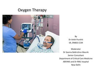
- 1. Oxygen Therapy By Dr Ankit Purohit SR, DNBSS CCM Moderator Dr Seema Balkrishna Wasnik Senior Consultant Department of Critical Care Medicine ABVIMS and Dr RML hospital New Delhi
- 2. Oxygen • Eighth element on the periodic table. • At ambient temperature and pressure (ATP), colourless, odourless, transparent and tasteless gas with the chemical symbol O2. • Derived from Greek Word “Oxys” , meaning sharp. “Oxygen sustains life and supports combustion. While there are many benefits to oxygen by inhalation, it is not without hazards and toxic effects. It is therefore important for persons who are responsible for oxygen administration to be familiar with its indications for use, potential hazards and equipment” (Kacmarek, Stoller & Heuer, 2013).
- 3. Fast Facts about O2 • Makes up 20.9% of air by volume and 23% air by weight. • except inert elements can oxidize all other elements. • Is a non-flammable gas. • Accelerates combustion. • At -182.9 C (-300◦ F) oxygen is a pale blue liquid. • Its critical temperature is -118.4 C (above the critical temperature oxygen exist as a gas regardless of the pressure).
- 4. Timeline • 1771 and 1772- the Swedish pharmacist Karl Scheele in experiment discovered gas augmenting combustion. 1777 findings published. • Joseph Priestly performed same experiment in 1774, published in 1775. • Priestly gave PHLOGISTON THEORY- fire released combustible material in air. The dephlogisticated air was oxygen. • 1778 Antoine Lavoisier named the gas oxygene, meaning acid former,. • 1798 -Thomas Beddoes (father of respiratory medicine) and James Watt opened PNEUMATIC institute and treated COPD Asthma. • 1885 – George Holtzapple, treated pneumonia using oxygen. • 1907 – Arbuthnot Lane devised red rubber nasal tubing, Haldane Developed oxygen mask. • Paul Bert and Adolph Fick Advanced Understanding of oxygen physiology, during first world war. • 1936 – Alvan Barach led to foundation of long term oxygen therapy. • 1950- Coats Pierce Gilson used oxygen cylinders to deliver oxygen therapy.
- 5. Overview of topic 1) Oxygen Transport 2) Indications for Oxygen Therapy , Special considerations. 3) Assessment of oxygen therapy. 4) Oxygen Toxicity. 5) Oxygen delivery at patient interface, Oxygen delivery devices 6) Types of Oxygen Delivery & storage Systems
- 6. Oxygen Disassociation curve • Most of oxygen is carried as reversible bind to hemoglobin. Remaining in dissolved form in plasma which reflects as PaO2. • Some of the factors affecting the loading and unloading of oxygen are: • Blood pH (Bohr effect) • Body temperature • Erythrocyte concentration of certain organic phosphates (e.g., 2,3diphosphoglycerate) • Rise in tempertaure,2,3 -DPG, Decrease in pH, shifts curve to right • Variation to the structure of the hemoglobin (Hb) molecules (e.g., sickle cells, methemoglobin (metHb) and fetal hemoglobin (HbF)) • Chemical combinations of Hb with other substances (e.g., carbon monoxide) • 50% SpO2- 26.5 mmHg of PaO2.
- 7. Madan et al; Correlation between the levels of SpO2 and PaO2, Lung India. 2017, May.
- 8. Gas exchange and Oxygen cascade • At capillary level exchange takes place by diffusion only. • Difference between atmospheric oxygen pressure and arterial pressure acts as driving force. • PAo2 – 100 mmHg, PVo2 – 40mmHg, • Usually there is small difference of 5-10 mmHg between PaO2 and PaO2 • 1) right to left shunt between pulmonary and cardiac circulation • 2) regional differences between pulmonary ventilation and blood flow. • Neonates have lower PaO2 50-80 mmHg due to more anatomical and physiological shunts. Alveolar Air Equation PAO2 = [(PB-PH2O) * FiO2] - PaCO2 /RQ
- 9. Ref -Doctor’s Podcast, FRCA
- 10. Fick’s law of diffusion • The movement of gas across the alveolar- capillary membrane is best described by Fick’s law of diffusion. • Directly proportional to surface area and inversely to thickness • At the tissue level, oxygen diffuses from the blood (Pcapillaries O2 = 40 mmHg ) across the microvasculature and interstitial space into the cell (Pintracellular O2= 5mmHg)where cellular respiration take place. Fick’s Law of Diffusion V = A x D (P1 - P2)/T Where the factors affecting gas exchange are: V = flow of gas (oxygen) A = cross sectional area available for diffusion D = diffusion coefficient P1 - P2 = the partial pressure gradient P1 = partial pressure of oxygen in the alveolus (PAO2) P2 = partial pressure of oxygen in the blood (PaO2) T = thickness of the membrane (alveolar-capillary membrane
- 11. Ref -Doctor’s Podcast, FRCA
- 13. Ref -Doctor’s Podcast, FRCA
- 14. Dead Space Physiology • Dead Space constitute the region with ventilation but no perfusion. • It start from nose, pharynx, mouth, down till Terminal bronchioles. • It constitute about 150 ml. • In mechanical ventilated patients, circuits, catheter mount, masks, T-piece constitute dead space. • Their volume is measured by simply filling water in them and measuring its quantity. • Christian Bhor gave initial equation to calculate dead space. • Enghoff modified it latter, replacing alveolar CO2 with arterial CO2. • Exercise cause progressive decrease in dead space, due to progressive increase in TV .
- 15. Robertson et al; Dead Space, Physiology of wasted ventilation,2015, series Physiology in respiratory medicine.
- 16. Robertson et al; Dead Space, Physiology of wasted ventilation,2015, series Physiology in respiratory medicine
- 17. Physiological shunt • Occurs due to mixing of deoxygenated blood with oxygenated blood. • Perfusion of non or poorly ventilated area leads to deoxygenated blood to get mixed in cardiac output. • Physiological shunt - is caused by mixing of blood from Thebesian veins & bronchial veins in pulmonary veins. Constitute 2% of whole blood. • Pathologically – Cyanotic heart diseases, Eisenmenger syndrome, ARDS, Pulmonary Hypertension.
- 19. BMJ 1998
- 21. BMJ 1998
- 23. Highlights • Inappropriate dose & failure of monitoring has serious consequences. • Adequate ventilation, diffusion & circulation; failure of any of these leads to tissue hypoxia in 4 minutes. • Mechanism include 2 main types- arterial hypoxemia & O2 Hb transport system without arterial hypoxemia. • PaO2 of 40 mmHg – Hyperventilation due to carotid Chemoreceptors. • PaO2 of 30 mmHg – Systemic Hypotension and coma. • Situation of acute hypoxemia in patients with previous chronic hypoxemia,PaO2 & PvO2 are unreliable. Assessed in conjunction with clinical state and ABG.
- 24. Indications for Oxygen Therapy
- 26. Chart 1 - Oxygen prescription for acutely hypoxaemic patients in hospital. B R O'Driscoll et al. Thorax 2017;72:ii1-ii90 Copyright © BMJ Publishing Group Ltd & British Thoracic Society. All rights reserved.
- 30. Chart 2 - Flow chart for oxygen administration on general wards in hospitals. *For Venturi masks, the higher flow rate is required if the respiratory rate is >30. B R O'Driscoll et al. Thorax 2017;72:ii1-ii90 Copyright © BMJ Publishing Group Ltd & British Thoracic Society. All rights reserved.
- 31. Radiopedia.
- 32. HIGHLIGHTS • A1 – aim to achieve near normal oxygen saturation for all acutely ill patients, except risk of hypercapnic respiratory failure. • A2 - 94 -98 % target saturation in the patients. • A3 – Hypercapnic respiratory failure, 88 – 92% saturation target. • A5 – Hypoxemia assessment is done in upright position. • B2 – oxygen saturation is fifth vital sign. • B4 – quick and precise medical history, never discontinue oxygen therapy for room air assessment . Urgent clinical examination. Measure SpO2 and ABG assessment. Track and Trigger system. NEWS chart . • C1 – in shock patients, ABG should always be first sample for assessment. Earlobe blood gas is also acceptable, but less accurate.
- 33. Highlights • C3 - ABG to be done in – Spo2 < 94% , decrease in Spo2 by 3% or Breathlessness, drowsiness, risk of hypercapnic oxygen failure. Any illness requiring ABG. • D1- breathless patient with SpO2 <85%, start on NRBM @ 15 l/min. Target 94 -98% . Gradually move to simple Oxygen mask or nasal cannula once target reached. For hypercapnic risk group 88-92% saturation • E1 – CPR patients after ROSC, 94-98%, mechanical ventilation. • E2 – critical ill, trauma NRBM with reservoir @15l/min.94-98% • E3 – drowning ,94-98%. E4- CO poisoning, 100%
- 34. Highlights • F1- Asthma, F2- Pneumonia, F3- Lung cancer, F4 – ILD, F5 – Pneumothorax, F7 – Pleural effusion, F8 – PE, F9 - cardiogenic pulmonary edema, F11- Anemia, F12- sickle cell crisis, F13 – MI,F14 – stroke, F15,16 – poisoning, F17 – met and renal disorders; Target 94-98% • G1 – Hypercapnic respiratory failure – 88-92%. Long term smokers, old aged should be treated as COPD. SPIROMETRY. Venturi mask at 28%, 2- 6 l/min. ABG on arrival, traiged as very urgent. • Acute exacerbation hypercapnic failure due to more oxygen, oxygen is withdrawn gradually or patient land in hypoxemia. • G2- neurological failure, high risk of death. Mechanical ventilation. • G6 – NIV for chronic patients with pH <7.35.
- 35. Highlights • H- Pregnancy – major trauma, sepsis, acute hypoxemia(CVS) 94 - 98%. • H5- more then 20weeks pregnant, Left lateral tilt or Manual uterine displacement, to increase venous flow. • K- Palliative care- Restricted to patients with SpO2<90%.No role for monitoring. • L,M- Heliox and Entonox – upper airway obstruction. No good evidence for exacerbation of Asthma & COPD. Entonox avoided for analgesia in patients of Type 2 failure. • N- CPAP and humidified HFNC – sleep disorders patients planned for surgery. CPAP with saturation target 88-92%.HFNC as alternative to NRBM in Respiratory Distress.(COVID) • P – Tracheostomy, laryngectomy- t piece with high flow O2.
- 36. Highlights • Q – humidification – in Acute set ups,Not required for low flow device. Beneficial for viscous secretions. • R – Nebulisation –Asthma , done by O2 at Flow rate 6 l/min. Type 2 Respiratory failure, Done by air jet. If to be done by O2, Duration limited for to 6 minutes. • S – Prescription – prescribe the device , route and target range of saturation on clinical notes and drug chart. • T- Monitoring and Adjusting – Pulse oximeters, ABG at designated intervals. Regular charting.
- 37. LTOT – Long Term Oxygen Therapy • provision of oxygen therapy for continuous use at home for patients with chronic hypoxemia (PaO2 at or below 7.3 kPa, (55mHg). 1) oxygen flow rate must be sufficient to raise the waking oxygen tension above 8 kPa, (60 mmHg) 2) LTOT is likely to be life long 3) LTOT is usually given for at least 15 hours daily, to include night time, in view of the presence of worsening arterial hypoxemia during sleep Reference – BTS guidelines 2006.
- 38. Indications • chronic hypoxaemia-long term oxygen therapy is indicated for the following conditions with chronic hypoxaemia: 1) chronic obstructive pulmonary disease 2) severe chronic asthma 3) interstitial lung disease 4) cystic fibrosis 5) bronchiectasis 6) pulmonary vascular disease 7) primary pulmonary hypertension 8) pulmonary malignancy 9) chronic heart failure
- 39. Indications PaO2 is consistently at or below 7.3 kPa (55 mmHg), a period of clinical stability (defined as the absence of exacerbation of chronic lung disease for the previous five weeks), patients breathing on room air. clinically stable PaO2 is between 55 mmHg to 60 mmHg , together with the presence of one of the following: • secondary polycythaemia. • clinical and or echocardiographic evidence of pulmonary hypertension LTOT should not be prescribed in patients with chronic hypoxaemia patients with a PaO2 value above 60 mmHg.
- 40. Indications Nocturnal hypoventilation 1) obesity Neuromuscular/spinal/chest wall disease. 2) obstructive sleep apnoea (with CPAP therapy) Palliative Use palliation of dyspnoea in malignancy and other causes of disabling dyspnoea due to terminal disease.
- 41. Assessment of oxygen concentration: Oximetry • Oximetry is the measurement of blood hemoglobin (Hb) saturations using spectrophotometry. • Trace obtained is Plethysmograph • Hemoximetry (also called CO-oximetry) – performed in arterial blood gas analysis. • Pulse Oximetry - portable, non-invasive monitoring technique
- 42. Jubran et al; critcal care.2015. 19-272
- 44. Oxygen toxicity • Paul Bert who, in 1878, demonstrated convulsions in larks( bird) exposed to 15-20 ATA (atmosphere absolute) air. • In 1899, J Lorraine Smith, while trying to reproduce 'Bert effect', noticed fatal pneumonia in rats after 4 days of exposure to 73% oxygen at 1 ATA. • ACUTE AND CHRONIC OXYGEN TOXICITY – in acute short duration, high concentrations; • in chronic, long duration and low concentrations. • In normal humans the first signs of toxicity appear after about 10 hours of oxygen at 1 ATA. • Oxygen concentration of 100% for 24 to 48 hours can be tolerated. • Oxygen at 2 ATA produces characteristic pulmonary signs and symptoms • beginning with mild carinal irritation on deep inspiration 3-6 hours into the exposure, • intense carinal irritation an uncontrolled cough after about 10 hours • finally chest pain and dyspnoea. • Symptoms subside 4 hours after cessation of exposure Chawala et al, Oxygen toxicity, MJAFI 2001; 57 : 131-133.
- 45. Signs of toxicity • CNS TOXICITY (PAUL BERT EFFECT) • twitching of perioral and small muscles of the hand is a fairly constant feature. • Facial pallor and 'cogwheel' breathing result of intense peripheral vasoconstriction due to hyperoxia and diaphragmatic twitching respectively. • If exposure is continued, vertigo and nausea, followed by altered behaviour, clumsiness, and finally convulsions result. • PULMONARY TOXICITY(LORRAINE SMITH EFFECT) • Symptoms appear after a latent period whose duration decreases with increase in P02. • In normal humans the first signs of toxicity appear after about 10 hours of oxygen at 1 ATA. • Clinical features can be divided into three Phases • (a) Tracheobronchitis (b) ARDS (c) Pulmonary • interstitial fibrosis. • Absorption atelectasis due to washout of N2 can lead to collapse of parts of the lung in the event of air trapping.
- 46. Oxygen delivery at Patient Interface • Classification of devices – • 1) fixed vs variable performance. • 2) Patient dependent vs patient independent • 3) Low vs high gas flows. • Fixed performance Devices –They give fixed Fio2 despite varying patient parameters. • Variable performance Devices- Patient dependent, give variable Fio2 depending on patient parameters. • When gas flow exceeds patient peak INSPIRATORY flow, oxygen concentration received is patient independent. • Low flow devices with tight face or nose fitting mask, deliver Oxygen concentration patient independent. Ely J., Clapham M.; Delivering oxygen to patients, BJM nov. 2003
- 48. Low flow oxygen delivery devices • provide a variable FiO2 depending on the patient’s/client’s inspiratory demands. • As the inspiratory demands increase, ambient air is entrained and the FiO2 is maintained. • O2 delivered by device is diluted by inspired air • Nasal Cannula • Simple Mask • Partial Rebreather Mask • Non-Rebreather Mask • Transtracheal Catheter Could a Low Flow Oxygen delivery devices could still deliver a high FiO2? Theoretically, a reservoir mask set at 10 -15 L/min, could provide an FiO2 of 1.0 if it fit properly to a patient’s face and met the patient’s inspiratory flow demands on every breath.
- 49. Low flow oxygen device performances Richard D Branson and Jay A Johannigman Respiratory Care January 2013, 58 (1) 86-97; DOI: https://doi.org/10.4187/respcare.02251
- 50. Nasal Cannula • Disposable plastic device with protrusions to be fitted in nose. • Efficacy from 24% to 45% of oxygen concentration with flows upto 6 litres. Low flow 24-44 % 1 Lmin=24% 2 Lmin=28% 3 Lmin=32% 4 Lmin=36% 5 Lmin=40% 6 Lmin=44% ADVANTAGE – Patient can be mobilized. Low cost. DISADVANTAGE - variable FiO2, variable performance.
- 51. Facemask • Hudson Mask – fits over nose and mouth and contain hole or expiratory port. • Held in place by elastic strap and has metal piece over nose to fit. • FiO2 – 40 to 60 % at flow rates of 6 to 10 l/min. • ADVANTAGE – higher oxygen concentrations. • DISADVANTAGE – claustrophobia, feeding not possible, • Variable performance.
- 52. Non re-breather mask • Consist of mask with reservoir bag. • Mask has series of non rebreathing valves. Between mask and bag, and the covers of exhalation port. • Connected to oxygen pipe. • Exhaled air doesnt enter the bag, exhaled trhough vlaves in mask. • FiO2 – 60 – 80% @ O2 flow of 10 – 15 l/min. • DISADVANTAGE – risk of suffocation, if bag deflates with no air flow.
- 53. Partial Rebreather mask • No one way valves • There is mixing of exhaled and inhaled air. • FiO2 0f 80 -90 % @ O2 rate of 10 -15 l/min. • Suffocation chances are minimal.
- 55. Transtracheal catheter Its post cricothyrotomy. Rescue procedure to provide oxygen in patients with upper airway obstruction.
- 56. Oxygen Concentrator • employ selective removal of nitrogen from room air to increase the concentration of oxygen in the delivered gas product. • electrically powered • Battery-operated portable oxygen concentrators • function in continuous flow mode and/or pulse dose or demand mode. • to separate and concentrate oxygen from the air, molecular sieves or semi-permeable membranes are used. • Molecular sieves use sodium-aluminium silicate crystals and employ Pressure Swing Adsorption (PSA) or Vacuum Pressure Swing Adsorption (VPSA) technology. • Semi-permeable membranes are thin plastic membranes that are selectively permeable to O2 molecules and water vapor.
- 57. High Flow Oxygen Delivery Devices • High flow oxygen delivery devices will provide a fixed FiO2 (0.24 - 1.0) regardless of the patient’s inspiratory demands. • High flow devices are following. 1) Air Entrainment Mask (Venturi); 2) Nasal High Flow Oxygen Therapy; 3) Mechanical Ventilators (invasive ) 4) NIV, BiPAP, CPAP/APAP Machines (non invasive) 5) Manual Resuscitation Bags 6) Hyperbaric Oxygen Chambers.
- 58. Venturi mask • 40 – 60 % concentration is achieved. • Fixed delivery of fio2. • Delivers 24-60% oxygen • Different colours deliver different rates • Flow rate: Varies with colour. The correct flow rate to use with each colour it is shown on mask, along with the percentage of oxygen delivered. • Types: – BLUE = 2-4L/min = 24% O2 – WHITE = 4-6L/min = 28% O2 – YELLOW = 8-10L/min = 35% O2 – RED = 10-12L/min = 40% O2 – GREEN = 12-15L/min = 60% O2
- 59. Principle of venturi mask
- 61. Nasal high flow devices • alternative to standard high- flow face mask (HFFM) oxygen therapy. It provides delivery of up to 60-70 L/min of heated and humidified, blended air and oxygen via wide-bore nasal cannula. • It is indicated in patients experiencing respiratory distress, high breathing rates. • Due to high flows(upto 70 l/min), it overcomes high peak inspiratory rates. • It is indicated as replacement of NIV, in many patients and is recommeneded as bridge to weaning post extubation.
- 62. HFN Advantage • 1) HFNC in comparison to venturi show better results post extubation. • 2) improve patient discomfort and decrease respiratory rate. • 3) fewer episode of interface displacement. • 4) decreased need of NIV and re intubation or intubation than venturi. • 5) high gas flow generate a Positive airway pressure of 2 – 5 mbar, help to recruit ateletatic lung. • 6) NHF decrease dead space- a) increased TV. b) improved inspiratory aerodynamic.
- 63. Dr Chris Nickson, Life in the Fastlane.
- 64. NIV(non invasive ventilation) • CPAP (continuous positive airways pressure) – High pressure air/oxygen with a tight-fitting mask – Positive pressure all the time to help keep airways open (split them) – Used in acute pulmonary oedema and sleep apnoea • BiPAP (bilevel positive airways pressure) – High positive pressure on inspiration and lower positive pressure on expiration – Used in exacerbations of COPD and ARDS
- 65. Invasive Ventilation • Provided by securing airway via devices creating a closed breathing system. • Endotracheal tube, Supraglottic airway devices and Tracheostomy. • They allow controlled ventilation, Pulmonary toileting. • ETT, tracheostomy protect airway from aspiration. • Recruitment and administration of PEEP is possible via ETT. • Positive airway pressure ventilation is possible with complete rest to exhausted patient. • Advent of invasive ventilation changed the course of critically ill patients.
- 66. Humidification • Ideally inspired gas should be humidified to 37◦ C and 44 cmH2O/L (Wattier & Ward,2011.) • Dry medical gas inhalation can lead to crusting , dryness of secretions and damage to mucousa. • Clinical Signs and Symptoms of Inadequate Airway Humidification (Cairo & Pilbeam, 2004) • Atelectasis • Dry, non-productive cough • Increased airway resistance • Increased incidence of infection • Increased work of breathing • Substernal pain • Thick dehydrated secretions
- 67. Type of humidifiers • PASSIVE- Heat moist exchangers (HME filters). • ACTIVE • Low Flow Oxygen Humidifiers/ UNheated • Molecular Humidity - bubble type humidifiers, bubble-diffuser type • humidifiers used with nasal cannula. • Humidity is not indicated at flows less than 4 L/min (Cairo & Pilbeam, 2004). • The use of humidity is not recommended with reservoir type masks as condensates may affect the function of the mask (parts stick together) . • High Flow Oxygen Humidifiers/Heated. • Molecular Humidity- Passover- type (+/- wick, +/- heater) (e.g., used to humidify tracH mask systems, incubators). • Aerosol Humidity air entraining jet nebulizers (+/- baffles, +/- heaters)
- 68. Hyperbaric oxygen therapy • Undersea and Hyperbaric Medical Society (UHMS) is an international, nonprofit organization, it is primary source of information for diving and hyperbaric oxygen. • The increased pressure inside the chamber, combined with the delivery of 100% oxygen (FiO2 = 1.0), drives the diffusion of oxygen into the blood plasma at up to 10 times normal concentration.
- 69. Physiological effects of hyperbaric oxygen • Based on DALTON law, BOYLES Law, HENRY’S law • Normal dissolved oxygen in plasma is 3ml/L • At 3 atmospheric pressure it plasma dissolved oxygen increase to 60 ml/L; • At 100% arterial PaO2 reaches 2000 mmHg. • stimulates the growth of new blood vessels to locations with reduced circulation, improving blood flow to areas with arterial blockage; • causes a rebound arterial dilation after HBOT, resulting in an increased blood vessel diameter greater than when therapy began, improving blood flow to compromised organs; • stimulates an adaptive increase in superoxide dismutase (SOD), • Enhance white blood cell action and potentiating germ-killing antibiotics.
- 70. Leach et al,Hyperbaric Oxygen therapy; BMJ october1998
- 71. Indications 1. Air or Gas Embolism 2. Carbon Monoxide Poisoning 3. Cyanide Poisoning 4. Clostridial Myositis and Myonecrosis (Gas Gangrene) 5. Crush Injury, Compartment Syndrome and Other Acute Traumatic 6. Ischemias 7. Decompression Sickness 8. Arterial Insufficiencies: 9. Central Retinal Artery Occlusion 10. Enhancement of Healing In Selected Problem Wounds 11. Severe Anemia 12. Intracranial Abscess 13. Necrotizing Soft Tissue Infections 14. Osteomyelitis (Refractory) 15. Delayed Radiation Injury (Soft Tissue and Bony Necrosis) 16. Compromised Grafts and Flaps 17. Acute Thermal Burn Injury 18. Idiopathic Sudden Sensorneural Hearing Loss
- 72. Complications Barotrauma: • Ear or sinus trauma • Tympanic membrane rupture • Alveolar over distension and pneumothorax • Gas embolism • Oxygen Toxicity: Central nervous system (CNS) toxic reaction (Early signs twitching, sweating, pallor and restlessness, followed by seizures or convulsions.) •Pulmonary toxic reaction Other: • Fire • Sudden decompression • Reversible visual changes • Claustrophobia • Decreased Cardiac Output (Cairo & Pilbeam,2004)
- 73. Oxygen delivery system There are three main types of oxygen delivery systems: • Compressed gas cylinders; • Liquid oxygen in cryogenic containers; • Oxygen concentrators for medical use. Systems are chosen based on size, weight , storage capacity and cost to company.
- 74. Compressed gas cylinders • BODY – Steel carbon fiber cylinder. Marked 3AA MRI – Aluminium alloy cylinders are used. Marked 3AL or 3ALM. Have flat concave base and tapered neck with tapered screw for valves. Color coded – White shoulder, black body. • VALVE – made of Bronze or brass. Components – port , stem , seat or diaphragm, nut. Type – packed and diaphragmatic • PACKED VALVE –TEFLON covers stem as resilient seal to prevent leak. Direct acting. Withstand high pressures • DIAPHRAGMATIC VALVE – metal disk which cylinder and act as diaphragm. can be opened fully at one half to three turns. • HANDLE WHEEL - used to open close cylinder.
- 75. Cylinder sizes
- 76. Pressure units(Dorsch and Dorsch)
- 79. Valve outlet connection(Dorsch and Dorsch)
- 80. Cylinder weight pressure(Dorsch and Dorsch)
- 81. Calculating flow rate of cylinder • the following formula can be used: • Duration of Flow in minutes =(gauge pressure psi - safe residual pressure psi) x cylinder factor. • Flow rate would be in Litres/minute. • Some examples of cylinder factors for different sized cylinders are: D cylinder 0.16 E cylinder 0.28 M cylinder 1.56
- 83. Pin Index system (Dorsch and Dorsch)
- 84. Safety measures (Dorsch and Dorsch)
- 85. Guidelines for safe use
- 86. Cryogenic containers • They contain compressed liquid oxygen. • manufactured by fractional distillation of air at an oxygen manufacturing plant. • oxygen is stored on site in large cryogenic vessels known as dewars. • Portable liquid oxygen units offer continuous flow or intermittent flow of oxygen to the patient/client. Oxygen Dewar
- 87. Manifold System
- 88. Shut off valves in pipelines(Dorsch and Dorsch)
- 91. Pressure in pipelines(Dorsch AND Dorsch)
- 92. THANK YOU