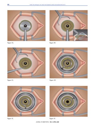
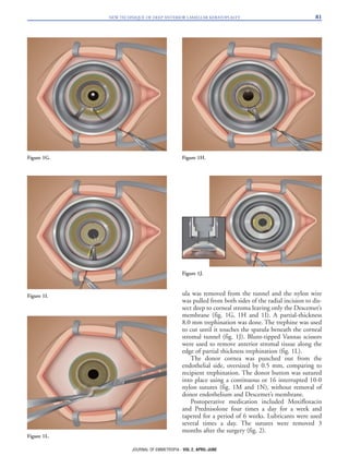
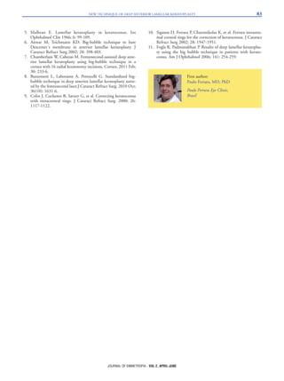
SlideShare a Scribd company logo
INTRODUCTION
Deep anterior lamellar keratoplasty (DALK) is a
surgical technique for cornea replacement, which the
healthy host corneal endothelium is preserved.
Keratoconus patients are those who benefit most from
a successful DALK procedure, once the endothelium of
these patients is usually normal and should therefore be
maintained.
Several studies comparing visual outcomes between
penetrating keratoplasty (PK) and DALK have shown
similar results1-4. Initially, DALK was performed using
manual corneal dissection5. The greatest limitation of
early DALK techniques was that they left variable
amounts of residual stroma. This provided a scaffold
for vascular ingrowth or led to variable amounts of
postoperative scarring in the interface due to the
remaining vessels. Also, the uneven or irregular dissec-
tion plane could impair visual acuity. The main disad-
vantage of DALK is the significant learning curve to
master the technique. Moreover, it is time-consuming
procedure, especially when the stroma has to be manu-
ally separated from the Descemet´s membrane.
Anwar and Teichmann’s6 big-bubble technique of
injecting air into the corneal stroma to isolate Descemet’s
membrane markedly improved DALK outcomes.
Removing the overlying stroma completely created a clear
graft interface without irregularities. Visual outcomes are
excellent, and postoperative interface problems from vas-
cularization of the recipient corneal bed are minimal.
A variant of the big-bubble technique in deep ante-
rior lamellar keratoplasty (DALK) assisted by the fem-
tosecond laser has been described, with good and
reproducible results7,8.
The present technique allows a deep and smooth
separation of corneal stroma from Descemet´s mem-
brane, by an interlamela cleavage plane created by a
nylon wire.
SURGICAL TECHNIQUE
The surgical procedure is done under peribulbar
anesthesia. The surgery shares some steps of intrastro-
mal corneal ring segments implantation. Initially, using
a diamond knife, set at 90% of corneal thickness (90
degrees meridian), at 8 mm optic zone (fig. 1A), a 0.9
mm radial incision was formed and corneal pockets
were created using the Ferrara spreader (fig. 1B and
1C). A 6.0 nylon preloaded spatula was then inserted
into the corneal pocket and in a counterclockwise
direction rotated in 3600 to create a deep stromal tun-
nel, in a similar manner as in the manual technique for
intrastromal corneal ring segments implantation9,10
(fig. 1D, 1E and 1F). After the tunnelization the spat-
79
TECHNICAL REPORT
New technique of Deep Anterior Lamellar Keratoplasty
Paulo Ferrara, MD, PhD1; Leonardo Torquetti, MD, PhD2; Leandro Cunha, MD2
We present a simple, effective and reproducible technique of deep anterior lamellar kerato-
plasty. Using a diamond knife, set at 90% of corneal thickness at (90 degrees meridian), at
8 mm optic zone, a radial incision was done and corneal pockets were created. A 6.0 nylon
preloaded spatula was then inserted into the corneal pocket and in a counterclockwise
direction rotated in 3600 to create a deep stromal tunnel. A partial-thickness 8 mm trephi-
nation was done. The spatula was removed from the tunnel and the nylon wire was pulled
from both sides of the radial incision to dissect deep to the corneal stroma leaving little
residual stroma over the Descemet’s membrane. The residual anterior stroma is removed
with a Vannas scissors. The donor button is sutured. The main advantages of this technique
are the very low learning curve and the regular stromal dissection with almost no residual
tissue over the Descemet’s membrane.
KEYWORDS: Deep anterior lamellar keratoplasty, keratoconus.
J Emmetropia 2011; 2: 79-83
Submitted: 2/13/2011
Revised: 4/6/2011
Accepted: 4/25/2011
1 Paulo Ferrara Eye Clinic. Director.
2 Paulo Ferrara Eye Clinic. Assistant.
No financial support was provided for this study.
Correspondence to: Paulo Ferrara, MD, PhD. Paulo Ferrara Eye
Clinic, Contorno Ave 4747, Suite 615, Lifecenter – Funcionários –
Belo Horizonte – MG - 30110-031 – Brasil – pferrara@ferrarar-
ing.com.br
© 2010 SECOIR
Sociedad Española de Cirugía Ocular Implanto-Refractiva
ISSN: 2171-4703
NEW TECHNIQUE OF DEEP ANTERIOR LAMELLAR KERATOPLASTY80
JOURNAL OF EMMETROPIA - VOL 2, APRIL-JUNE
Figure 1A.
Figure 1D.
Figure 1E. Figure 1F.
Figure 1B.
Figure 1C.
ula was removed from the tunnel and the nylon wire
was pulled from both sides of the radial incision to dis-
sect deep to corneal stroma leaving only the Descemet’s
membrane (fig. 1G, 1H and 1I). A partial-thickness
8.0 mm trephination was done. The trephine was used
to cut until it touches the spatula beneath the corneal
stromal tunnel (fig. 1J). Blunt-tipped Vannas scissors
were used to remove anterior stromal tissue along the
edge of partial thickness trephination (fig. 1L).
The donor cornea was punched out from the
endothelial side, oversized by 0.5 mm, comparing to
recipient trephination. The donor button was sutured
into place using a continuous or 16 interrupted 10-0
nylon sutures (fig. 1M and 1N), without removal of
donor endothelium and Descemet’s membrane.
Postoperative medication included Moxifloxacin
and Prednisolone four times a day for a week and
tapered for a period of 6 weeks. Lubricants were used
several times a day. The sutures were removed 3
months after the surgery (fig. 2).
NEW TECHNIQUE OF DEEP ANTERIOR LAMELLAR KERATOPLASTY 81
JOURNAL OF EMMETROPIA - VOL 2, APRIL-JUNE
Figure 1G. Figure 1H.
Figure 1L.
Figure 1I.
Figure 1J.
DISCUSSION
Several techniques of lamellar keratoplasty have
been described for keratoconus treatment. Anwar and
Teichmann described the big-bubble technique to
achieve separation of the Descemet´s membrane from
stroma after intrastromal air injection6. This technique
has been widely used as a technique which provides
rapid and satisfactory outcome that is comparable to
PK2. The technique allows a safe and direct access to
Descemet’s plane, with the advantages of shortening
the surgical time, reducing the risk for perforation, and
exposing a smooth, even surface of excellent optical
quality. However, a big bubble is not formed in all
cases6,11 and sometimes more than one injection of air
into the deep stroma is required before cleavage
between Descemet’s membrane and the stroma is
achieved. In these cases, repeated injections of air infil-
trating the corneal stroma may cause complete whiten-
ing of the central cornea within the area of trephina-
tion, making it difficult to recognize the line of separa-
tion between Descemet’s membrane and the stroma.
The increased safety of DALK and the potential for
better visual outcomes in expertly performed proce-
dures justifies the effort required for corneal surgeons
to master the procedure and the additional operating
room time. Another potential advantage is the utility of
using tissue that is less optimal for PKP with lower
endothelial cell counts or longer duration of death to
preservation and time in preservation to expand the
donor pool for optical keratoplasty.
The presented new technique of DALK eliminates
any potential difficulties in recognizing formation of
the big bubble during the surgery, which used to be the
main challenge in the most widely used technique
nowadays (big-bubble). Moreover, this new technique
has a shorter learning curve for the surgeon, which is
important as it can be a reliable technique when com-
pared to other DALK techniques or even the PK.
REFERENCES
1. Krumeich JH, Knülle A, Krumeich BM. Deep anterior lamel-
lar (DALK) vs. penetrating keratoplasty (PKP): a clinical and
statistical analysis. Klin Monbl Augenheilkd. 2008 Jul; 225:
637-648.
2. Javadi MA, Feizi S, Yazdani S, Mirbabaee F. Deep Anterior
Lamellar Keratoplasty Versus Penetrating Keratoplasty for
Keratoconus: A Clinical Trial. Cornea. 2010 Feb 17.
3. Jones MN, Armitage WJ, Ayliffe W, Larkin DF, Kaye SB;
NHSBT Ocular Tissue Advisory Group and Contributing
Ophthalmologists (OTAG Audit Study 5). Penetrating and
deep anterior lamellar keratoplasty for keratoconus: a compar-
ison of graft outcomes in the United kingdom. Invest
Ophthalmol Vis Sci. 2009 Dec; 50: 5625-5629.
4. Han DC, Mehta JS, Por YM, Htoon HM, Tan
DT.Comparison of outcomes of lamellar keratoplasty and
penetrating keratoplasty in keratoconus. Am J Ophthalmol.
2009 Nov; 148: 744-751.e1.
NEW TECHNIQUE OF DEEP ANTERIOR LAMELLAR KERATOPLASTY82
JOURNAL OF EMMETROPIA - VOL 2, APRIL-JUNE
Figure 1M. Figure 1N.
Figure 2.
5. Malbran E. Lamellar keratoplasty in keratoconus. Int
Ophthalmol Clin 1966; 6: 99-109.
6. Anwar M, Teichmann KD. Big-bubble technique to bare
Descemet´s membrane in anterior lamellar keratoplasty. J
Cataract Refract Surg 2002; 28: 398-403.
7. Chamberlain W, Cabezas M. Femtosecond-assisted deep ante-
rior lamellar keratoplasty using big-bubble technique in a
cornea with 16 radial keratotomy incisions. Cornea. 2011 Feb;
30: 233-6.
8. Buzzonetti L, Laborante A, Petrocelli G. Standardized big-
bubble technique in deep anterior lamellar keratoplasty assist-
ed by the femtosecond laser.J Cataract Refract Surg. 2010 Oct;
36(10): 1631-6.
9. Colin J, Cochener B, Savary G, et al. Correcting keratoconus
with intracorneal rings. J Cataract Refract Surg. 2000; 26:
1117-1122.
10. Siganos D, Ferrara P, Chatzinikolas K, et al. Ferrara intrastro-
mal corneal rings for the correction of keratoconus. J Cataract
Refract Surg 2002; 28: 1947-1951.
11. Fogla R, Padmanabhan P. Results of deep lamellar keratoplas-
ty using the big bubble technique in patients with kerato-
conus. Am J Ophthalmol 2006; 141: 254-259.
NEW TECHNIQUE OF DEEP ANTERIOR LAMELLAR KERATOPLASTY 83
JOURNAL OF EMMETROPIA - VOL 2, APRIL-JUNE
First author:
Paulo Ferrara, MD, PhD
Paulo Ferrara Eye Clinic,
Brasil

More Related Content

What's hot

Penetrating Keratoplasty
Penetrating Keratoplasty Penetrating Keratoplasty
Penetrating Keratoplasty
Jigyasa Sahu
 
FLACS ie Femtosecond Laser Assisted Cataract Surgery - Unbiased Review..
FLACS ie Femtosecond Laser Assisted Cataract Surgery - Unbiased Review..FLACS ie Femtosecond Laser Assisted Cataract Surgery - Unbiased Review..
FLACS ie Femtosecond Laser Assisted Cataract Surgery - Unbiased Review..
Rajesh Fogla
 
Optical Coherence Tomography
Optical Coherence TomographyOptical Coherence Tomography
Optical Coherence Tomography
Anuraag Singh
 
Mivs
MivsMivs
ARMD Management-Recent Advances
ARMD Management-Recent AdvancesARMD Management-Recent Advances
ARMD Management-Recent Advances
Amreen Deshmukh
 
Intraocular lenses
Intraocular lenses Intraocular lenses
Intraocular lenses
Sumeet Agrawal
 
Minimally invasive glaucoma surgery
Minimally invasive glaucoma surgery Minimally invasive glaucoma surgery
Minimally invasive glaucoma surgery
aditisingh77985
 
Vitrectomy Principles
Vitrectomy PrinciplesVitrectomy Principles
Vitrectomy Principles
Dr. Shah Noor Hassan
 
Minimally Invasive Glaucoma Surgery (MIGS)
Minimally Invasive Glaucoma Surgery (MIGS)Minimally Invasive Glaucoma Surgery (MIGS)
Minimally Invasive Glaucoma Surgery (MIGS)
Meironi Waimir
 
Keratoplasty
Keratoplasty Keratoplasty
Keratoplasty
Siva G
 
Corneal rings
Corneal ringsCorneal rings
Corneal rings
Doha
 
Just SMILE (small incision lenticule extraction )
Just SMILE (small incision lenticule extraction )Just SMILE (small incision lenticule extraction )
Just SMILE (small incision lenticule extraction )
DiyarAlzubaidy
 
Artificial anterior chamber
Artificial anterior chamberArtificial anterior chamber
Artificial anterior chamber
Dinesh Madduri
 
Update in intraocular lenses
Update in intraocular lensesUpdate in intraocular lenses
Update in intraocular lenses
Álvaro Rodríguez-Ratón
 
Tissue Adhesive In Ophthalmology
 Tissue Adhesive In Ophthalmology Tissue Adhesive In Ophthalmology
Tissue Adhesive In Ophthalmology
DiyarAlzubaidy
 
Phacodynamics basics!
Phacodynamics basics!Phacodynamics basics!
Phacodynamics basics!
Dr-Anjali Hiroli
 
Principles of optical coherence tomography
Principles of optical coherence tomographyPrinciples of optical coherence tomography
Principles of optical coherence tomography
Jagdish Dukre
 
Step by step IRIS clip
Step by step IRIS clipStep by step IRIS clip
Step by step IRIS clip
Laxmi Eye Institute
 
Minimally invasive Glaucoma surgery MIGS
Minimally invasive Glaucoma surgery MIGSMinimally invasive Glaucoma surgery MIGS
Minimally invasive Glaucoma surgery MIGS
ankita mahapatra
 

What's hot (20)

Penetrating Keratoplasty
Penetrating Keratoplasty Penetrating Keratoplasty
Penetrating Keratoplasty
 
FLACS ie Femtosecond Laser Assisted Cataract Surgery - Unbiased Review..
FLACS ie Femtosecond Laser Assisted Cataract Surgery - Unbiased Review..FLACS ie Femtosecond Laser Assisted Cataract Surgery - Unbiased Review..
FLACS ie Femtosecond Laser Assisted Cataract Surgery - Unbiased Review..
 
Optical Coherence Tomography
Optical Coherence TomographyOptical Coherence Tomography
Optical Coherence Tomography
 
Mivs
MivsMivs
Mivs
 
DSAEK
DSAEKDSAEK
DSAEK
 
ARMD Management-Recent Advances
ARMD Management-Recent AdvancesARMD Management-Recent Advances
ARMD Management-Recent Advances
 
Intraocular lenses
Intraocular lenses Intraocular lenses
Intraocular lenses
 
Minimally invasive glaucoma surgery
Minimally invasive glaucoma surgery Minimally invasive glaucoma surgery
Minimally invasive glaucoma surgery
 
Vitrectomy Principles
Vitrectomy PrinciplesVitrectomy Principles
Vitrectomy Principles
 
Minimally Invasive Glaucoma Surgery (MIGS)
Minimally Invasive Glaucoma Surgery (MIGS)Minimally Invasive Glaucoma Surgery (MIGS)
Minimally Invasive Glaucoma Surgery (MIGS)
 
Keratoplasty
Keratoplasty Keratoplasty
Keratoplasty
 
Corneal rings
Corneal ringsCorneal rings
Corneal rings
 
Just SMILE (small incision lenticule extraction )
Just SMILE (small incision lenticule extraction )Just SMILE (small incision lenticule extraction )
Just SMILE (small incision lenticule extraction )
 
Artificial anterior chamber
Artificial anterior chamberArtificial anterior chamber
Artificial anterior chamber
 
Update in intraocular lenses
Update in intraocular lensesUpdate in intraocular lenses
Update in intraocular lenses
 
Tissue Adhesive In Ophthalmology
 Tissue Adhesive In Ophthalmology Tissue Adhesive In Ophthalmology
Tissue Adhesive In Ophthalmology
 
Phacodynamics basics!
Phacodynamics basics!Phacodynamics basics!
Phacodynamics basics!
 
Principles of optical coherence tomography
Principles of optical coherence tomographyPrinciples of optical coherence tomography
Principles of optical coherence tomography
 
Step by step IRIS clip
Step by step IRIS clipStep by step IRIS clip
Step by step IRIS clip
 
Minimally invasive Glaucoma surgery MIGS
Minimally invasive Glaucoma surgery MIGSMinimally invasive Glaucoma surgery MIGS
Minimally invasive Glaucoma surgery MIGS
 

Viewers also liked

Keratoplasty
KeratoplastyKeratoplasty
Keratoplasty
Karan Bhatia
 
Keratoplasty update 2016
Keratoplasty update 2016Keratoplasty update 2016
Keratoplasty update 2016
DINESH and SONALEE
 
Keratoplasty patient education
Keratoplasty patient educationKeratoplasty patient education
Keratoplasty patient education
Please hit like if you really liked my PPTs
 
Penetrating keratoplasty by pushkar dhir
Penetrating keratoplasty by pushkar dhirPenetrating keratoplasty by pushkar dhir
Penetrating keratoplasty by pushkar dhir
Pushkar Dhir
 
Keratoplasty
KeratoplastyKeratoplasty
Keratoplasty
Nitish Narang
 
Painful Ophthalmoplegia
Painful Ophthalmoplegia   Painful Ophthalmoplegia
Painful Ophthalmoplegia
Junaid Naina
 
Lamellar Keratoplasty by Michael Duplessie
Lamellar Keratoplasty by Michael DuplessieLamellar Keratoplasty by Michael Duplessie
Lamellar Keratoplasty by Michael Duplessie
Michael Duplessie
 

Viewers also liked (8)

Keratoplasty
KeratoplastyKeratoplasty
Keratoplasty
 
Keratoplasty update 2016
Keratoplasty update 2016Keratoplasty update 2016
Keratoplasty update 2016
 
Keratoplasty patient education
Keratoplasty patient educationKeratoplasty patient education
Keratoplasty patient education
 
Penetrating keratoplasty by pushkar dhir
Penetrating keratoplasty by pushkar dhirPenetrating keratoplasty by pushkar dhir
Penetrating keratoplasty by pushkar dhir
 
Keratoplasty
KeratoplastyKeratoplasty
Keratoplasty
 
Painful Ophthalmoplegia
Painful Ophthalmoplegia   Painful Ophthalmoplegia
Painful Ophthalmoplegia
 
Lamellar Keratoplasty by Michael Duplessie
Lamellar Keratoplasty by Michael DuplessieLamellar Keratoplasty by Michael Duplessie
Lamellar Keratoplasty by Michael Duplessie
 
subjective refraction
  subjective refraction  subjective refraction
subjective refraction
 

Similar to 11 DALK

keratoprosthesis
keratoprosthesiskeratoprosthesis
keratoprosthesis
Sivateja Challa
 
KERATOPROSTHESIS
KERATOPROSTHESISKERATOPROSTHESIS
KERATOPROSTHESIS
SSSIHMS-PG
 
Dalk
DalkDalk
Lamellar keratoplasty
Lamellar keratoplastyLamellar keratoplasty
Lamellar keratoplasty
SubhraSarita
 
Advances in glaucoma surgeries
Advances in glaucoma surgeriesAdvances in glaucoma surgeries
Advances in glaucoma surgeries
Kriti Chandra
 
Ferrara ring review (2017)
Ferrara ring review (2017)Ferrara ring review (2017)
Ferrara ring review (2017)
Ferrara Ophthalmics
 
Lamellar keratoplasty
Lamellar keratoplastyLamellar keratoplasty
Lamellar keratoplasty
Akshay Nayak
 
Ferrara Ring after DALK
Ferrara Ring after DALKFerrara Ring after DALK
Ferrara Ring after DALK
Ferrara Ophthalmics
 
320º Six Month Follow up
320º Six Month Follow up320º Six Month Follow up
320º Six Month Follow up
Ferrara Ophthalmics
 
Otoplasty: New Modification of the Mustardé technique
Otoplasty: New Modification of the Mustardé techniqueOtoplasty: New Modification of the Mustardé technique
Otoplasty: New Modification of the Mustardé technique
Dr. Mohamed El-Rouby دكتور محمد الروبي
 
09 clinical outcomes _ 210º
09 clinical outcomes _ 210º09 clinical outcomes _ 210º
09 clinical outcomes _ 210º
Ferrara Ophthalmics
 
Tessuti per DSAEK: le aspettative del chirurgo
Tessuti per DSAEK: le aspettative del chirurgoTessuti per DSAEK: le aspettative del chirurgo
Tessuti per DSAEK: le aspettative del chirurgo
Luca Avoni
 
Keratoprosthesis
KeratoprosthesisKeratoprosthesis
Keratoprosthesis
peterroy90
 
LASIK Surgery is Safe in the Long-Term
LASIK Surgery is Safe in the Long-TermLASIK Surgery is Safe in the Long-Term
LASIK Surgery is Safe in the Long-TermLondon Vision Clinic
 
lamellar keratoplasty, DMEK, DSAEK, Cornea Transplant
lamellar keratoplasty, DMEK, DSAEK, Cornea Transplantlamellar keratoplasty, DMEK, DSAEK, Cornea Transplant
lamellar keratoplasty, DMEK, DSAEK, Cornea Transplant
Michael Duplessie
 
Applications of ultrasonics in endodontics
Applications of ultrasonics in endodonticsApplications of ultrasonics in endodontics
Applications of ultrasonics in endodontics
MettinaAngela
 
Dr Prakash Bam MSICS.pptx
Dr Prakash Bam MSICS.pptxDr Prakash Bam MSICS.pptx
Dr Prakash Bam MSICS.pptx
PrakashBam
 
Keratoprsthesis
KeratoprsthesisKeratoprsthesis
Keratoprsthesis
Nikhil Rp
 
Una nuova variante di cheratoplastica perforante: la PK decagonale con laser ...
Una nuova variante di cheratoplastica perforante: la PK decagonale con laser ...Una nuova variante di cheratoplastica perforante: la PK decagonale con laser ...
Una nuova variante di cheratoplastica perforante: la PK decagonale con laser ...Merqurio
 

Similar to 11 DALK (20)

keratoprosthesis
keratoprosthesiskeratoprosthesis
keratoprosthesis
 
KERATOPROSTHESIS
KERATOPROSTHESISKERATOPROSTHESIS
KERATOPROSTHESIS
 
Dalk
DalkDalk
Dalk
 
Lamellar keratoplasty
Lamellar keratoplastyLamellar keratoplasty
Lamellar keratoplasty
 
Advances in glaucoma surgeries
Advances in glaucoma surgeriesAdvances in glaucoma surgeries
Advances in glaucoma surgeries
 
Ferrara ring review (2017)
Ferrara ring review (2017)Ferrara ring review (2017)
Ferrara ring review (2017)
 
Lamellar keratoplasty
Lamellar keratoplastyLamellar keratoplasty
Lamellar keratoplasty
 
Ferrara Ring after DALK
Ferrara Ring after DALKFerrara Ring after DALK
Ferrara Ring after DALK
 
320º Six Month Follow up
320º Six Month Follow up320º Six Month Follow up
320º Six Month Follow up
 
Otoplasty: New Modification of the Mustardé technique
Otoplasty: New Modification of the Mustardé techniqueOtoplasty: New Modification of the Mustardé technique
Otoplasty: New Modification of the Mustardé technique
 
09 clinical outcomes _ 210º
09 clinical outcomes _ 210º09 clinical outcomes _ 210º
09 clinical outcomes _ 210º
 
poster final
poster finalposter final
poster final
 
Tessuti per DSAEK: le aspettative del chirurgo
Tessuti per DSAEK: le aspettative del chirurgoTessuti per DSAEK: le aspettative del chirurgo
Tessuti per DSAEK: le aspettative del chirurgo
 
Keratoprosthesis
KeratoprosthesisKeratoprosthesis
Keratoprosthesis
 
LASIK Surgery is Safe in the Long-Term
LASIK Surgery is Safe in the Long-TermLASIK Surgery is Safe in the Long-Term
LASIK Surgery is Safe in the Long-Term
 
lamellar keratoplasty, DMEK, DSAEK, Cornea Transplant
lamellar keratoplasty, DMEK, DSAEK, Cornea Transplantlamellar keratoplasty, DMEK, DSAEK, Cornea Transplant
lamellar keratoplasty, DMEK, DSAEK, Cornea Transplant
 
Applications of ultrasonics in endodontics
Applications of ultrasonics in endodonticsApplications of ultrasonics in endodontics
Applications of ultrasonics in endodontics
 
Dr Prakash Bam MSICS.pptx
Dr Prakash Bam MSICS.pptxDr Prakash Bam MSICS.pptx
Dr Prakash Bam MSICS.pptx
 
Keratoprsthesis
KeratoprsthesisKeratoprsthesis
Keratoprsthesis
 
Una nuova variante di cheratoplastica perforante: la PK decagonale con laser ...
Una nuova variante di cheratoplastica perforante: la PK decagonale con laser ...Una nuova variante di cheratoplastica perforante: la PK decagonale con laser ...
Una nuova variante di cheratoplastica perforante: la PK decagonale con laser ...
 

More from Ferrara Ophthalmics

Double Rings - New approachs to fine tune clinical outcomes.pdf
Double Rings - New approachs to fine tune clinical outcomes.pdfDouble Rings - New approachs to fine tune clinical outcomes.pdf
Double Rings - New approachs to fine tune clinical outcomes.pdf
Ferrara Ophthalmics
 
ICRS as a Refractive Tool.pdf
ICRS as a Refractive Tool.pdfICRS as a Refractive Tool.pdf
ICRS as a Refractive Tool.pdf
Ferrara Ophthalmics
 
Anel HM é capa da Oftalmologia em Foco.pdf
Anel HM é capa da Oftalmologia em Foco.pdfAnel HM é capa da Oftalmologia em Foco.pdf
Anel HM é capa da Oftalmologia em Foco.pdf
Ferrara Ophthalmics
 
apostila-refrativa-rio-ingles-final-pdf-revisado.pdf
apostila-refrativa-rio-ingles-final-pdf-revisado.pdfapostila-refrativa-rio-ingles-final-pdf-revisado.pdf
apostila-refrativa-rio-ingles-final-pdf-revisado.pdf
Ferrara Ophthalmics
 
apostila-refrativa-rio-final-pdf-revisado.pdf
apostila-refrativa-rio-final-pdf-revisado.pdfapostila-refrativa-rio-final-pdf-revisado.pdf
apostila-refrativa-rio-final-pdf-revisado.pdf
Ferrara Ophthalmics
 
Resultados preliminares do implante de um novo anel associado ao PRK para pre...
Resultados preliminares do implante de um novo anel associado ao PRK para pre...Resultados preliminares do implante de um novo anel associado ao PRK para pre...
Resultados preliminares do implante de um novo anel associado ao PRK para pre...
Ferrara Ophthalmics
 
320º Intra Corneal Ring Segment - Ferrara Ring™
320º Intra Corneal Ring Segment - Ferrara Ring™320º Intra Corneal Ring Segment - Ferrara Ring™
320º Intra Corneal Ring Segment - Ferrara Ring™
Ferrara Ophthalmics
 
Como planejo minha cirurgia simposio ferrara
Como planejo minha cirurgia   simposio ferraraComo planejo minha cirurgia   simposio ferrara
Como planejo minha cirurgia simposio ferrara
Ferrara Ophthalmics
 
Ferrara Ring - 30yo - 1998 - ceratocone
Ferrara Ring - 30yo - 1998 - ceratoconeFerrara Ring - 30yo - 1998 - ceratocone
Ferrara Ring - 30yo - 1998 - ceratocone
Ferrara Ophthalmics
 
O Filtro Amarelo
O Filtro AmareloO Filtro Amarelo
O Filtro Amarelo
Ferrara Ophthalmics
 
30yo Ferrara Ring
30yo Ferrara Ring30yo Ferrara Ring
30yo Ferrara Ring
Ferrara Ophthalmics
 
Análise da tomografia corneana pós Anel de Ferrara
Análise da tomografia corneana pós Anel de FerraraAnálise da tomografia corneana pós Anel de Ferrara
Análise da tomografia corneana pós Anel de Ferrara
Ferrara Ophthalmics
 
Segmentos de Anel de Ferrara com arco de 140º
Segmentos de Anel de Ferrara com arco de 140ºSegmentos de Anel de Ferrara com arco de 140º
Segmentos de Anel de Ferrara com arco de 140º
Ferrara Ophthalmics
 
Anel de Ferrara em Crianças
Anel de Ferrara em Crianças Anel de Ferrara em Crianças
Anel de Ferrara em Crianças
Ferrara Ophthalmics
 
A Tomografia e o Implante de Anel de Ferrara
A Tomografia e o Implante de Anel de FerraraA Tomografia e o Implante de Anel de Ferrara
A Tomografia e o Implante de Anel de Ferrara
Ferrara Ophthalmics
 
Ferrara ICRS in Mild Keratoconus
Ferrara ICRS in Mild KeratoconusFerrara ICRS in Mild Keratoconus
Ferrara ICRS in Mild Keratoconus
Ferrara Ophthalmics
 
Book Chapter
Book ChapterBook Chapter
Book Chapter
Ferrara Ophthalmics
 
Mecanismo de ação e nomograma
Mecanismo de ação e nomogramaMecanismo de ação e nomograma
Mecanismo de ação e nomograma
Ferrara Ophthalmics
 
Ferrara Ring 320º
Ferrara Ring 320ºFerrara Ring 320º
Ferrara Ring 320º
Ferrara Ophthalmics
 
Asphericity in ring selection
Asphericity in ring selectionAsphericity in ring selection
Asphericity in ring selection
Ferrara Ophthalmics
 

More from Ferrara Ophthalmics (20)

Double Rings - New approachs to fine tune clinical outcomes.pdf
Double Rings - New approachs to fine tune clinical outcomes.pdfDouble Rings - New approachs to fine tune clinical outcomes.pdf
Double Rings - New approachs to fine tune clinical outcomes.pdf
 
ICRS as a Refractive Tool.pdf
ICRS as a Refractive Tool.pdfICRS as a Refractive Tool.pdf
ICRS as a Refractive Tool.pdf
 
Anel HM é capa da Oftalmologia em Foco.pdf
Anel HM é capa da Oftalmologia em Foco.pdfAnel HM é capa da Oftalmologia em Foco.pdf
Anel HM é capa da Oftalmologia em Foco.pdf
 
apostila-refrativa-rio-ingles-final-pdf-revisado.pdf
apostila-refrativa-rio-ingles-final-pdf-revisado.pdfapostila-refrativa-rio-ingles-final-pdf-revisado.pdf
apostila-refrativa-rio-ingles-final-pdf-revisado.pdf
 
apostila-refrativa-rio-final-pdf-revisado.pdf
apostila-refrativa-rio-final-pdf-revisado.pdfapostila-refrativa-rio-final-pdf-revisado.pdf
apostila-refrativa-rio-final-pdf-revisado.pdf
 
Resultados preliminares do implante de um novo anel associado ao PRK para pre...
Resultados preliminares do implante de um novo anel associado ao PRK para pre...Resultados preliminares do implante de um novo anel associado ao PRK para pre...
Resultados preliminares do implante de um novo anel associado ao PRK para pre...
 
320º Intra Corneal Ring Segment - Ferrara Ring™
320º Intra Corneal Ring Segment - Ferrara Ring™320º Intra Corneal Ring Segment - Ferrara Ring™
320º Intra Corneal Ring Segment - Ferrara Ring™
 
Como planejo minha cirurgia simposio ferrara
Como planejo minha cirurgia   simposio ferraraComo planejo minha cirurgia   simposio ferrara
Como planejo minha cirurgia simposio ferrara
 
Ferrara Ring - 30yo - 1998 - ceratocone
Ferrara Ring - 30yo - 1998 - ceratoconeFerrara Ring - 30yo - 1998 - ceratocone
Ferrara Ring - 30yo - 1998 - ceratocone
 
O Filtro Amarelo
O Filtro AmareloO Filtro Amarelo
O Filtro Amarelo
 
30yo Ferrara Ring
30yo Ferrara Ring30yo Ferrara Ring
30yo Ferrara Ring
 
Análise da tomografia corneana pós Anel de Ferrara
Análise da tomografia corneana pós Anel de FerraraAnálise da tomografia corneana pós Anel de Ferrara
Análise da tomografia corneana pós Anel de Ferrara
 
Segmentos de Anel de Ferrara com arco de 140º
Segmentos de Anel de Ferrara com arco de 140ºSegmentos de Anel de Ferrara com arco de 140º
Segmentos de Anel de Ferrara com arco de 140º
 
Anel de Ferrara em Crianças
Anel de Ferrara em Crianças Anel de Ferrara em Crianças
Anel de Ferrara em Crianças
 
A Tomografia e o Implante de Anel de Ferrara
A Tomografia e o Implante de Anel de FerraraA Tomografia e o Implante de Anel de Ferrara
A Tomografia e o Implante de Anel de Ferrara
 
Ferrara ICRS in Mild Keratoconus
Ferrara ICRS in Mild KeratoconusFerrara ICRS in Mild Keratoconus
Ferrara ICRS in Mild Keratoconus
 
Book Chapter
Book ChapterBook Chapter
Book Chapter
 
Mecanismo de ação e nomograma
Mecanismo de ação e nomogramaMecanismo de ação e nomograma
Mecanismo de ação e nomograma
 
Ferrara Ring 320º
Ferrara Ring 320ºFerrara Ring 320º
Ferrara Ring 320º
 
Asphericity in ring selection
Asphericity in ring selectionAsphericity in ring selection
Asphericity in ring selection
 

Recently uploaded

Ozempic: Preoperative Management of Patients on GLP-1 Receptor Agonists
Ozempic: Preoperative Management of Patients on GLP-1 Receptor Agonists  Ozempic: Preoperative Management of Patients on GLP-1 Receptor Agonists
Ozempic: Preoperative Management of Patients on GLP-1 Receptor Agonists
Saeid Safari
 
Pharynx and Clinical Correlations BY Dr.Rabia Inam Gandapore.pptx
Pharynx and Clinical Correlations BY Dr.Rabia Inam Gandapore.pptxPharynx and Clinical Correlations BY Dr.Rabia Inam Gandapore.pptx
Pharynx and Clinical Correlations BY Dr.Rabia Inam Gandapore.pptx
Dr. Rabia Inam Gandapore
 
Non-respiratory Functions of the Lungs.pdf
Non-respiratory Functions of the Lungs.pdfNon-respiratory Functions of the Lungs.pdf
Non-respiratory Functions of the Lungs.pdf
MedicoseAcademics
 
Prix Galien International 2024 Forum Program
Prix Galien International 2024 Forum ProgramPrix Galien International 2024 Forum Program
Prix Galien International 2024 Forum Program
Levi Shapiro
 
Knee anatomy and clinical tests 2024.pdf
Knee anatomy and clinical tests 2024.pdfKnee anatomy and clinical tests 2024.pdf
Knee anatomy and clinical tests 2024.pdf
vimalpl1234
 
Flu Vaccine Alert in Bangalore Karnataka
Flu Vaccine Alert in Bangalore KarnatakaFlu Vaccine Alert in Bangalore Karnataka
Flu Vaccine Alert in Bangalore Karnataka
addon Scans
 
How to Give Better Lectures: Some Tips for Doctors
How to Give Better Lectures: Some Tips for DoctorsHow to Give Better Lectures: Some Tips for Doctors
How to Give Better Lectures: Some Tips for Doctors
LanceCatedral
 
Phone Us ❤85270-49040❤ #ℂall #gIRLS In Surat By Surat @ℂall @Girls Hotel With...
Phone Us ❤85270-49040❤ #ℂall #gIRLS In Surat By Surat @ℂall @Girls Hotel With...Phone Us ❤85270-49040❤ #ℂall #gIRLS In Surat By Surat @ℂall @Girls Hotel With...
Phone Us ❤85270-49040❤ #ℂall #gIRLS In Surat By Surat @ℂall @Girls Hotel With...
Savita Shen $i11
 
New Directions in Targeted Therapeutic Approaches for Older Adults With Mantl...
New Directions in Targeted Therapeutic Approaches for Older Adults With Mantl...New Directions in Targeted Therapeutic Approaches for Older Adults With Mantl...
New Directions in Targeted Therapeutic Approaches for Older Adults With Mantl...
i3 Health
 
Pulmonary Thromboembolism - etilogy, types, medical- Surgical and nursing man...
Pulmonary Thromboembolism - etilogy, types, medical- Surgical and nursing man...Pulmonary Thromboembolism - etilogy, types, medical- Surgical and nursing man...
Pulmonary Thromboembolism - etilogy, types, medical- Surgical and nursing man...
VarunMahajani
 
BENIGN PROSTATIC HYPERPLASIA.BPH. BPHpdf
BENIGN PROSTATIC HYPERPLASIA.BPH. BPHpdfBENIGN PROSTATIC HYPERPLASIA.BPH. BPHpdf
BENIGN PROSTATIC HYPERPLASIA.BPH. BPHpdf
DR SETH JOTHAM
 
Maxilla, Mandible & Hyoid Bone & Clinical Correlations by Dr. RIG.pptx
Maxilla, Mandible & Hyoid Bone & Clinical Correlations by Dr. RIG.pptxMaxilla, Mandible & Hyoid Bone & Clinical Correlations by Dr. RIG.pptx
Maxilla, Mandible & Hyoid Bone & Clinical Correlations by Dr. RIG.pptx
Dr. Rabia Inam Gandapore
 
ANATOMY AND PHYSIOLOGY OF URINARY SYSTEM.pptx
ANATOMY AND PHYSIOLOGY OF URINARY SYSTEM.pptxANATOMY AND PHYSIOLOGY OF URINARY SYSTEM.pptx
ANATOMY AND PHYSIOLOGY OF URINARY SYSTEM.pptx
Swetaba Besh
 
BRACHYTHERAPY OVERVIEW AND APPLICATORS
BRACHYTHERAPY OVERVIEW  AND  APPLICATORSBRACHYTHERAPY OVERVIEW  AND  APPLICATORS
BRACHYTHERAPY OVERVIEW AND APPLICATORS
Krishan Murari
 
Surat @ℂall @Girls ꧁❤8527049040❤꧂@ℂall @Girls Service Vip Top Model Safe
Surat @ℂall @Girls ꧁❤8527049040❤꧂@ℂall @Girls Service Vip Top Model SafeSurat @ℂall @Girls ꧁❤8527049040❤꧂@ℂall @Girls Service Vip Top Model Safe
Surat @ℂall @Girls ꧁❤8527049040❤꧂@ℂall @Girls Service Vip Top Model Safe
Savita Shen $i11
 
Alcohol_Dr. Jeenal Mistry MD Pharmacology.pdf
Alcohol_Dr. Jeenal Mistry MD Pharmacology.pdfAlcohol_Dr. Jeenal Mistry MD Pharmacology.pdf
Alcohol_Dr. Jeenal Mistry MD Pharmacology.pdf
Dr Jeenal Mistry
 
Couples presenting to the infertility clinic- Do they really have infertility...
Couples presenting to the infertility clinic- Do they really have infertility...Couples presenting to the infertility clinic- Do they really have infertility...
Couples presenting to the infertility clinic- Do they really have infertility...
Sujoy Dasgupta
 
Hemodialysis: Chapter 3, Dialysis Water Unit - Dr.Gawad
Hemodialysis: Chapter 3, Dialysis Water Unit - Dr.GawadHemodialysis: Chapter 3, Dialysis Water Unit - Dr.Gawad
Hemodialysis: Chapter 3, Dialysis Water Unit - Dr.Gawad
NephroTube - Dr.Gawad
 
ARTHROLOGY PPT NCISM SYLLABUS AYURVEDA STUDENTS
ARTHROLOGY PPT NCISM SYLLABUS AYURVEDA STUDENTSARTHROLOGY PPT NCISM SYLLABUS AYURVEDA STUDENTS
ARTHROLOGY PPT NCISM SYLLABUS AYURVEDA STUDENTS
Dr. Vinay Pareek
 
micro teaching on communication m.sc nursing.pdf
micro teaching on communication m.sc nursing.pdfmicro teaching on communication m.sc nursing.pdf
micro teaching on communication m.sc nursing.pdf
Anurag Sharma
 

Recently uploaded (20)

Ozempic: Preoperative Management of Patients on GLP-1 Receptor Agonists
Ozempic: Preoperative Management of Patients on GLP-1 Receptor Agonists  Ozempic: Preoperative Management of Patients on GLP-1 Receptor Agonists
Ozempic: Preoperative Management of Patients on GLP-1 Receptor Agonists
 
Pharynx and Clinical Correlations BY Dr.Rabia Inam Gandapore.pptx
Pharynx and Clinical Correlations BY Dr.Rabia Inam Gandapore.pptxPharynx and Clinical Correlations BY Dr.Rabia Inam Gandapore.pptx
Pharynx and Clinical Correlations BY Dr.Rabia Inam Gandapore.pptx
 
Non-respiratory Functions of the Lungs.pdf
Non-respiratory Functions of the Lungs.pdfNon-respiratory Functions of the Lungs.pdf
Non-respiratory Functions of the Lungs.pdf
 
Prix Galien International 2024 Forum Program
Prix Galien International 2024 Forum ProgramPrix Galien International 2024 Forum Program
Prix Galien International 2024 Forum Program
 
Knee anatomy and clinical tests 2024.pdf
Knee anatomy and clinical tests 2024.pdfKnee anatomy and clinical tests 2024.pdf
Knee anatomy and clinical tests 2024.pdf
 
Flu Vaccine Alert in Bangalore Karnataka
Flu Vaccine Alert in Bangalore KarnatakaFlu Vaccine Alert in Bangalore Karnataka
Flu Vaccine Alert in Bangalore Karnataka
 
How to Give Better Lectures: Some Tips for Doctors
How to Give Better Lectures: Some Tips for DoctorsHow to Give Better Lectures: Some Tips for Doctors
How to Give Better Lectures: Some Tips for Doctors
 
Phone Us ❤85270-49040❤ #ℂall #gIRLS In Surat By Surat @ℂall @Girls Hotel With...
Phone Us ❤85270-49040❤ #ℂall #gIRLS In Surat By Surat @ℂall @Girls Hotel With...Phone Us ❤85270-49040❤ #ℂall #gIRLS In Surat By Surat @ℂall @Girls Hotel With...
Phone Us ❤85270-49040❤ #ℂall #gIRLS In Surat By Surat @ℂall @Girls Hotel With...
 
New Directions in Targeted Therapeutic Approaches for Older Adults With Mantl...
New Directions in Targeted Therapeutic Approaches for Older Adults With Mantl...New Directions in Targeted Therapeutic Approaches for Older Adults With Mantl...
New Directions in Targeted Therapeutic Approaches for Older Adults With Mantl...
 
Pulmonary Thromboembolism - etilogy, types, medical- Surgical and nursing man...
Pulmonary Thromboembolism - etilogy, types, medical- Surgical and nursing man...Pulmonary Thromboembolism - etilogy, types, medical- Surgical and nursing man...
Pulmonary Thromboembolism - etilogy, types, medical- Surgical and nursing man...
 
BENIGN PROSTATIC HYPERPLASIA.BPH. BPHpdf
BENIGN PROSTATIC HYPERPLASIA.BPH. BPHpdfBENIGN PROSTATIC HYPERPLASIA.BPH. BPHpdf
BENIGN PROSTATIC HYPERPLASIA.BPH. BPHpdf
 
Maxilla, Mandible & Hyoid Bone & Clinical Correlations by Dr. RIG.pptx
Maxilla, Mandible & Hyoid Bone & Clinical Correlations by Dr. RIG.pptxMaxilla, Mandible & Hyoid Bone & Clinical Correlations by Dr. RIG.pptx
Maxilla, Mandible & Hyoid Bone & Clinical Correlations by Dr. RIG.pptx
 
ANATOMY AND PHYSIOLOGY OF URINARY SYSTEM.pptx
ANATOMY AND PHYSIOLOGY OF URINARY SYSTEM.pptxANATOMY AND PHYSIOLOGY OF URINARY SYSTEM.pptx
ANATOMY AND PHYSIOLOGY OF URINARY SYSTEM.pptx
 
BRACHYTHERAPY OVERVIEW AND APPLICATORS
BRACHYTHERAPY OVERVIEW  AND  APPLICATORSBRACHYTHERAPY OVERVIEW  AND  APPLICATORS
BRACHYTHERAPY OVERVIEW AND APPLICATORS
 
Surat @ℂall @Girls ꧁❤8527049040❤꧂@ℂall @Girls Service Vip Top Model Safe
Surat @ℂall @Girls ꧁❤8527049040❤꧂@ℂall @Girls Service Vip Top Model SafeSurat @ℂall @Girls ꧁❤8527049040❤꧂@ℂall @Girls Service Vip Top Model Safe
Surat @ℂall @Girls ꧁❤8527049040❤꧂@ℂall @Girls Service Vip Top Model Safe
 
Alcohol_Dr. Jeenal Mistry MD Pharmacology.pdf
Alcohol_Dr. Jeenal Mistry MD Pharmacology.pdfAlcohol_Dr. Jeenal Mistry MD Pharmacology.pdf
Alcohol_Dr. Jeenal Mistry MD Pharmacology.pdf
 
Couples presenting to the infertility clinic- Do they really have infertility...
Couples presenting to the infertility clinic- Do they really have infertility...Couples presenting to the infertility clinic- Do they really have infertility...
Couples presenting to the infertility clinic- Do they really have infertility...
 
Hemodialysis: Chapter 3, Dialysis Water Unit - Dr.Gawad
Hemodialysis: Chapter 3, Dialysis Water Unit - Dr.GawadHemodialysis: Chapter 3, Dialysis Water Unit - Dr.Gawad
Hemodialysis: Chapter 3, Dialysis Water Unit - Dr.Gawad
 
ARTHROLOGY PPT NCISM SYLLABUS AYURVEDA STUDENTS
ARTHROLOGY PPT NCISM SYLLABUS AYURVEDA STUDENTSARTHROLOGY PPT NCISM SYLLABUS AYURVEDA STUDENTS
ARTHROLOGY PPT NCISM SYLLABUS AYURVEDA STUDENTS
 
micro teaching on communication m.sc nursing.pdf
micro teaching on communication m.sc nursing.pdfmicro teaching on communication m.sc nursing.pdf
micro teaching on communication m.sc nursing.pdf
 

11 DALK

  • 1. INTRODUCTION Deep anterior lamellar keratoplasty (DALK) is a surgical technique for cornea replacement, which the healthy host corneal endothelium is preserved. Keratoconus patients are those who benefit most from a successful DALK procedure, once the endothelium of these patients is usually normal and should therefore be maintained. Several studies comparing visual outcomes between penetrating keratoplasty (PK) and DALK have shown similar results1-4. Initially, DALK was performed using manual corneal dissection5. The greatest limitation of early DALK techniques was that they left variable amounts of residual stroma. This provided a scaffold for vascular ingrowth or led to variable amounts of postoperative scarring in the interface due to the remaining vessels. Also, the uneven or irregular dissec- tion plane could impair visual acuity. The main disad- vantage of DALK is the significant learning curve to master the technique. Moreover, it is time-consuming procedure, especially when the stroma has to be manu- ally separated from the Descemet´s membrane. Anwar and Teichmann’s6 big-bubble technique of injecting air into the corneal stroma to isolate Descemet’s membrane markedly improved DALK outcomes. Removing the overlying stroma completely created a clear graft interface without irregularities. Visual outcomes are excellent, and postoperative interface problems from vas- cularization of the recipient corneal bed are minimal. A variant of the big-bubble technique in deep ante- rior lamellar keratoplasty (DALK) assisted by the fem- tosecond laser has been described, with good and reproducible results7,8. The present technique allows a deep and smooth separation of corneal stroma from Descemet´s mem- brane, by an interlamela cleavage plane created by a nylon wire. SURGICAL TECHNIQUE The surgical procedure is done under peribulbar anesthesia. The surgery shares some steps of intrastro- mal corneal ring segments implantation. Initially, using a diamond knife, set at 90% of corneal thickness (90 degrees meridian), at 8 mm optic zone (fig. 1A), a 0.9 mm radial incision was formed and corneal pockets were created using the Ferrara spreader (fig. 1B and 1C). A 6.0 nylon preloaded spatula was then inserted into the corneal pocket and in a counterclockwise direction rotated in 3600 to create a deep stromal tun- nel, in a similar manner as in the manual technique for intrastromal corneal ring segments implantation9,10 (fig. 1D, 1E and 1F). After the tunnelization the spat- 79 TECHNICAL REPORT New technique of Deep Anterior Lamellar Keratoplasty Paulo Ferrara, MD, PhD1; Leonardo Torquetti, MD, PhD2; Leandro Cunha, MD2 We present a simple, effective and reproducible technique of deep anterior lamellar kerato- plasty. Using a diamond knife, set at 90% of corneal thickness at (90 degrees meridian), at 8 mm optic zone, a radial incision was done and corneal pockets were created. A 6.0 nylon preloaded spatula was then inserted into the corneal pocket and in a counterclockwise direction rotated in 3600 to create a deep stromal tunnel. A partial-thickness 8 mm trephi- nation was done. The spatula was removed from the tunnel and the nylon wire was pulled from both sides of the radial incision to dissect deep to the corneal stroma leaving little residual stroma over the Descemet’s membrane. The residual anterior stroma is removed with a Vannas scissors. The donor button is sutured. The main advantages of this technique are the very low learning curve and the regular stromal dissection with almost no residual tissue over the Descemet’s membrane. KEYWORDS: Deep anterior lamellar keratoplasty, keratoconus. J Emmetropia 2011; 2: 79-83 Submitted: 2/13/2011 Revised: 4/6/2011 Accepted: 4/25/2011 1 Paulo Ferrara Eye Clinic. Director. 2 Paulo Ferrara Eye Clinic. Assistant. No financial support was provided for this study. Correspondence to: Paulo Ferrara, MD, PhD. Paulo Ferrara Eye Clinic, Contorno Ave 4747, Suite 615, Lifecenter – Funcionários – Belo Horizonte – MG - 30110-031 – Brasil – pferrara@ferrarar- ing.com.br © 2010 SECOIR Sociedad Española de Cirugía Ocular Implanto-Refractiva ISSN: 2171-4703
  • 2. NEW TECHNIQUE OF DEEP ANTERIOR LAMELLAR KERATOPLASTY80 JOURNAL OF EMMETROPIA - VOL 2, APRIL-JUNE Figure 1A. Figure 1D. Figure 1E. Figure 1F. Figure 1B. Figure 1C.
  • 3. ula was removed from the tunnel and the nylon wire was pulled from both sides of the radial incision to dis- sect deep to corneal stroma leaving only the Descemet’s membrane (fig. 1G, 1H and 1I). A partial-thickness 8.0 mm trephination was done. The trephine was used to cut until it touches the spatula beneath the corneal stromal tunnel (fig. 1J). Blunt-tipped Vannas scissors were used to remove anterior stromal tissue along the edge of partial thickness trephination (fig. 1L). The donor cornea was punched out from the endothelial side, oversized by 0.5 mm, comparing to recipient trephination. The donor button was sutured into place using a continuous or 16 interrupted 10-0 nylon sutures (fig. 1M and 1N), without removal of donor endothelium and Descemet’s membrane. Postoperative medication included Moxifloxacin and Prednisolone four times a day for a week and tapered for a period of 6 weeks. Lubricants were used several times a day. The sutures were removed 3 months after the surgery (fig. 2). NEW TECHNIQUE OF DEEP ANTERIOR LAMELLAR KERATOPLASTY 81 JOURNAL OF EMMETROPIA - VOL 2, APRIL-JUNE Figure 1G. Figure 1H. Figure 1L. Figure 1I. Figure 1J.
  • 4. DISCUSSION Several techniques of lamellar keratoplasty have been described for keratoconus treatment. Anwar and Teichmann described the big-bubble technique to achieve separation of the Descemet´s membrane from stroma after intrastromal air injection6. This technique has been widely used as a technique which provides rapid and satisfactory outcome that is comparable to PK2. The technique allows a safe and direct access to Descemet’s plane, with the advantages of shortening the surgical time, reducing the risk for perforation, and exposing a smooth, even surface of excellent optical quality. However, a big bubble is not formed in all cases6,11 and sometimes more than one injection of air into the deep stroma is required before cleavage between Descemet’s membrane and the stroma is achieved. In these cases, repeated injections of air infil- trating the corneal stroma may cause complete whiten- ing of the central cornea within the area of trephina- tion, making it difficult to recognize the line of separa- tion between Descemet’s membrane and the stroma. The increased safety of DALK and the potential for better visual outcomes in expertly performed proce- dures justifies the effort required for corneal surgeons to master the procedure and the additional operating room time. Another potential advantage is the utility of using tissue that is less optimal for PKP with lower endothelial cell counts or longer duration of death to preservation and time in preservation to expand the donor pool for optical keratoplasty. The presented new technique of DALK eliminates any potential difficulties in recognizing formation of the big bubble during the surgery, which used to be the main challenge in the most widely used technique nowadays (big-bubble). Moreover, this new technique has a shorter learning curve for the surgeon, which is important as it can be a reliable technique when com- pared to other DALK techniques or even the PK. REFERENCES 1. Krumeich JH, Knülle A, Krumeich BM. Deep anterior lamel- lar (DALK) vs. penetrating keratoplasty (PKP): a clinical and statistical analysis. Klin Monbl Augenheilkd. 2008 Jul; 225: 637-648. 2. Javadi MA, Feizi S, Yazdani S, Mirbabaee F. Deep Anterior Lamellar Keratoplasty Versus Penetrating Keratoplasty for Keratoconus: A Clinical Trial. Cornea. 2010 Feb 17. 3. Jones MN, Armitage WJ, Ayliffe W, Larkin DF, Kaye SB; NHSBT Ocular Tissue Advisory Group and Contributing Ophthalmologists (OTAG Audit Study 5). Penetrating and deep anterior lamellar keratoplasty for keratoconus: a compar- ison of graft outcomes in the United kingdom. Invest Ophthalmol Vis Sci. 2009 Dec; 50: 5625-5629. 4. Han DC, Mehta JS, Por YM, Htoon HM, Tan DT.Comparison of outcomes of lamellar keratoplasty and penetrating keratoplasty in keratoconus. Am J Ophthalmol. 2009 Nov; 148: 744-751.e1. NEW TECHNIQUE OF DEEP ANTERIOR LAMELLAR KERATOPLASTY82 JOURNAL OF EMMETROPIA - VOL 2, APRIL-JUNE Figure 1M. Figure 1N. Figure 2.
  • 5. 5. Malbran E. Lamellar keratoplasty in keratoconus. Int Ophthalmol Clin 1966; 6: 99-109. 6. Anwar M, Teichmann KD. Big-bubble technique to bare Descemet´s membrane in anterior lamellar keratoplasty. J Cataract Refract Surg 2002; 28: 398-403. 7. Chamberlain W, Cabezas M. Femtosecond-assisted deep ante- rior lamellar keratoplasty using big-bubble technique in a cornea with 16 radial keratotomy incisions. Cornea. 2011 Feb; 30: 233-6. 8. Buzzonetti L, Laborante A, Petrocelli G. Standardized big- bubble technique in deep anterior lamellar keratoplasty assist- ed by the femtosecond laser.J Cataract Refract Surg. 2010 Oct; 36(10): 1631-6. 9. Colin J, Cochener B, Savary G, et al. Correcting keratoconus with intracorneal rings. J Cataract Refract Surg. 2000; 26: 1117-1122. 10. Siganos D, Ferrara P, Chatzinikolas K, et al. Ferrara intrastro- mal corneal rings for the correction of keratoconus. J Cataract Refract Surg 2002; 28: 1947-1951. 11. Fogla R, Padmanabhan P. Results of deep lamellar keratoplas- ty using the big bubble technique in patients with kerato- conus. Am J Ophthalmol 2006; 141: 254-259. NEW TECHNIQUE OF DEEP ANTERIOR LAMELLAR KERATOPLASTY 83 JOURNAL OF EMMETROPIA - VOL 2, APRIL-JUNE First author: Paulo Ferrara, MD, PhD Paulo Ferrara Eye Clinic, Brasil