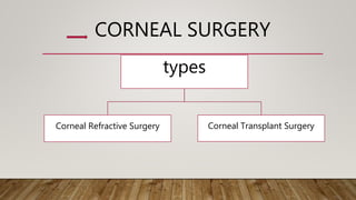
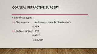
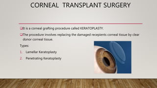
CORNEAL SURGERY
SHREESAGAR.B.V
4424
7TH TERM
CORNEALPHYSIOLOGY
 Transparent, dome shaped, outermost layer that covers the front of the eye.
Provides greatest amount refractive power to the eye.
Avascular, relies on atmosphere for oxygen and aqueous humor for its nutritional
needs.
Corneal surface is kept smooth by
constant moistening action of tears.
LAYERS OF CORNEA
1 Epithelium (55µm).
 Stratified squamous epithelium,5-6 layers
 It maintains stromal dehydration.
2 Bowmans Membrane(12µm):acellular,collagen fibrils,resistant
and do not regenerate
3 Stroma (470µm)
 Collagen fibers arrangement are responsible
for corneal strength, optical characters.90%thickness.
4 Dua’s Membrane
5 Descemets Membrane: highly resistant and regenerates.
6 Endothelium (5µm)
 It contains single layer of polygonal cells.
CORNEAL SURGERY
types
Corneal Refractive Surgery Corneal Transplant Surgery
CORNEAL REFRACTIVE SURGERY
• It is of two types:
>Flap surgery: -Automated Lamellar Keratoplasty
-LASIK
>Surface surgery: -PRK
-LASEK
-epi LASIK
FLAP SURGERY
• ALK
 The surgeon uses a instrument called a microkeratome to cut a thin flap of the
corneal tissue.
The flap is then lifted like a hinged door and target corneal tissue is removed
from microkeratome and flap is replaced
LASIK
Lamellar assisted in situ keratomileusis
Microkeratome or femtosecond laser
Procedure is similar to ALK
The corneal tissue here is removed through excimer laser
LASIK
Advantages:
 Pain free recovery.
 Quick restoration of eyesight.
 Better result for severe
short sight.
Disadvantages:
 Dry eyes.
 Halos, starburst.
 Loss of contrast sensitivity.
 Thick corneal flap (100-180 microns).
SURFACE PROCEDURES
It differs from flap method wherein only the epithelium of the corneal epithelium
is ablated rather than the partial thickness of the stroma.
PRK-Photo Refractive Keratectomy
It is a outpatient procedure performed with local anesthetic eye drops.it is atype of refractive surgery which reshapes the cornea by
removing microscopic amount of tissue.Done using excimer laser.
Used to correct mild to moderate
 Myopia.
 Hyperopia.
 Astigmatism.
Advantages:
 Highly accurate for myopia.
 80% patients have 20/20 vision
Disadvantages:
 Mild discomfort including minor
eye irritation.
 Dry eyes.
LASEK
Laser assisted sub –epithelium keratomileusis
Epithelium is kept intact here.
The surgeon uses an alcohol solution to loosen then lift a thin layer of the
epithelium with a trephine blade.
Advantages:
 No flap complications.
 Causes less dry eyes than
LASIK.
 Finer trephine blade is used
instead of microkeratome.
 Good for patient with
thin cornea.
Disadvantages:
 May cause more pain and
discomfort than LASIK.
 Dry eyes.
 Longer recovery time than
LASIK.
EPI-LASIK
Similar to LASEK.
Epi-keratome is used rather than a trephine blade and alcohol
EPI-LASIK
Advantages:
Less complications to corneal nerves.
Disadvantages:
Dry eyes.
Poor night vision.
Newer techniques:C-TEN(Customised
Transepithelial No-touch surgery)
CORNEAL TRANSPLANT SURGERY
It is a corneal grafting procedure called KERATOPLASTY.
The procedure involves replacing the damaged recepients corneal tissue by clear
donor corneal tissue.
Types:
1. Lamellar Keratoplasty
2. Penetrating Keratoplasty
INDICATIONS
1. Optical: to improve visual acuity in conditions such as pseudophakic bullous
keratopathy,keratoconus,corneal degeneration etc.
2. Tectonic/Reconstuctive: to preserve corneal anatomy in cases of thinning of
stroma and as in descemetoceles or reconstruction of eye as after corneal
perforation.
3. Therapeutic: to remove inflamed corneal tissue unresponsive to drugs.
4. Cosmetic: to improve patients appearance.
DONOR CORNEA
The donor cornea is removed within 6 hours of death and stored under sterile
conditions.
Evaluation:biomicroscopic examination of whole globe is done before considering
the media storage.
Corneal transplants are rarely a failure,as it is avascular.
METHODS OF PRESERVATION
1. Short term: 48 hours,whole globe is preserved at 4c in moist chamber.
2. Intermediate term:2 weeks,in MK media(Mc-Carey Kaufman) or Optisol medium
3. Long term:35 days,by organ culture method
SURGERY TECHNIQUE
A-Normal cornea
B-Penetrating Keratoplasty
C-Deep Anterior Lamellar Keratoplasty
D-Descemets Stripping Automated Endothelial Keratoplasty
E-Descemet’s Membrane Endothelial Keratoplasty
A knife called trephine is used to cut and remove
a circular piece from the recipient’s scarred
cornea
A similar knife is used to cut and remove a piece
from the donor cornea.
The donor cornea then is placed where the
recipient’s cornea was removed.
It is sewn into place with very fine sutures which
are smaller in diameter than a human hair
POSSIBLE COMPLICATIONS OF SURGERY
• Graft Rejection
• Wound Separation
• Loose sutures
• Astigmatism
• Infection
• Secondary glaucoma
• Iris prolapse
• Flat anterior chamber
SIGNS OF GRAFT REJECTION
R
S
V
P
Redness
Sensitivity to light
Vision Changes
Pain
A loose suture may give a foreign body sensation.
Wound separation may lead to prolapse ,infection many complications
secondarily.
ASTIGMATISM: it can occur as a result of distorted shape of the graft(oblong
shaped).
it is usually treated by contacts or spectacles.
AFTER SURGERY
• Following surgery, your eye most likely will be red, irritated, and sensitive to light.
• You may experience increased tearing and a slight discharge.
• Discomfort can be controlled with medication eye drops.
•
• Eye drops are used to reduce inflammation and graft rejection.
• Activities are restrained to prevent any blow to eye.
Your eye will be covered with a patch the day of
surgery
Your surgeon most likely will remove the patch at
your follow-up appointment the next day.
You must wear the patch and shield over your eye
while sleeping or showering
VISION AFTER SURGEY
• Vision usually is blurred after surgery. It gradually improves as healing takes
place.
• As the eye heals and the sutures are removed, the shape of the cornea changes
• Therefore, your surgeon usually will wait between 3 and 12 months before
prescribing a new lens for your glasses
• If needed, a contact lens may be prescribed.
PENETRATING KERATOPLASTY
•THANK YOUTHANK YOU

Corneal surgery

  • 1.
  • 2.
    CORNEALPHYSIOLOGY  Transparent, domeshaped, outermost layer that covers the front of the eye. Provides greatest amount refractive power to the eye. Avascular, relies on atmosphere for oxygen and aqueous humor for its nutritional needs. Corneal surface is kept smooth by constant moistening action of tears.
  • 3.
    LAYERS OF CORNEA 1Epithelium (55µm).  Stratified squamous epithelium,5-6 layers  It maintains stromal dehydration. 2 Bowmans Membrane(12µm):acellular,collagen fibrils,resistant and do not regenerate 3 Stroma (470µm)  Collagen fibers arrangement are responsible for corneal strength, optical characters.90%thickness. 4 Dua’s Membrane 5 Descemets Membrane: highly resistant and regenerates. 6 Endothelium (5µm)  It contains single layer of polygonal cells.
  • 4.
    CORNEAL SURGERY types Corneal RefractiveSurgery Corneal Transplant Surgery
  • 5.
    CORNEAL REFRACTIVE SURGERY •It is of two types: >Flap surgery: -Automated Lamellar Keratoplasty -LASIK >Surface surgery: -PRK -LASEK -epi LASIK
  • 6.
    FLAP SURGERY • ALK The surgeon uses a instrument called a microkeratome to cut a thin flap of the corneal tissue. The flap is then lifted like a hinged door and target corneal tissue is removed from microkeratome and flap is replaced
  • 8.
    LASIK Lamellar assisted insitu keratomileusis Microkeratome or femtosecond laser Procedure is similar to ALK The corneal tissue here is removed through excimer laser
  • 9.
    LASIK Advantages:  Pain freerecovery.  Quick restoration of eyesight.  Better result for severe short sight. Disadvantages:  Dry eyes.  Halos, starburst.  Loss of contrast sensitivity.  Thick corneal flap (100-180 microns).
  • 10.
    SURFACE PROCEDURES It differsfrom flap method wherein only the epithelium of the corneal epithelium is ablated rather than the partial thickness of the stroma.
  • 11.
    PRK-Photo Refractive Keratectomy Itis a outpatient procedure performed with local anesthetic eye drops.it is atype of refractive surgery which reshapes the cornea by removing microscopic amount of tissue.Done using excimer laser. Used to correct mild to moderate  Myopia.  Hyperopia.  Astigmatism. Advantages:  Highly accurate for myopia.  80% patients have 20/20 vision Disadvantages:  Mild discomfort including minor eye irritation.  Dry eyes.
  • 13.
    LASEK Laser assisted sub–epithelium keratomileusis Epithelium is kept intact here. The surgeon uses an alcohol solution to loosen then lift a thin layer of the epithelium with a trephine blade.
  • 14.
    Advantages:  No flapcomplications.  Causes less dry eyes than LASIK.  Finer trephine blade is used instead of microkeratome.  Good for patient with thin cornea. Disadvantages:  May cause more pain and discomfort than LASIK.  Dry eyes.  Longer recovery time than LASIK.
  • 15.
    EPI-LASIK Similar to LASEK. Epi-keratomeis used rather than a trephine blade and alcohol
  • 16.
    EPI-LASIK Advantages: Less complications tocorneal nerves. Disadvantages: Dry eyes. Poor night vision. Newer techniques:C-TEN(Customised Transepithelial No-touch surgery)
  • 17.
    CORNEAL TRANSPLANT SURGERY Itis a corneal grafting procedure called KERATOPLASTY. The procedure involves replacing the damaged recepients corneal tissue by clear donor corneal tissue. Types: 1. Lamellar Keratoplasty 2. Penetrating Keratoplasty
  • 18.
    INDICATIONS 1. Optical: toimprove visual acuity in conditions such as pseudophakic bullous keratopathy,keratoconus,corneal degeneration etc. 2. Tectonic/Reconstuctive: to preserve corneal anatomy in cases of thinning of stroma and as in descemetoceles or reconstruction of eye as after corneal perforation. 3. Therapeutic: to remove inflamed corneal tissue unresponsive to drugs. 4. Cosmetic: to improve patients appearance.
  • 19.
    DONOR CORNEA The donorcornea is removed within 6 hours of death and stored under sterile conditions. Evaluation:biomicroscopic examination of whole globe is done before considering the media storage. Corneal transplants are rarely a failure,as it is avascular.
  • 20.
    METHODS OF PRESERVATION 1.Short term: 48 hours,whole globe is preserved at 4c in moist chamber. 2. Intermediate term:2 weeks,in MK media(Mc-Carey Kaufman) or Optisol medium 3. Long term:35 days,by organ culture method
  • 21.
    SURGERY TECHNIQUE A-Normal cornea B-PenetratingKeratoplasty C-Deep Anterior Lamellar Keratoplasty D-Descemets Stripping Automated Endothelial Keratoplasty E-Descemet’s Membrane Endothelial Keratoplasty
  • 22.
    A knife calledtrephine is used to cut and remove a circular piece from the recipient’s scarred cornea A similar knife is used to cut and remove a piece from the donor cornea. The donor cornea then is placed where the recipient’s cornea was removed. It is sewn into place with very fine sutures which are smaller in diameter than a human hair
  • 23.
    POSSIBLE COMPLICATIONS OFSURGERY • Graft Rejection • Wound Separation • Loose sutures • Astigmatism • Infection • Secondary glaucoma • Iris prolapse • Flat anterior chamber
  • 24.
    SIGNS OF GRAFTREJECTION R S V P Redness Sensitivity to light Vision Changes Pain
  • 25.
    A loose suturemay give a foreign body sensation. Wound separation may lead to prolapse ,infection many complications secondarily. ASTIGMATISM: it can occur as a result of distorted shape of the graft(oblong shaped). it is usually treated by contacts or spectacles.
  • 26.
    AFTER SURGERY • Followingsurgery, your eye most likely will be red, irritated, and sensitive to light. • You may experience increased tearing and a slight discharge. • Discomfort can be controlled with medication eye drops. • • Eye drops are used to reduce inflammation and graft rejection. • Activities are restrained to prevent any blow to eye. Your eye will be covered with a patch the day of surgery Your surgeon most likely will remove the patch at your follow-up appointment the next day. You must wear the patch and shield over your eye while sleeping or showering
  • 27.
    VISION AFTER SURGEY •Vision usually is blurred after surgery. It gradually improves as healing takes place. • As the eye heals and the sutures are removed, the shape of the cornea changes • Therefore, your surgeon usually will wait between 3 and 12 months before prescribing a new lens for your glasses • If needed, a contact lens may be prescribed.
  • 28.
  • 29.