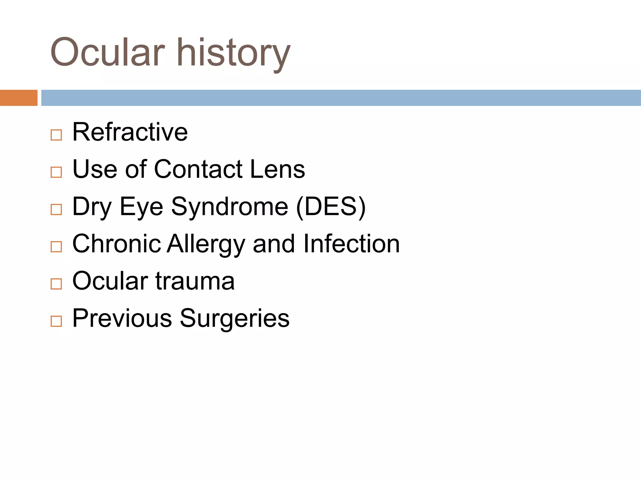
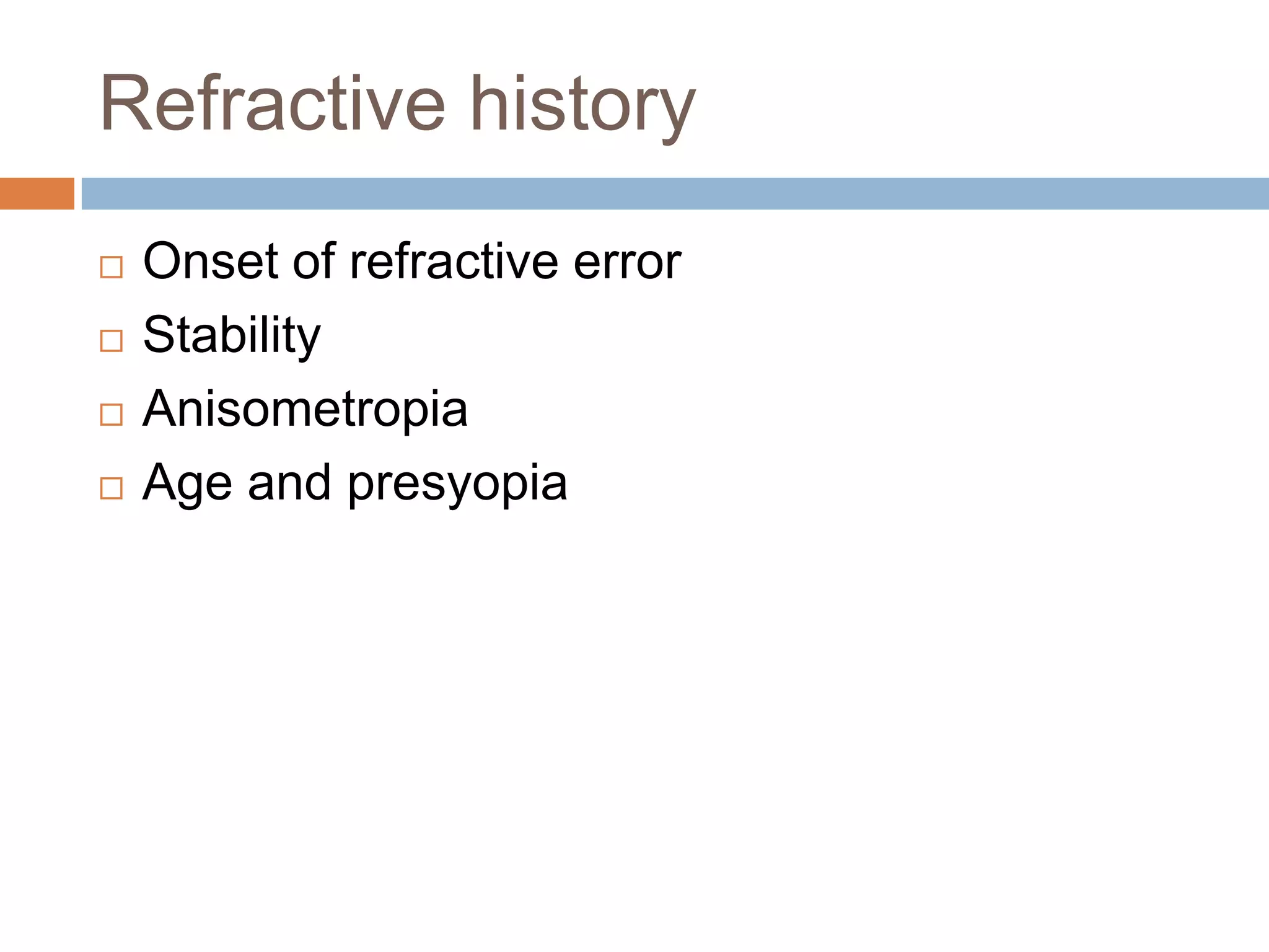
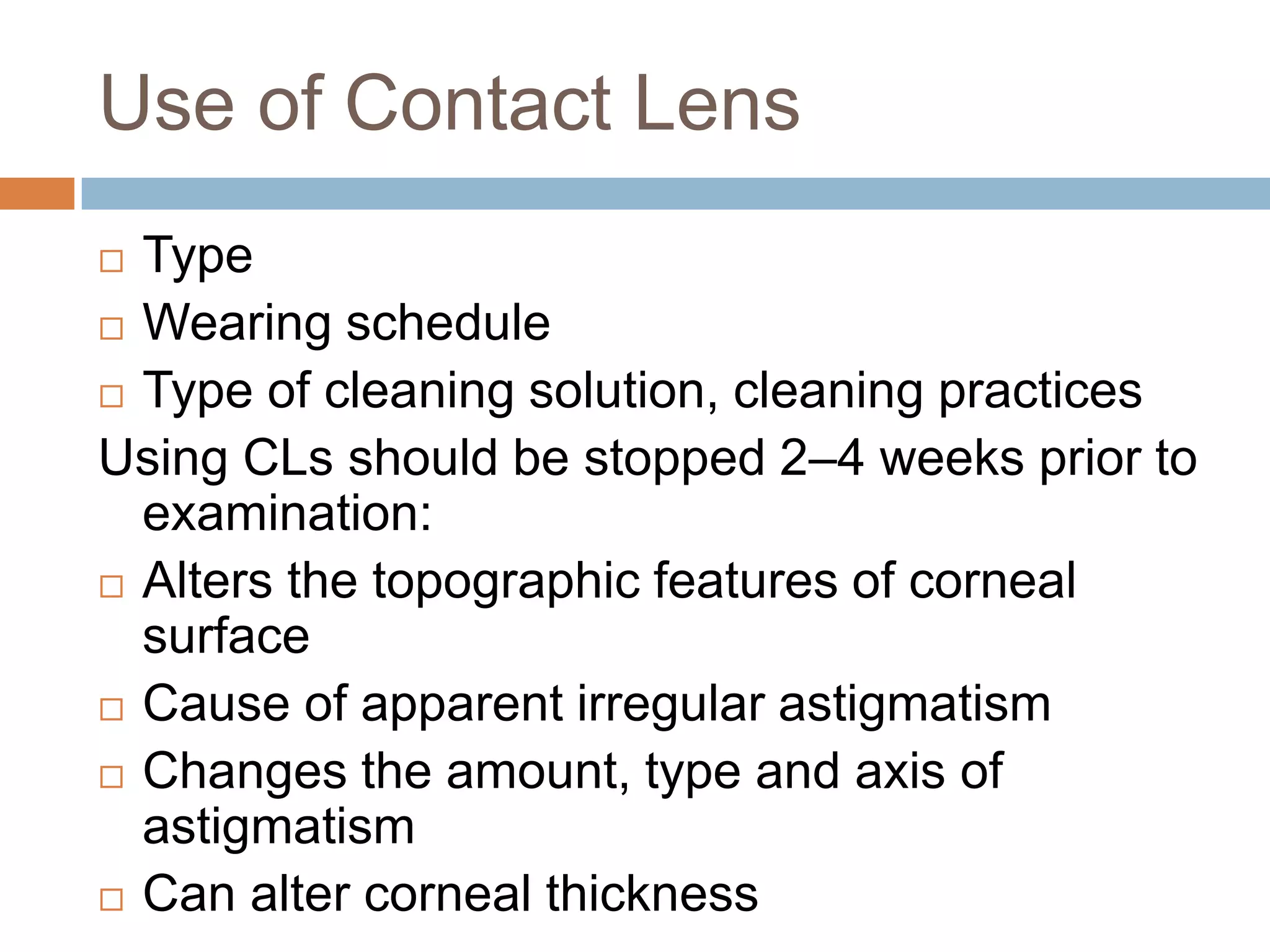
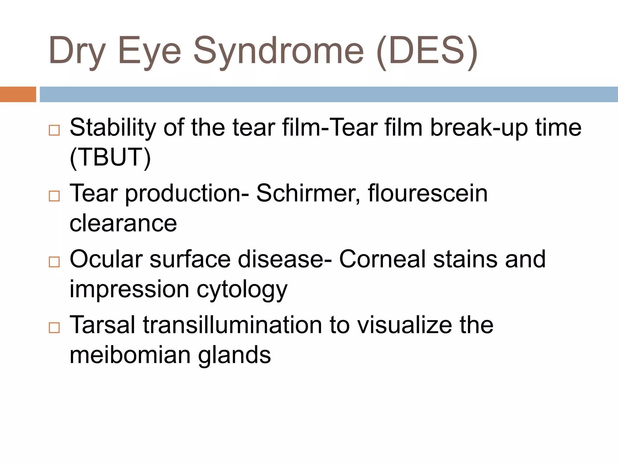
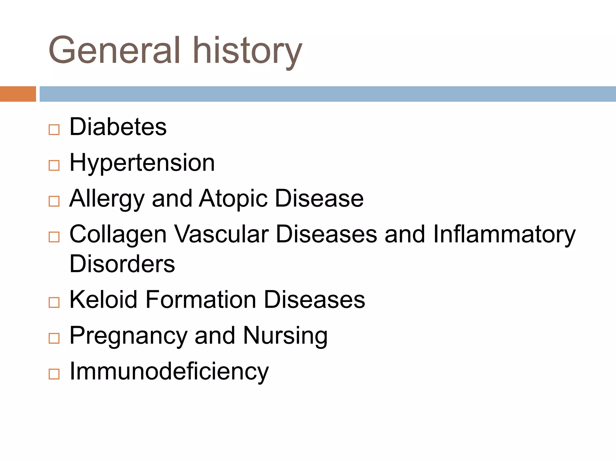
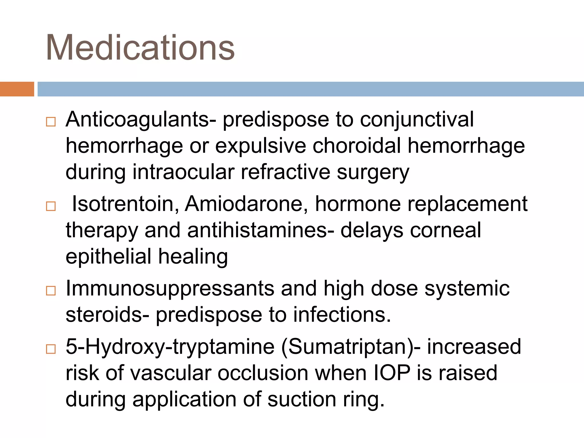
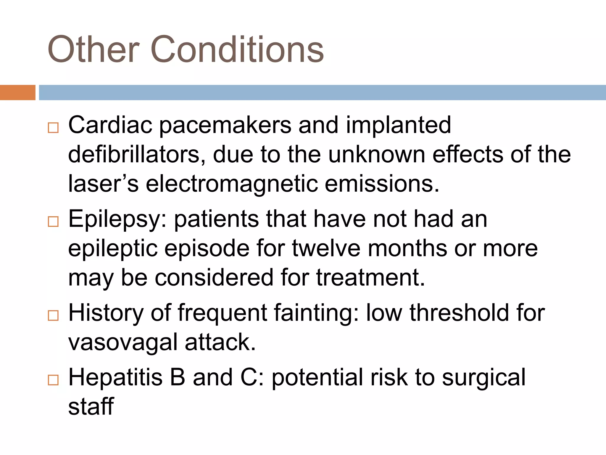
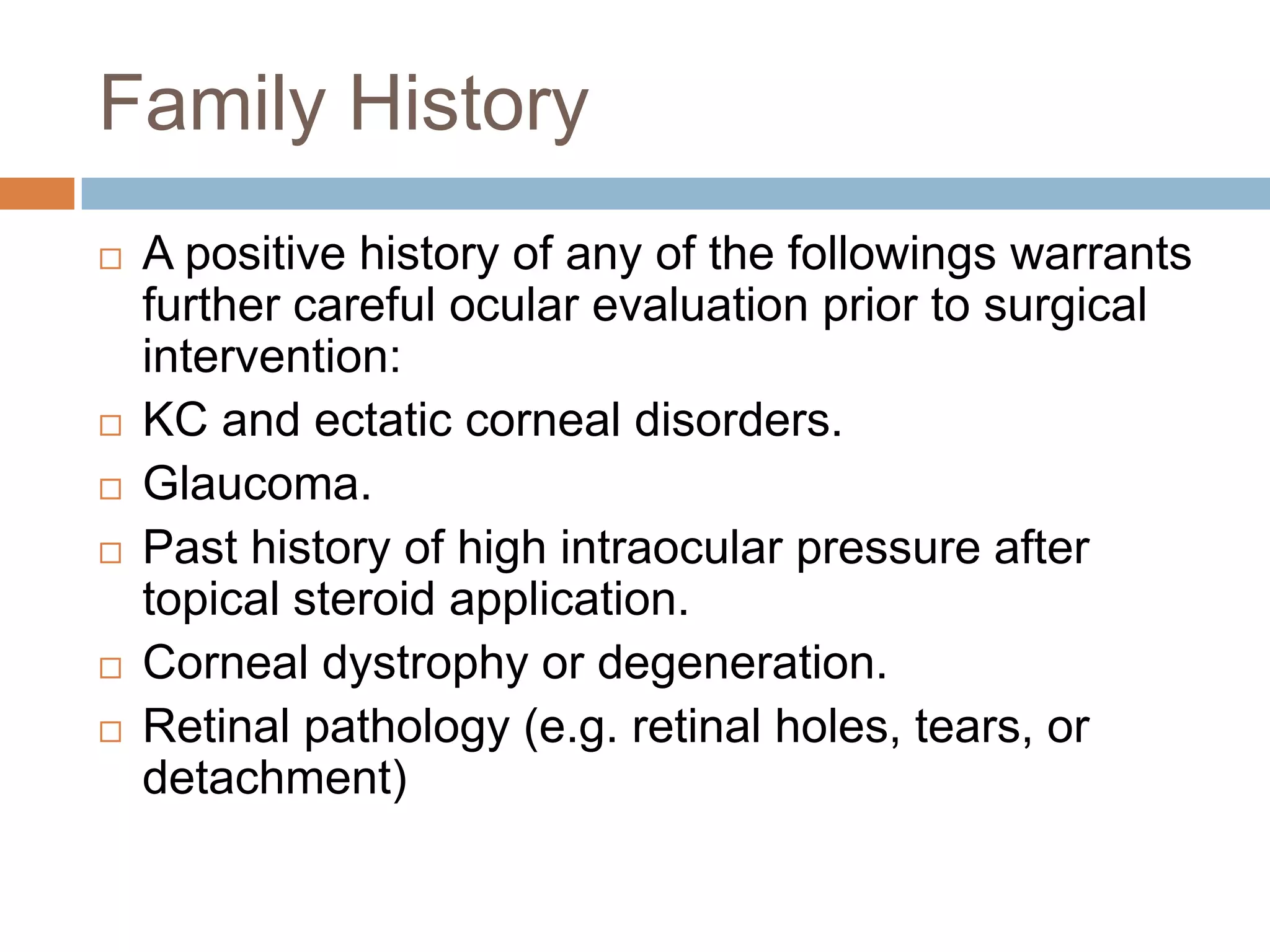
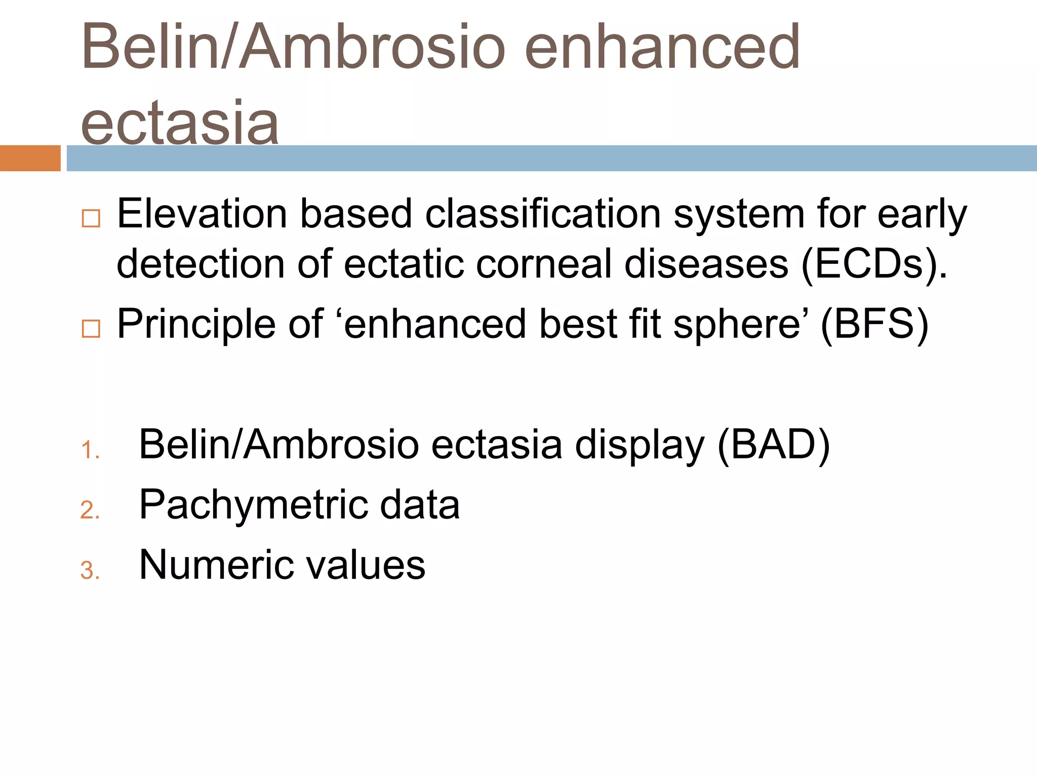
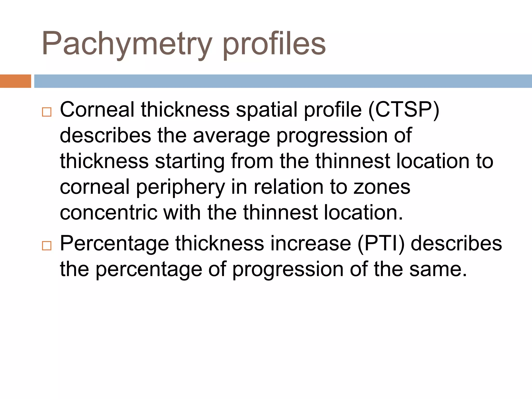
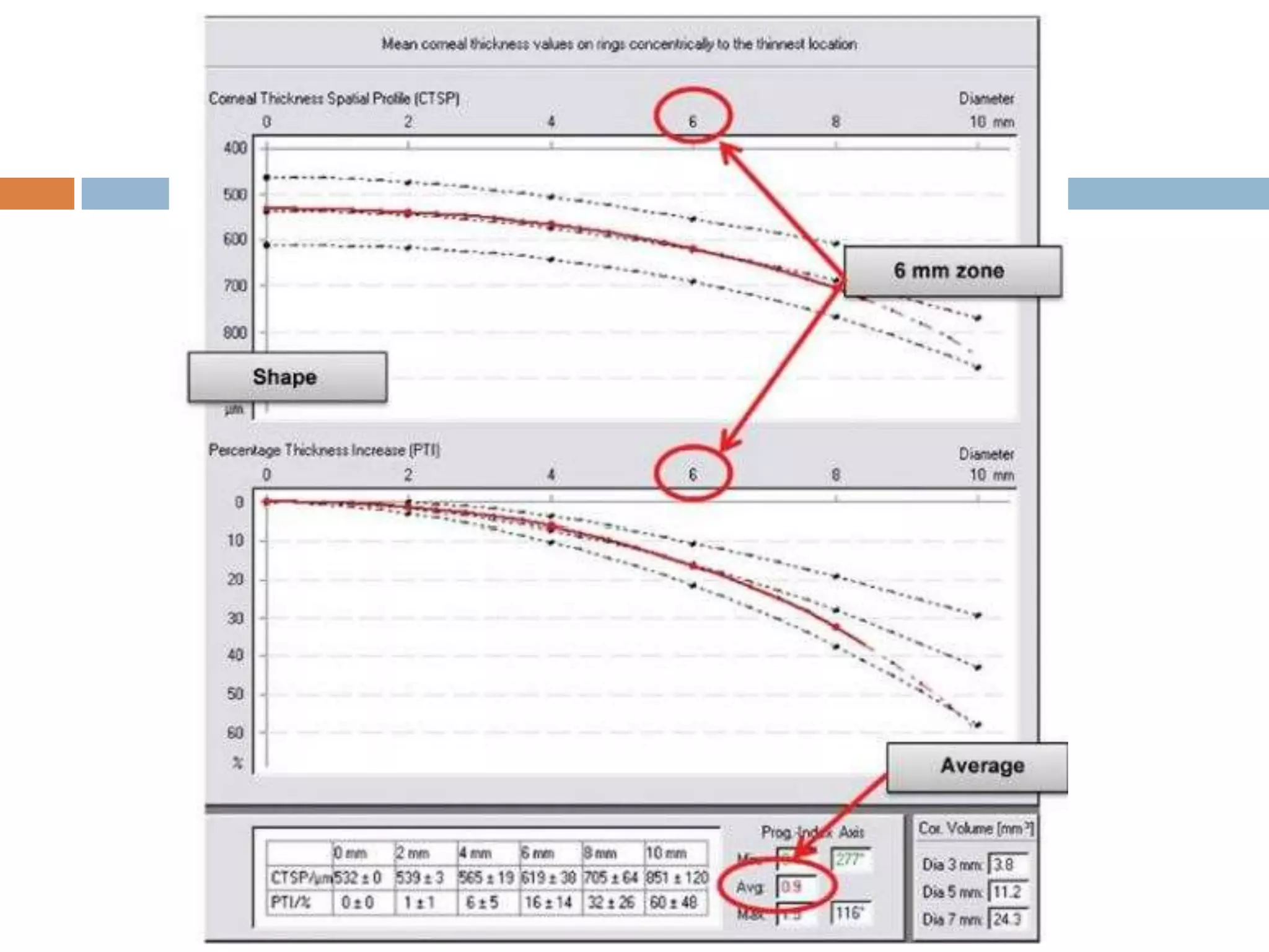
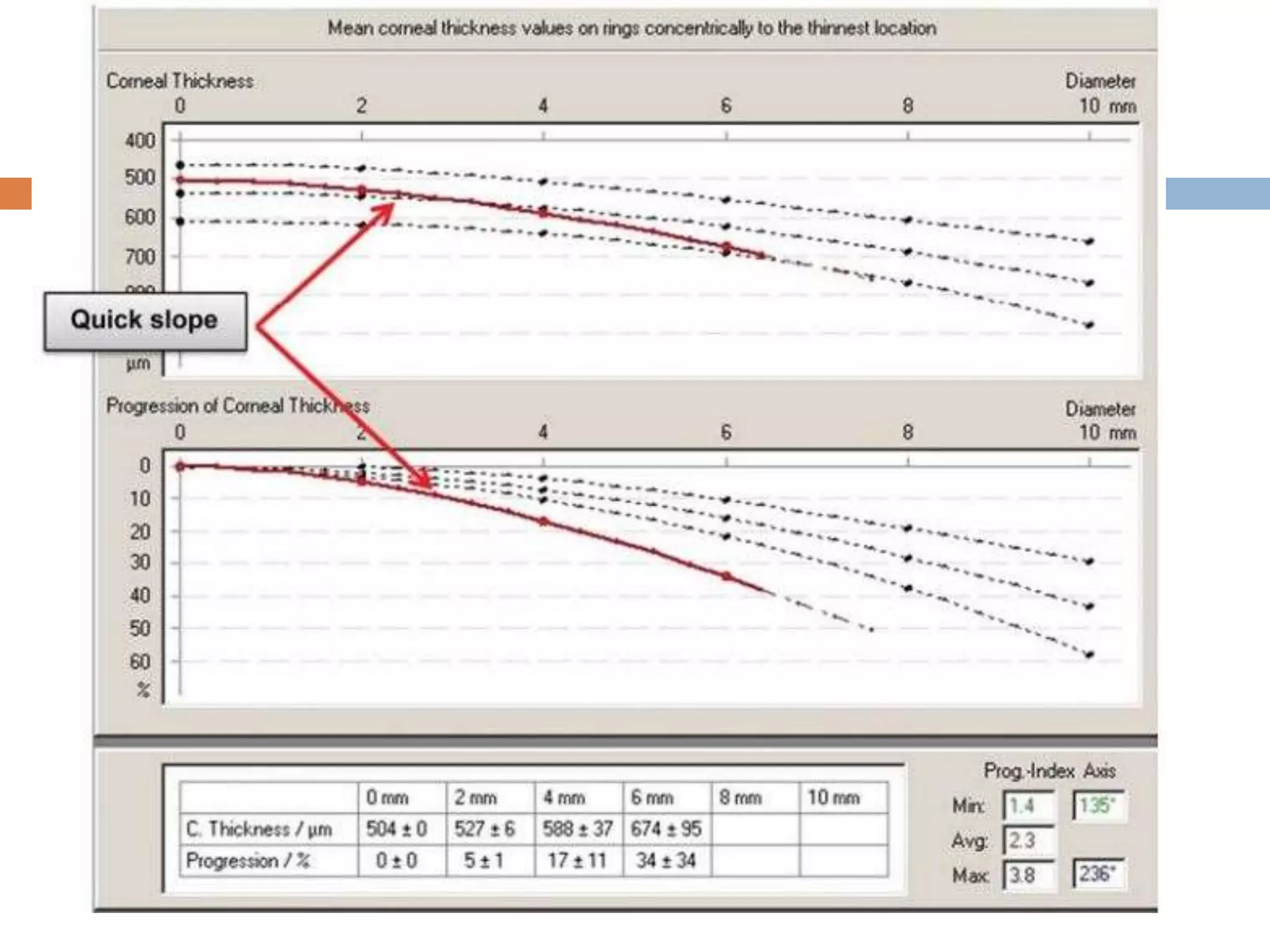
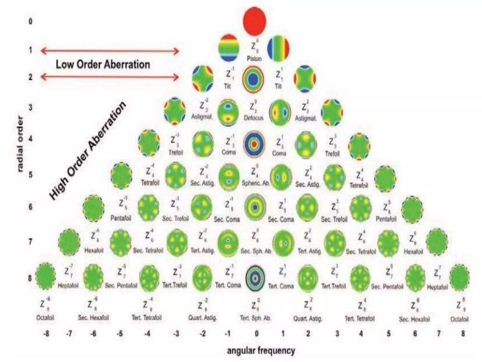
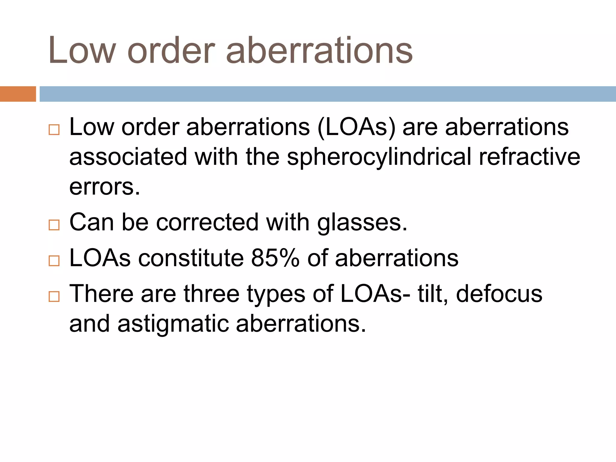
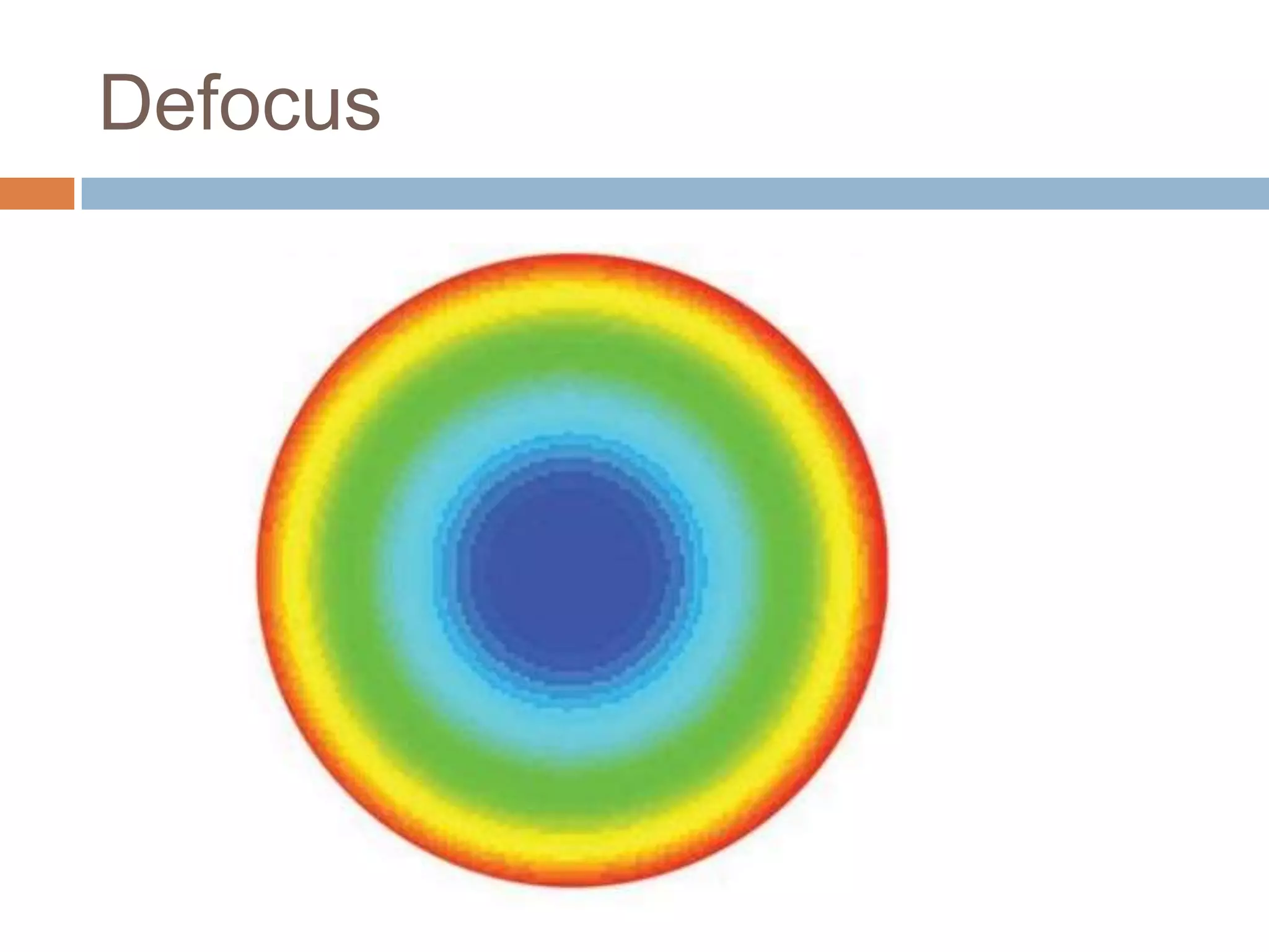
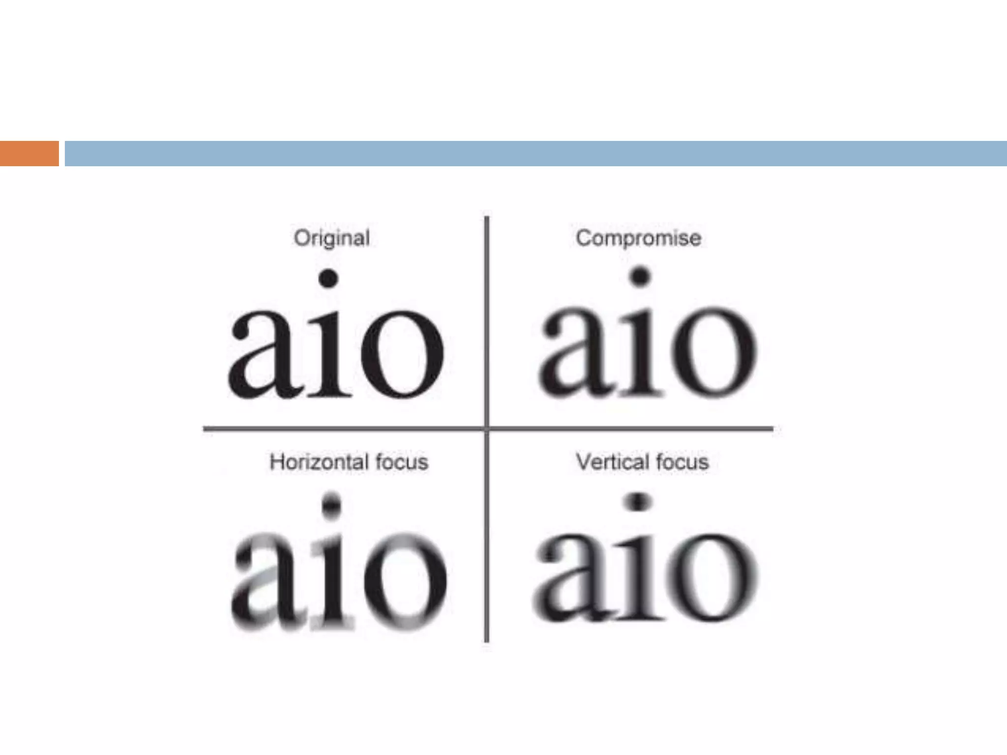
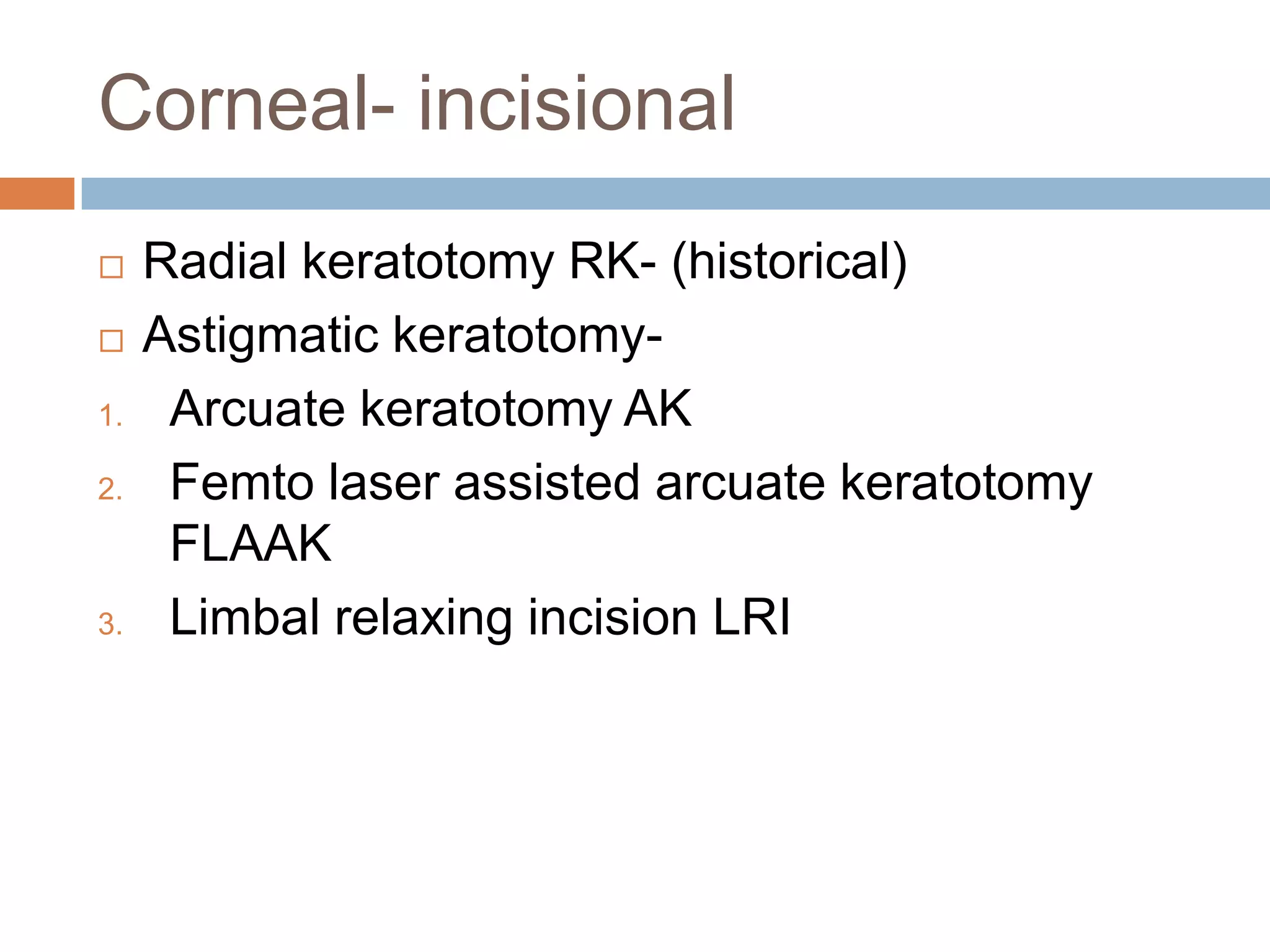
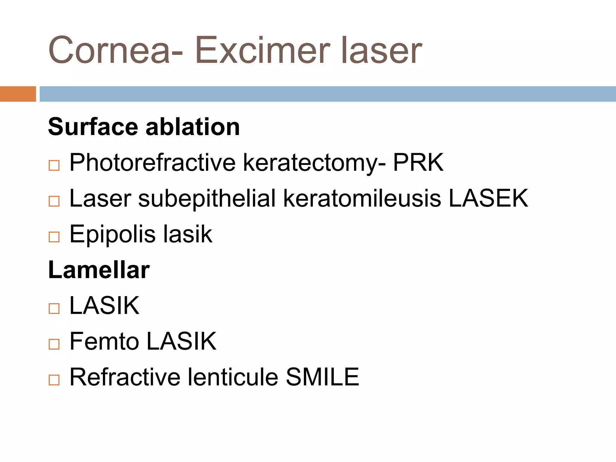
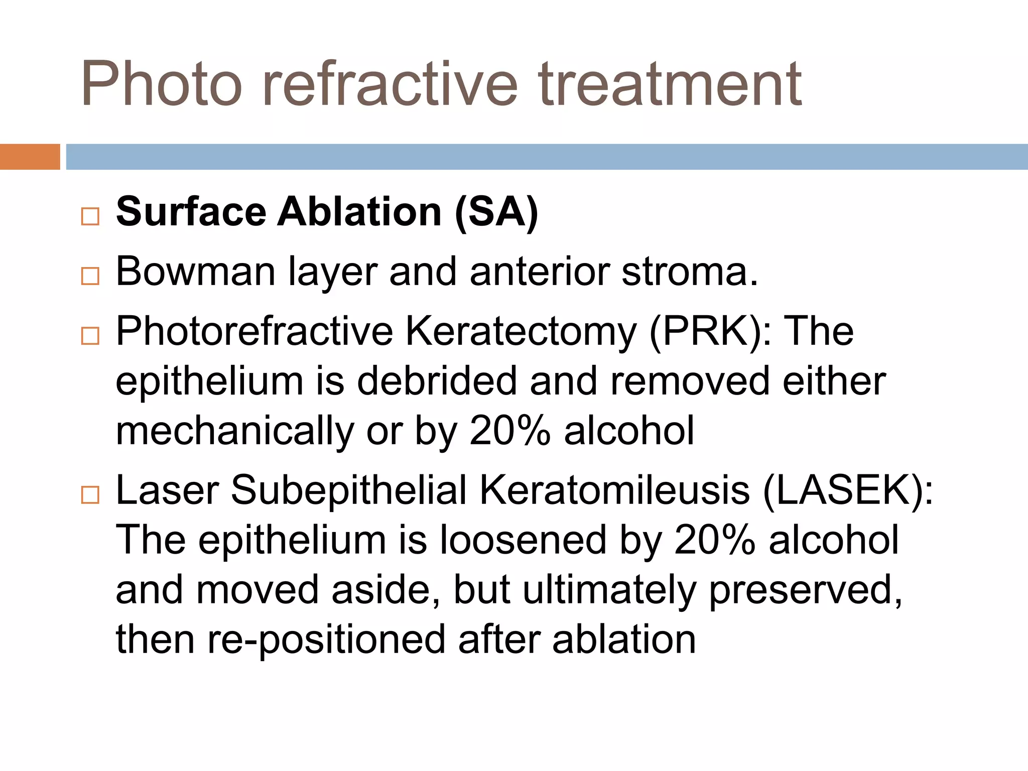
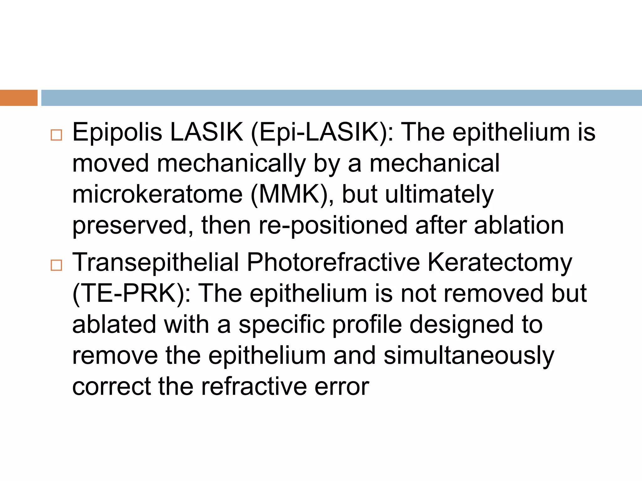
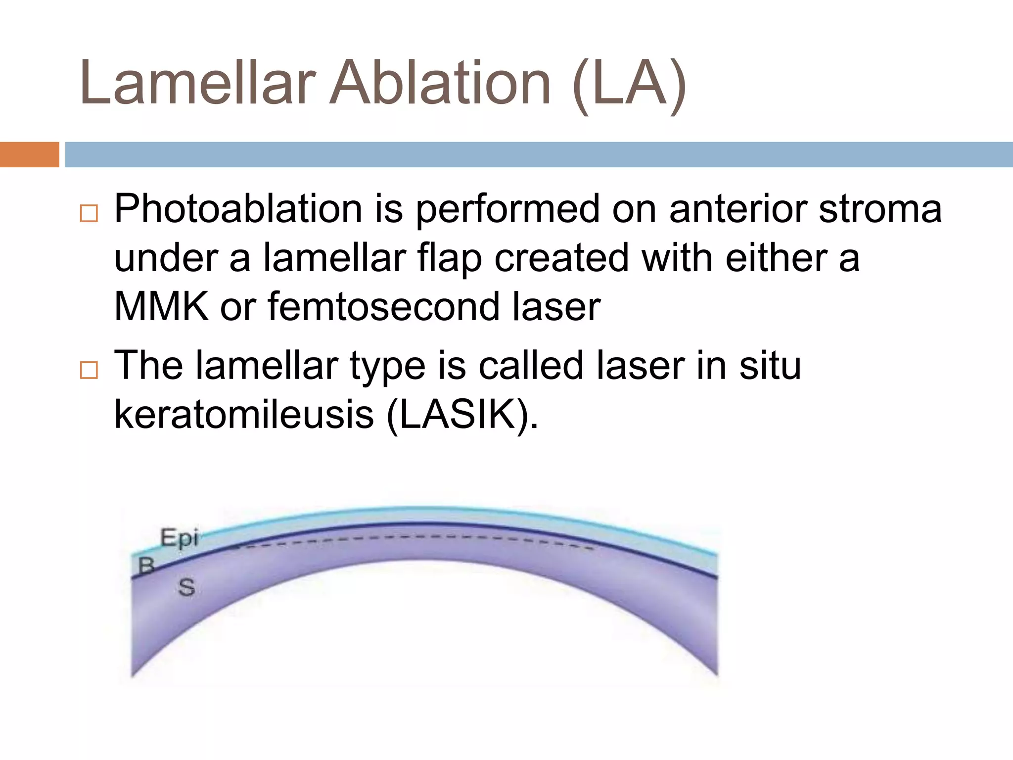
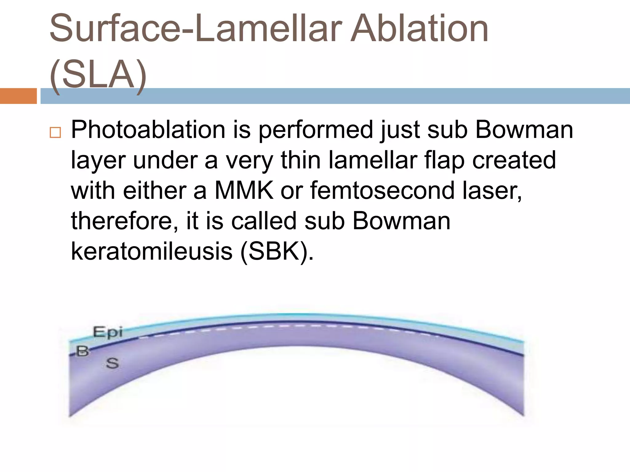
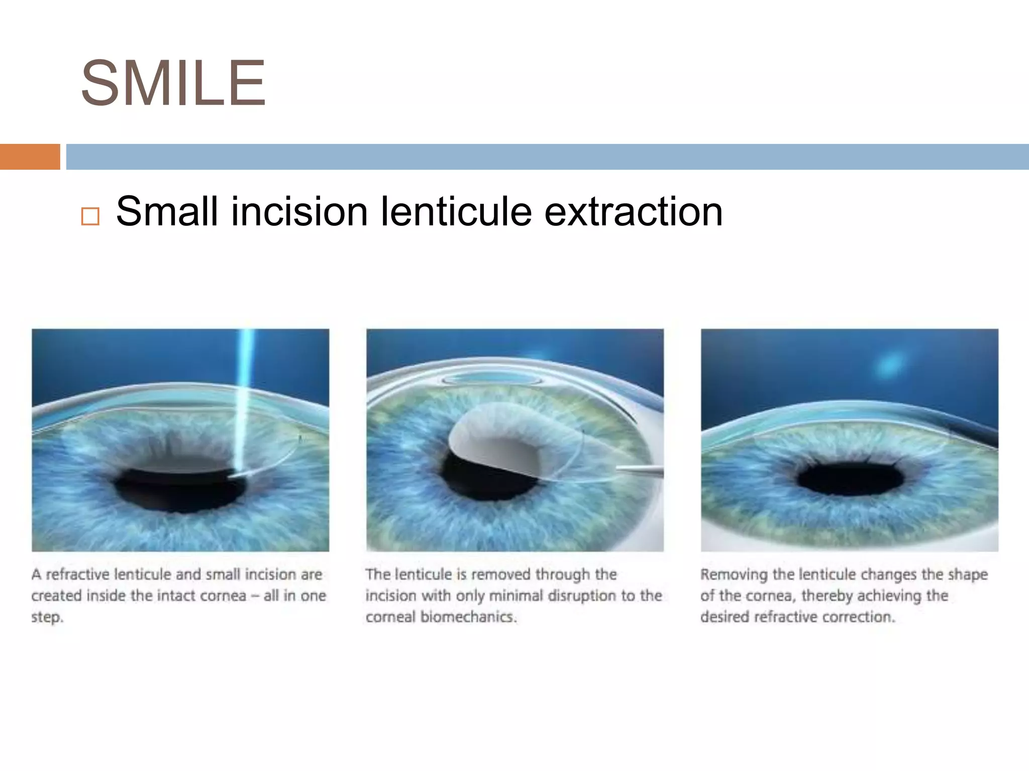
REFRACTIVE
SURGERIES
-Shreya
 The goal of refractive surgery is to reduce
dependence on contact lenses or glasses for
use in routine daily activities.
 A wide variety of surgical techniques and
technologies are available, and all require an
appropriate preoperative evaluation to
determine the best technique and ensure the
optimal outcome for each patient individually.
PATIENT EVALUATION
Psychosocial approach
 Patients with unreasonable high expectations and
those with type-A and compulsive personalities
are not good candidates for refractive surgery
 A good candidate is that who understands that the
refractive surgery is not risk-free, follows
instructions, is willing to wear glasses after the
surgery
 A good surgeon is that who does not give unreal
promises, tells that patient the reasonable
expected visual acuity and visual function and
discusses the issue of enhancement.
Social History
 Profession
 Patient expectations
 Suitability for post-operative care
Ocular history
 Refractive
 Use of Contact Lens
 Dry Eye Syndrome (DES)
 Chronic Allergy and Infection
 Ocular trauma
 Previous Surgeries
Refractive history
 Onset of refractive error
 Stability
 Anisometropia
 Age and presyopia
Use of Contact Lens
 Type
 Wearing schedule
 Type of cleaning solution, cleaning practices
Using CLs should be stopped 2–4 weeks prior to
examination:
 Alters the topographic features of corneal
surface
 Cause of apparent irregular astigmatism
 Changes the amount, type and axis of
astigmatism
 Can alter corneal thickness
Dry Eye Syndrome (DES)
 Stability of the tear film-Tear film break-up time
(TBUT)
 Tear production- Schirmer, flourescein
clearance
 Ocular surface disease- Corneal stains and
impression cytology
 Tarsal transillumination to visualize the
meibomian glands
Chronic Allergy and Infection
 Constant rubbing and development of
keratoconus.
 Constant rubbing and post LASIK
complications such as flap dislocation.
 Chronic infections and post surgery
complications
Ocular trauma
 Pathological findings such as retinal tears or
atrophy or optic nerve head atrophy
 Corneal opacities and scars
 Iridodenesis
 Lens subluxation
Previous Surgeries
 Refractive surgery
 Cataract surgery- IOL, technique, date of
surgery
 Retinal surgery
 Squint surgery
 Pterygium surgery
 Glaucoma and Previous Glaucoma Surgery
 Keratoconus and ectatic Corneal Disorders
Other Pathologies-
 Recurrent corneal erosion, corneal ulceration
or ocular infections
General history
 Diabetes
 Hypertension
 Allergy and Atopic Disease
 Collagen Vascular Diseases and Inflammatory
Disorders
 Keloid Formation Diseases
 Pregnancy and Nursing
 Immunodeficiency
Medications
 Anticoagulants- predispose to conjunctival
hemorrhage or expulsive choroidal hemorrhage
during intraocular refractive surgery
 Isotrentoin, Amiodarone, hormone replacement
therapy and antihistamines- delays corneal
epithelial healing
 Immunosuppressants and high dose systemic
steroids- predispose to infections.
 5-Hydroxy-tryptamine (Sumatriptan)- increased
risk of vascular occlusion when IOP is raised
during application of suction ring.
Other Conditions
 Cardiac pacemakers and implanted
defibrillators, due to the unknown effects of the
laser’s electromagnetic emissions.
 Epilepsy: patients that have not had an
epileptic episode for twelve months or more
may be considered for treatment.
 History of frequent fainting: low threshold for
vasovagal attack.
 Hepatitis B and C: potential risk to surgical
staff
Family History
 A positive history of any of the followings warrants
further careful ocular evaluation prior to surgical
intervention:
 KC and ectatic corneal disorders.
 Glaucoma.
 Past history of high intraocular pressure after
topical steroid application.
 Corneal dystrophy or degeneration.
 Retinal pathology (e.g. retinal holes, tears, or
detachment)
Examination
Examination
 Visual acuity- BCVA, UCVA
 Clinical Refraction- Manifest and cycloplegic
refraction
 Pupillometry
 Tear film tests
 IOP measurements
 Ocular motility
 Orbital anatomy
External and Slit lamp
examination
 Blepharitis and meibomitis
 Keratitis and punctate keratopathy, scarring,
dystrophies
 Conjunctival scars, bleb, buckle
 Cataract
Fundoscopy
 Peripheral retinal pathology
 Diabetic retinopathy
 Macular edema, epiretinal membranes and
degenerative changes in the macula
 ONH abnormality
Corneal imaging
Topography and tomography
sciences
 Corneal topography- Placido-based machines consisting
of two maps: anterior sagittal (axial) and anterior
tangential (instantaneous) curvature maps.
 Corneal tomography-
 maps and images given by Scheimpflug-based machines
 topographic maps with more maps and profiles of both
corneal surfaces and corneal pachymetry map.
 most important screening test for refractive surgery to
detect abnormalities, diagnose early cases of ectatic
corneal diseases and classify these diseases, diagnose
post keratorefractive ectasia and put the plan for the best
choice in refractive surgery.
Enantiomorphism
Instantaneous power and
curvature
 Also called meridional or tangential power
 Better sensitivity to peripheral changes
 less “smoothing” of the curvature
 In these maps, diopters are relative units of
curvature and are not the equivalent of
diopters of corneal power.
 Tangential maps are more susceptible to local
curvature changes, because it depends on
circles
 Each point on the tangential map is
independent of any reference axis- less
affected by misalignments.
 Determination of morphologic patterns of the
cone in ectatic corneal disorders
 Depending on this map, there are three
patterns of the cone:
 a. Nipple
 b. Oval
 c. Globus
Elevation maps
 Elevation maps describe the height details of
the measured corneal surface by matching it
with a reference surface(RS) above which
points are considered elevations and expressed
in plus values and below which points are
considered depressions and expressed in
minus values.
Position of the reference sphere
 Float mode - RS is adjusted with the corneal
surface such that all elevations are equal to all
depressions.
 Non-float mode – RS touches the apex of
cornea
Parameters
 Radius- mean central radii of measured
corneal surface
 Diameter - diameter of the used zone of the
cornea
Types of RS
 Best fit sphere (BFS)
 Best fit ellipsoid (BFE)
 Best fit toric ellipsoid (BFTE)
Best fit sphere
CONE LOCATION
PACHYMETRY MAPS
There are three main land marks on the
pachymetry map:
 cornea apex
 thinnest location
 two opposing points on the vertical meridian at
the central 5 mm circle
The two opposing points are superior (S) and
inferior (I); the normal S-I difference is <30
microns
Belin/Ambrosio enhanced
ectasia
 Elevation based classification system for early
detection of ectatic corneal diseases (ECDs).
 Principle of ‘enhanced best fit sphere’ (BFS)
1. Belin/Ambrosio ectasia display (BAD)
2. Pachymetric data
3. Numeric values
Principle
Progression index
 Min- Pachymetric progression index minimum
Half meridian with smallest progression index.
(green)
 Max- Pachymetric progression index maximum
Half meridian with largest progression index. (blue)
 Avg- Pachymetric progression index average
Ratio of individual progression to normative
progression.
 ARTmax- Ambrosio Relational Thickness
maximum
Ratio of thinnest corneal thickness to pachymetric
Deviation parameters
 Df- deviation of front elevation difference map
 Db- deviation of back elevation difference map
 Dp- deviation of average pacymetric
progression
 Dt- deviation of minimum thickness
 Da- deviation of ARTmax
 D- total deviation value
Pachymetry profiles
 Corneal thickness spatial profile (CTSP)
describes the average progression of
thickness starting from the thinnest location to
corneal periphery in relation to zones
concentric with the thinnest location.
 Percentage thickness increase (PTI) describes
the percentage of progression of the same.
Corneal topometry
 Measures the slope of the cornea
 Affected by keratorefractive surgery
 Abnormal corneal topometry is the main cause
of spherical aberrations
Corneal surface may take one of four main
shapes:
 Spheric
 Aspheric oblate
 Aspheric prolate
 Aspheric hyperprolate.
 Corneal topometry is expressed by Q-value
 Normal Q-value is –1 to 0
 Q-value is positive (>0) in oblate cornea
 Q-value is negative (<-1) in prolate and
hyperprolate cornea
 Q-value is plano (=0) in spherical cornea
 Abnormal Q-value results in spherical
aberrations
Wavefront science
Refractive Error: Optical Principles
and Wavefront Analysis
 One of the major applications of the wave theory of
light is in wavefront analysis.
 Currently, wavefront analysis can be performed
clinically by 4 methods:
1. Hartmann-Shack
2. Tscherning
3. Thin-beam single-ray tracing
4. Optical path difference
 Measures lower-order aberrations (sphere and
cylinder) and higher-order aberrations (spherical
aberration, coma, trefoil).
 Used in calculating custom ablations to enhance
vision.
Hartmann-Shack
 In an aberration-free eye, all the rays would emerge in
parallel, and the reflected wavefront would be a flat
plane (piston).
 In reality, the wavefront is not flat.
 To determine the shape of the reflected wavefront, an
array of lenses samples parts of the wavefront and
focuses light on a detector
 The extent of the divergence of the lenslet images from
their expected focal points determines the wavefront
error.
 Optical aberrations measured by the aberrometer can
be resolved into a variety of basic shapes, the
combination of which represents the total aberration of
the patient’s ocular system.
Low order aberrations
 Low order aberrations (LOAs) are aberrations
associated with the spherocylindrical refractive
errors.
 Can be corrected with glasses.
 LOAs constitute 85% of aberrations
 There are three types of LOAs- tilt, defocus
and astigmatic aberrations.
Tilt
Defocus
Astigmatic aberration
Higher-Order Aberrations
 May or may not be associated with refractive
errors
 HOAs result from media irregularities or
opacities
 Although there is a wide range and types of
HOAs, the main types are coma, trefoil and
spherical aberrations
 HOAs impact vision more severe than LOAs
 Coma is a central aberration and affects
central vision, whereas trefoils and spherical
are peripheral aberrations
Spherical aberration
Trefoil
 HOAs cannot be corrected with classic optics
 Coma results from a variation of magnification
(refractive power) over the entrance pupil. It
affects central vision
 Trefoil aberration results from regular
alternating variation in magnification along the
meridians in corneal periphery. It affects
peripheral vision.
 Spherical aberration results from abnormal Q
value. It affects peripheral vision
 Other higher-order aberrations
 There are numerous other higher-order
aberrations, of which only a small number
are of clinical interest.
Measurement metrics
 Objective visual function is measured at
corneal level (corneal wavefront) and at
pupillary level (ocular wavefront) by root mean
square method (RMS)
 Deviation averaged over the entire wavefront
in reference to the perfect wavefront.
 Quantitative value
 For large deviations
 Normal emmetropic eyes- ≤0.4µm.
RMS= √(+0.25)2+(-0.40)2+(+0.30)2
=0.559µm
Types of refractive surgeries
Types
 Corneal
 Intraocular
Corneal
 Incisional
 Excimer laser
 Non laser lamellar
 Collagen shrinkage
 Collagen cross linking
Corneal- incisional
 Radial keratotomy RK- (historical)
 Astigmatic keratotomy-
1. Arcuate keratotomy AK
2. Femto laser assisted arcuate keratotomy
FLAAK
3. Limbal relaxing incision LRI
Cornea- Excimer laser
Surface ablation
 Photorefractive keratectomy- PRK
 Laser subepithelial keratomileusis LASEK
 Epipolis lasik
Lamellar
 LASIK
 Femto LASIK
 Refractive lenticule SMILE
Photo refractive treatment
 Surface Ablation (SA)
 Bowman layer and anterior stroma.
 Photorefractive Keratectomy (PRK): The
epithelium is debrided and removed either
mechanically or by 20% alcohol
 Laser Subepithelial Keratomileusis (LASEK):
The epithelium is loosened by 20% alcohol
and moved aside, but ultimately preserved,
then re-positioned after ablation
 Epipolis LASIK (Epi-LASIK): The epithelium is
moved mechanically by a mechanical
microkeratome (MMK), but ultimately
preserved, then re-positioned after ablation
 Transepithelial Photorefractive Keratectomy
(TE-PRK): The epithelium is not removed but
ablated with a specific profile designed to
remove the epithelium and simultaneously
correct the refractive error
Diagrammatic comparison of single and multizone
keratectomies.
A. Depth of ablation required to correct 12.00 D of myopia
in a single pass.
B. Depiction of how the use of multiple zones reduces the
Lamellar Ablation (LA)
 Photoablation is performed on anterior stroma
under a lamellar flap created with either a
MMK or femtosecond laser
 The lamellar type is called laser in situ
keratomileusis (LASIK).
Surface-Lamellar Ablation
(SLA)
 Photoablation is performed just sub Bowman
layer under a very thin lamellar flap created
with either a MMK or femtosecond laser,
therefore, it is called sub Bowman
keratomileusis (SBK).
SMILE
 Small incision lenticule extraction
Cornea- Non laser lamellar
 Epikeratophakia (historical)
 Epikeratoplasty (historical)
 Intra stromal corneal ring segments
-keratoconus
-Post LASIK ectasia
Collagen Shrinkage Techniques
 Alteration in corneal biomechanics can also be
achieved by collagen shrinkage.
 Heating collagen to a critical temperature of
58°–76°C causes it to shrink, inducing
changes in the corneal curvature.
 Thermokeratoplasty and conductive
keratoplasty (CK) are avoided in the central
cornea because of scarring but can be used in
the midperiphery to cause local collagen
contraction with concurrent central corneal
steepening
Collagen shrinkage
 Laser thermokeratoplasty
 Conductive keratoplasty
Corneal collagen cross linking
Conductive keratoplasty
Intra ocular
 Phakic
 Pseudophakic
Phakic IOLs
 Phakic IOLs (PIOLs) are additive lenses
implanted into the anterior or posterior
chambers of the eye to compensate for
refractive errors and at the same time
preserving the crystalline lens and
accommodation
 Three types of PIOLs are currently available:
 1. Angle-Supported (anterior chamber)
 2. Iris-Fixated (anterior chamber)
 3. Sulcus-Supported (posterior chamber)
 The power of a phakic lens is independent of
the axial length of the eye. Rather, it depends
on central corneal power (K-readings), ACD
and patient refraction.
Indications
 PIOLs are usually indicated where
photoablation is relatively or absolutely
contraindicated.
 PIOLs are implanted in case of:
 1. High refractive errors
 2. Thin corneas.
 3. Abnormal (suspicious) corneal tomography.
 4. Dry eye.
Refractive lens exchange (RLE)
 Refractive lens exchange (RLE) is extracting
the clear crystalline lens and implantation of
an IOL to compensate for refractive errors
 It can be considered as one of the refractive
options to treat refractive errors, but has very
limited indications and carries almost the same
risks of cataract extraction
 Monofocal IOL
 Toric IOL
 Multifocal IOL
 Accomodating IOL
THANK YOU!

Work-up of Refractive surgeries

  • 1.
  • 2.
     The goalof refractive surgery is to reduce dependence on contact lenses or glasses for use in routine daily activities.  A wide variety of surgical techniques and technologies are available, and all require an appropriate preoperative evaluation to determine the best technique and ensure the optimal outcome for each patient individually.
  • 3.
  • 4.
    Psychosocial approach  Patientswith unreasonable high expectations and those with type-A and compulsive personalities are not good candidates for refractive surgery  A good candidate is that who understands that the refractive surgery is not risk-free, follows instructions, is willing to wear glasses after the surgery  A good surgeon is that who does not give unreal promises, tells that patient the reasonable expected visual acuity and visual function and discusses the issue of enhancement.
  • 5.
    Social History  Profession Patient expectations  Suitability for post-operative care
  • 6.
    Ocular history  Refractive Use of Contact Lens  Dry Eye Syndrome (DES)  Chronic Allergy and Infection  Ocular trauma  Previous Surgeries
  • 7.
    Refractive history  Onsetof refractive error  Stability  Anisometropia  Age and presyopia
  • 8.
    Use of ContactLens  Type  Wearing schedule  Type of cleaning solution, cleaning practices Using CLs should be stopped 2–4 weeks prior to examination:  Alters the topographic features of corneal surface  Cause of apparent irregular astigmatism  Changes the amount, type and axis of astigmatism  Can alter corneal thickness
  • 9.
    Dry Eye Syndrome(DES)  Stability of the tear film-Tear film break-up time (TBUT)  Tear production- Schirmer, flourescein clearance  Ocular surface disease- Corneal stains and impression cytology  Tarsal transillumination to visualize the meibomian glands
  • 10.
    Chronic Allergy andInfection  Constant rubbing and development of keratoconus.  Constant rubbing and post LASIK complications such as flap dislocation.  Chronic infections and post surgery complications
  • 11.
    Ocular trauma  Pathologicalfindings such as retinal tears or atrophy or optic nerve head atrophy  Corneal opacities and scars  Iridodenesis  Lens subluxation
  • 12.
    Previous Surgeries  Refractivesurgery  Cataract surgery- IOL, technique, date of surgery  Retinal surgery  Squint surgery  Pterygium surgery  Glaucoma and Previous Glaucoma Surgery
  • 13.
     Keratoconus andectatic Corneal Disorders Other Pathologies-  Recurrent corneal erosion, corneal ulceration or ocular infections
  • 14.
    General history  Diabetes Hypertension  Allergy and Atopic Disease  Collagen Vascular Diseases and Inflammatory Disorders  Keloid Formation Diseases  Pregnancy and Nursing  Immunodeficiency
  • 15.
    Medications  Anticoagulants- predisposeto conjunctival hemorrhage or expulsive choroidal hemorrhage during intraocular refractive surgery  Isotrentoin, Amiodarone, hormone replacement therapy and antihistamines- delays corneal epithelial healing  Immunosuppressants and high dose systemic steroids- predispose to infections.  5-Hydroxy-tryptamine (Sumatriptan)- increased risk of vascular occlusion when IOP is raised during application of suction ring.
  • 16.
    Other Conditions  Cardiacpacemakers and implanted defibrillators, due to the unknown effects of the laser’s electromagnetic emissions.  Epilepsy: patients that have not had an epileptic episode for twelve months or more may be considered for treatment.  History of frequent fainting: low threshold for vasovagal attack.  Hepatitis B and C: potential risk to surgical staff
  • 17.
    Family History  Apositive history of any of the followings warrants further careful ocular evaluation prior to surgical intervention:  KC and ectatic corneal disorders.  Glaucoma.  Past history of high intraocular pressure after topical steroid application.  Corneal dystrophy or degeneration.  Retinal pathology (e.g. retinal holes, tears, or detachment)
  • 18.
  • 19.
    Examination  Visual acuity-BCVA, UCVA  Clinical Refraction- Manifest and cycloplegic refraction  Pupillometry  Tear film tests  IOP measurements  Ocular motility  Orbital anatomy
  • 20.
    External and Slitlamp examination  Blepharitis and meibomitis  Keratitis and punctate keratopathy, scarring, dystrophies  Conjunctival scars, bleb, buckle  Cataract
  • 21.
    Fundoscopy  Peripheral retinalpathology  Diabetic retinopathy  Macular edema, epiretinal membranes and degenerative changes in the macula  ONH abnormality
  • 22.
  • 23.
    Topography and tomography sciences Corneal topography- Placido-based machines consisting of two maps: anterior sagittal (axial) and anterior tangential (instantaneous) curvature maps.  Corneal tomography-  maps and images given by Scheimpflug-based machines  topographic maps with more maps and profiles of both corneal surfaces and corneal pachymetry map.  most important screening test for refractive surgery to detect abnormalities, diagnose early cases of ectatic corneal diseases and classify these diseases, diagnose post keratorefractive ectasia and put the plan for the best choice in refractive surgery.
  • 46.
  • 47.
    Instantaneous power and curvature Also called meridional or tangential power  Better sensitivity to peripheral changes  less “smoothing” of the curvature  In these maps, diopters are relative units of curvature and are not the equivalent of diopters of corneal power.
  • 50.
     Tangential mapsare more susceptible to local curvature changes, because it depends on circles  Each point on the tangential map is independent of any reference axis- less affected by misalignments.
  • 51.
     Determination ofmorphologic patterns of the cone in ectatic corneal disorders  Depending on this map, there are three patterns of the cone:  a. Nipple  b. Oval  c. Globus
  • 55.
    Elevation maps  Elevationmaps describe the height details of the measured corneal surface by matching it with a reference surface(RS) above which points are considered elevations and expressed in plus values and below which points are considered depressions and expressed in minus values.
  • 57.
    Position of thereference sphere  Float mode - RS is adjusted with the corneal surface such that all elevations are equal to all depressions.  Non-float mode – RS touches the apex of cornea
  • 59.
    Parameters  Radius- meancentral radii of measured corneal surface  Diameter - diameter of the used zone of the cornea
  • 61.
    Types of RS Best fit sphere (BFS)  Best fit ellipsoid (BFE)  Best fit toric ellipsoid (BFTE)
  • 62.
  • 69.
  • 70.
    PACHYMETRY MAPS There arethree main land marks on the pachymetry map:  cornea apex  thinnest location  two opposing points on the vertical meridian at the central 5 mm circle The two opposing points are superior (S) and inferior (I); the normal S-I difference is <30 microns
  • 76.
    Belin/Ambrosio enhanced ectasia  Elevationbased classification system for early detection of ectatic corneal diseases (ECDs).  Principle of ‘enhanced best fit sphere’ (BFS) 1. Belin/Ambrosio ectasia display (BAD) 2. Pachymetric data 3. Numeric values
  • 78.
  • 81.
    Progression index  Min-Pachymetric progression index minimum Half meridian with smallest progression index. (green)  Max- Pachymetric progression index maximum Half meridian with largest progression index. (blue)  Avg- Pachymetric progression index average Ratio of individual progression to normative progression.  ARTmax- Ambrosio Relational Thickness maximum Ratio of thinnest corneal thickness to pachymetric
  • 82.
    Deviation parameters  Df-deviation of front elevation difference map  Db- deviation of back elevation difference map  Dp- deviation of average pacymetric progression  Dt- deviation of minimum thickness  Da- deviation of ARTmax  D- total deviation value
  • 83.
    Pachymetry profiles  Cornealthickness spatial profile (CTSP) describes the average progression of thickness starting from the thinnest location to corneal periphery in relation to zones concentric with the thinnest location.  Percentage thickness increase (PTI) describes the percentage of progression of the same.
  • 89.
    Corneal topometry  Measuresthe slope of the cornea  Affected by keratorefractive surgery  Abnormal corneal topometry is the main cause of spherical aberrations
  • 90.
    Corneal surface maytake one of four main shapes:  Spheric  Aspheric oblate  Aspheric prolate  Aspheric hyperprolate.
  • 96.
     Corneal topometryis expressed by Q-value  Normal Q-value is –1 to 0  Q-value is positive (>0) in oblate cornea  Q-value is negative (<-1) in prolate and hyperprolate cornea  Q-value is plano (=0) in spherical cornea  Abnormal Q-value results in spherical aberrations
  • 97.
  • 98.
    Refractive Error: OpticalPrinciples and Wavefront Analysis  One of the major applications of the wave theory of light is in wavefront analysis.  Currently, wavefront analysis can be performed clinically by 4 methods: 1. Hartmann-Shack 2. Tscherning 3. Thin-beam single-ray tracing 4. Optical path difference  Measures lower-order aberrations (sphere and cylinder) and higher-order aberrations (spherical aberration, coma, trefoil).  Used in calculating custom ablations to enhance vision.
  • 99.
  • 100.
     In anaberration-free eye, all the rays would emerge in parallel, and the reflected wavefront would be a flat plane (piston).  In reality, the wavefront is not flat.  To determine the shape of the reflected wavefront, an array of lenses samples parts of the wavefront and focuses light on a detector  The extent of the divergence of the lenslet images from their expected focal points determines the wavefront error.  Optical aberrations measured by the aberrometer can be resolved into a variety of basic shapes, the combination of which represents the total aberration of the patient’s ocular system.
  • 104.
    Low order aberrations Low order aberrations (LOAs) are aberrations associated with the spherocylindrical refractive errors.  Can be corrected with glasses.  LOAs constitute 85% of aberrations  There are three types of LOAs- tilt, defocus and astigmatic aberrations.
  • 105.
  • 106.
  • 107.
  • 109.
    Higher-Order Aberrations  Mayor may not be associated with refractive errors  HOAs result from media irregularities or opacities  Although there is a wide range and types of HOAs, the main types are coma, trefoil and spherical aberrations  HOAs impact vision more severe than LOAs  Coma is a central aberration and affects central vision, whereas trefoils and spherical are peripheral aberrations
  • 111.
  • 112.
  • 113.
     HOAs cannotbe corrected with classic optics  Coma results from a variation of magnification (refractive power) over the entrance pupil. It affects central vision  Trefoil aberration results from regular alternating variation in magnification along the meridians in corneal periphery. It affects peripheral vision.  Spherical aberration results from abnormal Q value. It affects peripheral vision
  • 114.
     Other higher-orderaberrations  There are numerous other higher-order aberrations, of which only a small number are of clinical interest.
  • 115.
    Measurement metrics  Objectivevisual function is measured at corneal level (corneal wavefront) and at pupillary level (ocular wavefront) by root mean square method (RMS)  Deviation averaged over the entire wavefront in reference to the perfect wavefront.  Quantitative value  For large deviations  Normal emmetropic eyes- ≤0.4µm.
  • 116.
  • 117.
  • 118.
  • 119.
    Corneal  Incisional  Excimerlaser  Non laser lamellar  Collagen shrinkage  Collagen cross linking
  • 120.
    Corneal- incisional  Radialkeratotomy RK- (historical)  Astigmatic keratotomy- 1. Arcuate keratotomy AK 2. Femto laser assisted arcuate keratotomy FLAAK 3. Limbal relaxing incision LRI
  • 121.
    Cornea- Excimer laser Surfaceablation  Photorefractive keratectomy- PRK  Laser subepithelial keratomileusis LASEK  Epipolis lasik Lamellar  LASIK  Femto LASIK  Refractive lenticule SMILE
  • 122.
    Photo refractive treatment Surface Ablation (SA)  Bowman layer and anterior stroma.  Photorefractive Keratectomy (PRK): The epithelium is debrided and removed either mechanically or by 20% alcohol  Laser Subepithelial Keratomileusis (LASEK): The epithelium is loosened by 20% alcohol and moved aside, but ultimately preserved, then re-positioned after ablation
  • 123.
     Epipolis LASIK(Epi-LASIK): The epithelium is moved mechanically by a mechanical microkeratome (MMK), but ultimately preserved, then re-positioned after ablation  Transepithelial Photorefractive Keratectomy (TE-PRK): The epithelium is not removed but ablated with a specific profile designed to remove the epithelium and simultaneously correct the refractive error
  • 124.
    Diagrammatic comparison ofsingle and multizone keratectomies. A. Depth of ablation required to correct 12.00 D of myopia in a single pass. B. Depiction of how the use of multiple zones reduces the
  • 125.
    Lamellar Ablation (LA) Photoablation is performed on anterior stroma under a lamellar flap created with either a MMK or femtosecond laser  The lamellar type is called laser in situ keratomileusis (LASIK).
  • 126.
    Surface-Lamellar Ablation (SLA)  Photoablationis performed just sub Bowman layer under a very thin lamellar flap created with either a MMK or femtosecond laser, therefore, it is called sub Bowman keratomileusis (SBK).
  • 127.
    SMILE  Small incisionlenticule extraction
  • 128.
    Cornea- Non laserlamellar  Epikeratophakia (historical)  Epikeratoplasty (historical)  Intra stromal corneal ring segments -keratoconus -Post LASIK ectasia
  • 130.
    Collagen Shrinkage Techniques Alteration in corneal biomechanics can also be achieved by collagen shrinkage.  Heating collagen to a critical temperature of 58°–76°C causes it to shrink, inducing changes in the corneal curvature.  Thermokeratoplasty and conductive keratoplasty (CK) are avoided in the central cornea because of scarring but can be used in the midperiphery to cause local collagen contraction with concurrent central corneal steepening
  • 131.
    Collagen shrinkage  Laserthermokeratoplasty  Conductive keratoplasty Corneal collagen cross linking
  • 132.
  • 133.
  • 134.
    Phakic IOLs  PhakicIOLs (PIOLs) are additive lenses implanted into the anterior or posterior chambers of the eye to compensate for refractive errors and at the same time preserving the crystalline lens and accommodation
  • 135.
     Three typesof PIOLs are currently available:  1. Angle-Supported (anterior chamber)  2. Iris-Fixated (anterior chamber)  3. Sulcus-Supported (posterior chamber)  The power of a phakic lens is independent of the axial length of the eye. Rather, it depends on central corneal power (K-readings), ACD and patient refraction.
  • 136.
    Indications  PIOLs areusually indicated where photoablation is relatively or absolutely contraindicated.  PIOLs are implanted in case of:  1. High refractive errors  2. Thin corneas.  3. Abnormal (suspicious) corneal tomography.  4. Dry eye.
  • 137.
    Refractive lens exchange(RLE)  Refractive lens exchange (RLE) is extracting the clear crystalline lens and implantation of an IOL to compensate for refractive errors  It can be considered as one of the refractive options to treat refractive errors, but has very limited indications and carries almost the same risks of cataract extraction
  • 138.
     Monofocal IOL Toric IOL  Multifocal IOL  Accomodating IOL
  • 139.

Editor's Notes

  • #6 highly myopic jeweller, who is used to examining objects without glasses a few inches from the eyes, may not be happy with postoperative emmetropia a SA may be preferable to a LA for a patient who wrestles, boxes, or rides horses and is at high risk of ocular trauma.
  • #8 around puberty, think of KC. 2. If started after 30, think of PMD. n 0.50D within one year >1D aniso; kc- f>1Dissignificant and ectatic corneal diseases Glasses for near; monovision The non-dominant eye can be targeted to be –0.75 D (mini monovision), –1.5 D (monovision), or –2.5 D (high monovision)
  • #13 The refractive index is altered by the silicone oil, if it is inside. The kind of the silicon oil should be known and whether there is a future plan to extract it. On the other hand, the buckle may interfere with suction ring application. e risk for phoria to become tropia, limbal conjunctival scars may be the cause of irregular corneal astigmatism Pterygium- recurrence; irr astig Using steroids after refractive surgery may impact the IOP, especially when used for a long period (after PRK for example). The bleb may be the cause of irregular astigmatism and may interfere with suction ring application.
  • #20 refractive amblyopia, In myopia, the MR should not differ from the CR by more than 0.50 D, and the axis of cylinder should not differ by more than 15 degrees. In hyperopia, the difference should not be more than 0.75 D,
  • #21 lid abnormalities such as ectropion, entropion, trichiasis, dystichiasis and ptosis
  • #22 l ischemia may be at risk for an ischemic event related to extreme IOP elevation.
  • #25 Corneal parameters in the left, 4 composite maps on the right
  • #26 Qs: Quality specification. Itspecifiesthe quality of the tomographic capture; itshould be“OK,” otherwise there is some missed information which was virtually reproduced (extrapolated) by the computer and the capture should preferably be repeated. • Q-val:Value ofQ, which representsthe asphericity of the anteriorsurface of the cornea. The ideal value is measured within the central 6 mm zone asshown between two brackets.Normal value is (–1 to 0). Plus Q (>0) isfound in oblate corneas(e.g. after > – 4D myopic photoablation and after radial keratotomy (RK). Extra minus Q (< –1) is found in hyperprolate corneas (e.g. after >+3 D hyperopicphotoablationandinkeratoconus(KC). Bothoblate andhyperprolate corneasproduce spherical aberrations K1: Curvature power ofthe flat meridian ofthe anteriorsurface ofthe cornea measured within the central 3 mm zone and expressed in diopters (D). Normal K1 is >34 D. It is important for myopic ablations: each –1 D correction reduces flat K by 0.75 D, final flat K should be >34 D. Refer to K-reading rules in chapter 6. • K2: Curvature power of the steep meridian of the anterior surface of the cornea measured within the central 3 mm zone and expressed in diopters(D). Normal K2 is 46 D may result in button hole. Refer to K-reading rules in chapter 6. • K-max: Maximum curvature power of the whole anterior surface of the cornea expressed in diopters (D). Normal K-max is
  • #29 Bowtie pattern consisting of two lobes,“a” and“b.”In symmetric bowtie (SB),“a” equals“b”in shape and values. Vertically oriented SB represents with-the-rule (WTR) astigmatism
  • #33 Round hot spot (R). A round area of relatively high K-readings.
  • #34 Superior steep pattern (SS). A superior area of relatively high K-readings.
  • #37 Abnormal symmetric bowtie (SB). It is abnormal due to abnormal high K-readings.
  • #46 Vortex pattern. Red segments (steep) and blue segments (flat) are distributed in a vortex pattern
  • #47 Enantiomorphism. Each cornea is a mirror image of the other cornea in same subject
  • #49 Principle of the sagittal (axial) method. A tangent to the point “a” is drawn, the normal of that tangent is taken, it intersects with the anatomical axis (reference axis) in point “b”; segment “ab” is considered as the radius of curvature Principle of the tangential (local) method. The radius of the tangential circle, which best fits the point to be measured, is taken as the radius of curvature
  • #50 The anterior curvature sagittal and tangential maps of the same cornea. The tangential map is more noisy and bettor to show irregularities. Notice the pattern on both maps; it seems as symmetric bowtie (SB) on left while it is claw pattern on the right.
  • #54 Oval hot spot (O). An oval area of relatively high K-readings.
  • #57 Principle of the elevation map. A reference body is matched to the measured corneal surface. Parts that are above the reference body are considered elevations and plotted with hot colors and plus values, whereas parts that are below the reference body are considered depressions and plotted with cold colors and minus values.
  • #61 Large dia- more sensit less specific more false positives Small dia- less sensi more speci more false negatives (hidesthe cone) Standard- 8 mm
  • #64 With-the-rule (WTR) astigmatism. The vertical meridian of the cornea is displayed in minus values and cold colors since it lies below the reference surface.
  • #65 Colours are reversed in sagittal andfront elevation maps
  • #66 Elevation map with best fit sphere reference body. It describes the shape. The normal shape is the symmetric sandy watch pattern representing WTR astigmatism.
  • #67 Skewed sandy watch -it can be normally seen with large angle Kappa, misalignment during capture or in abnormal distorted corneas
  • #68 Tongue-like extension. It can be considered as severely skewed sandy watch indicating an abnormal distorted cornea.
  • #69 Isolated island. It is an indicator of an abnormal surface with central or paracentral protrusion.
  • #71 Elevation map with BFTE reference body. It describes values. On this map, elevations take plus values and coded with yellow, whereas depressions take minus values and coded with blue. On BFTE mode, normal elevation values within the central 5 mm zone are: – ≤ 12 µm on the anterior surface – ≤ 15 µm on the posterior surface
  • #74 ON BFS, he cone is central, paracentral or peripheral when its apex falls within central 3 mm, 3–5 mm or out of 5 mm, respectively
  • #76 The pachymetry map with the three main landmarks, thinnest location, corneal apex and the two opposing points at the central 5 mm circle.
  • #77 Abn- misalignment; large angle kappa
  • #78 Ectatic disorders Due to rubbing
  • #79 pmd
  • #83 A- computer adjusted bfs blue dotted line (acc to a+b=c) B- exclude steepest 3.4-4mm- new bfs red dottedline C- superimpose with steep area onnew bfs Therefore cone elvation is more prominent
  • #84 Enhanced ectasia on bfsenhanced (bad)
  • #85 1st- bfs 2nd enhanced bfs 3rd difference (green- normal;yellow- suspicious; red- dangerous)
  • #86 Avg- 0.8-1.1 If less- corneal oedema, guttata, fuch’s If more - ecd
  • #87 D valyes differ for myopic/emmetropic and hyperopic/cylinder Therefore should be interpreted fter refracting thepatient D-value 2.69 99 spec 100 sensi
  • #89 hickness profiles. Normal profiles (red) follow the course of the standard (dotted black) curves, do not leave the course before the 6 mm central zone and take an average less than 1.2
  • #90 . Quick Slope (Fig. 1.50).The red curve leaves its course before the 6 mm zone. Itis encountered in forme fruste keratoconus (FFKC) and ectatic disorders
  • #91 . S-shape.The red curve takes the shape of an “S.” It is encountered in FFKC and ectatic disorders
  • #92 Flat shape The red curve takes a straight course. It is encountered in diseased thickened (oedematous) corneas such as Fuch’s dystrophy and cornea Guttata
  • #93 The red curve takes an upward course. It is encountered in PMD.
  • #96 the incident rays are focused in multifocal points since the peripheral incident and refractive angles are larger than the paracentral ones. This is called eccentricity where the surface acts as a multifocal lens.
  • #97 In the aspheric oblate refractive surface (Fig. 1.55), the eccentricity is more severe since the peripheralpart is steeperthan the centralpart,which exaggeratesthe incident andrefractive angles.
  • #98 In aspheric prolate surface (Fig. 1.56), the peripheral part is flatter than the central part, which compensates for the peripheral angles of incidence and refraction, resulting in one focal point for the incident rays.
  • #99 In aspheric hyperprolate surface (Fig. 1.57), eccentricity appears again but with a different type known as depth of focus (DOF). This is the principle of a type of presbyopic management known as Q-adjustment
  • #100 Negative spherical aberration induced by hyperprolate cornea- peri rays behind central rays Positive spherical aberration induced by spheric or oblate cornea-in front
  • #103 EP- entrance pupil (pupil centre); los- line of sight; n- nodal point; f- foveola; va- visual axis; oa-optical axis; pa- pupillary axis; fp- focal point; Vk- video keratoscope
  • #106 A low-power laser beam is focused on the retina. A point on the retina acts as a point source The reflected light is then propagated back (anteriorly) through the optical elements of the eye to a detector.
  • #108 Wavefront principle. In perfect refractive surface, the produced wavefronts are symmetric, parallel and take the shape of the refractive surface.
  • #109 Aberrated wavefront. Imperfect wavefronts are deviated from ideal reference wavefront.
  • #111 Zernike pyramid. Orders and shapes of LOAs and HOAs.
  • #114 Tilt. It is a LOA due to decentered optics causing a prismatic effect
  • #115 Defocus. It is a LOA associated with sphere refractive errors (myopia and hyperopia).
  • #116 Astigmatic aberration. It is a LOA associated with astigmatism. If an optical system with regular astigmatism is used to form an image of a cross, the vertical and horizontal lines will be in sharp focus at two different distances.
  • #117 According to which focal point is nearer to or on the retina, the image will be blurred horizontally if it is vertically focused (the horizontal is out of focus), blurred vertically if it is horizontally focused (the vertical is out of focus) or compromised (both are out of focus)
  • #118 . People with larger pupilsizes generally may have more visualsymptoms related to HOAs, particularly in low lighting conditions. But even people with small or moderate pupils can have significant visualsymptoms whenHOAs are caused by conditionssuch as corneal scars or cataracts.
  • #119 The coma results from central and paracentral asymmetry in ocular optical components, which affects central vision. asymmetric cornealsurface due to decentered ablation zone causing asymmetric refractive power along the entrance pupil which induces coma.
  • #120 Spherical aberration. It is a HOA produced by difference in curvature between corneal central zone and peripheral zone; such as in oblate and hyperprolate corneas. e responsible for halos around oncoming lights. The amount and shape of halos differ according to severity of aberrations and pupil size; the larger the pupil the bigger the amount of halos and the larger the number of halo rings.
  • #121 Trefoil. It is a HOA resulting from regular alternating variation in magnification along meridians in corneal periphery.
  • #141 EPIkeratophakia- +lens intrastromally for hyperopia/presbyopia (homo/alloplastic) K’plasty-suture lenticule overbowmans