SlideShare a Scribd company logo
802
·Clinical Research·
Clinical outcomes after implantation of a new intrastromal
corneal ring with 140-degree of arc in patients with
corneal ectasia
Jordana Sandes1
, Larissa R. S. Stival1
, Marcos Pereira de Ávila1
, Paulo Ferrara2
, Guilherme Ferrara2
,
Leopoldo Magacho1
, Luana P. N. Araújo3
, Leonardo Torquetti4
1
Center of Reference in Ophthalmology (CEROF), Goiânia
64605-020, Brazil
2
Paulo Ferrara Eye Clinic, Belo Horizonte-MG 30110-921, Brazil
3
Fundação Altino Ventura, Recife-PE 50070-040, Brazil
4
Center of Excellence in Ophthalmology, Pará de Minas
35660-017, Brazil
Correspondence to: Jordana Sandes. Street T-15, Sector Bueno,
Number 715, Apartment 2601, Goiânia-GO 74230-010, Brazil.
jordana.oftalmo@gmail.com
Received: 2017-09-08 Accepted: 2017-11-14
Abstract
● AIM: To evaluate the clinical and tomographic outcomes
after implantation of a new intrastromal corneal ring segment
(ICRS) with 140-degrees of arc in eyes with corneal ectasia.
● METHODS: We evaluated patients with corneal ectasia
implanted with Ferrara 140° ICRS from April 2010 to February
2015. Outcome measures included preoperative and
postoperative corrected distance visual acuity (CDVA),
keratometry simulated (K) reading, tomographic astigmatism
and asphericity. All patients were evaluated using the
Pentacam Scheimpflug system.
● RESULTS: The study evaluated 58 eyes. The mean follow-
up was 16.81±10.8mo. The CDVA (logMAR) improved from
0.5±0.20 (20/60) to 0.3±0.21 (20/40) (P<0.01). The average
K reduced from 49.87±7.01 to 47.34±4.90 D (P<0.01). The
asphericity changed from -0.60±0.86 to -0.23±0.67 D (P<0.01).
The mean preoperative tomographic astigmatism decreased
from -8.0±3.45 to -4.53±2.52 D (P<0.01).
● CONCLUSION: The new ICRS model with 140-degrees of
arc effectively improve the visual acuity and reduce the high
astigmatism usually found in patients with corneal ectasia.
● KEYWORDS: keratoconus; intrastromal corneal ring
segment; corneal ectasia
DOI:10.18240/ijo.2018.05.14
Citation: Sandes J, Stival LRS, Ávila MP, Ferrara P, Ferrara G, Magacho
L, Araújo LPN, Torquetti L. Clinical outcomes after implantation of
a new intrastromal corneal ring with 140-degree of arc in patients with
corneal ectasia. Int J Ophthalmol 2018;11(5):802-806
INTRODUCTION
Intrastromal corneal ring segments (ICRS) are an efficacious
alternative in patients with clear corneas who have
unsatisfactory corrected visual acuity with glasses or contact
lens and contact lens intolerance[1-5]
. It acts according to
Barraquer’s postulate which states that tissue addition
in corneal periphery leads to corneal flattening. The ring
diameter (optical zone) determines how much the cornea
will be flattened. The thicker and smaller the optical zone
of the implanted segment, the more flattening effect and
myopic reduction is achieved[6-8]
. The main advantages of this
procedure are its safety, reversibility, and stability, as well as
the fact that the segments do not affect the visual axis[9-10]
.
There are different models of ICRS with varying sizes and arc
thicknesses. Theoretically, a shorter segment induces a greater
astigmatic correction and induces lesser asphericity change,
comparing with long arch segments. The new model presents
a short arc length of 140 degrees (140-ICRS) with the main
advantage of astigmatism reduction. The primary indication is
in keratoconus patients with high astigmatism[11-12]
.
The intrastromal tunnel for ring implantation was initially
manually constructed; however, complications such as
epithelial defects, depth asymmetry and perforation, were
reported[13]
. Femtosecond laser has recently been used to create
the tunnel for ring implantation. This technique reportedly
creates a tunnel with precise depth, width and location
leading to faster visual recovery and less incidence of surgical
complications[14-15]
.
The main purpose of this study is to evaluate the clinical
results of the implantation of 140-ICRS regarding its efficacy
and capacity of corneal astigmatism reduction. Moreover we
compared the clinical outcomes of patients implanted with the
Ferrara 140-ICRS using the manual and femtosecond laser
assisted technique.
SUBJECTS AND METHODS
This retrospective study included keratoconus eyes with
high astigmatism that were intolerant to contact lens or
disease progression and presented visual acuity worse than
0.2 logMAR (20/30). An informed consent was given to all
eligible patients prior to data collection, requesting permission
A new intrastromal corneal ring with 140-degree arc in corneal ectasia
Int J Ophthalmol, Vol. 11, No. 5, May 18, 2018 www.ijo.cn
Tel:8629-82245172 8629-82210956 Email:ijopress@163.com
803
for the research and use of data from their medical records
relating to the pre and postoperative periods. All bioethical
principles were considered in accordance with the Declaration
of Helsinki and Brazilian regulations.
The same surgeon (Paulo Ferrara) performed all surgical
procedures using the manual or femtosecond laser-assisted
technique for ICRS implantation. Both techniques have
been widely described in the literature[2,5,13-15]
. Patients were
randomized to receive a manual or laser-assisted surgical
technique.
The corneal depth of ICRS were 80% for all cases (manual
and laser). In the manual technique, the surgery was performed
under topical anesthesia, and the visual axis is marked; a 5-mm
optical zone and incision side were aligned with the desired
axis in which the incision would be made, in the steepest
axis. The diamond blade was set at 80% of corneal depth at
the incision site. After the incison, a stromal spreader creates
a pocket on each side of the incision. Two semi-circular
dissecting spatulas were consecutively inserted through the
incision and gently pushed with some quick rotary back
and forth movements. Following channel creation, the ring
segments were implanted.
Using the femtosecond laser, LDV Z4 (Ziemer, Switzerland),
tunnel depth is set at 80% of the thinnest corneal thickness
within the probable ring track. The channel’s inner diameter
is set to 4.4 mm, the outer diameter 5.6 mm and the entry cut
thickness was 1 μm (at the steepest topographic axis). The ring
energy used for channel creation is 1.3 mJ. The femtosecond
laser takes approximately 15s to create the channel. The
segments are implanted immediately after channel creation
before the disappearing of the bubbles, which reveals the exact
tunnel location. The segments were placed in the final position
with a Sinskey hook through a dialing hole at both ends of the
segment[14-16]
.
According to the Ferrara Ring nomogram, for 140-ICRS,
the selection of the thickness of the segment to be implanted
varies with the preoperative tomographic astigmatism. For
asymmetric keratoconus a single segment was implanted and
for symmetrical keratoconus 2 segments were implanted.
Asymmetry means that, by dividing the cornea into two halves
from the more curved meridian considering the anterior sagittal
map of the pentacam, asymmetric corneas are those that the
halves are unequal, and symmetrical corneas are those that the
halves are very similar.
Descriptive analysis such as age, sex, technique and follow-
up was collected for all patients. Statistical analysis included
preoperative and postoperative, corrected distance visual acuity
(CDVA), refractive astigmatism, tomographic astigmatism,
keratometry simulated (K) readings, mean flattest axis (K1), mean
steepest axis (K2) and asphericity. Corneal tomography and
pachymetry were obtained using the software included within
the Pentacam rotation Scheimpflug camera (Oculus Pentacam,
Wetzlar, Germany). Statistical analysis was carried out using
the Minitab software (Minitab Inc., Chicago, USA).
Analysis of Astigmatism The astigmatism results were
analyzed arithmetically (nonvector analysis) and using vector
analysis when concerning the cylindrical axis. Although
observed changes in cylinders were commonly reported, they
do not accurately reflect the actual nature of the change in the
cylinder. The magnitude and axis of the cylinder are related to
the spherical power. The vector analysis used for calculating
surgically induced astigmatism change was a Doubled-Angle
polar plot.
Due to astigmatism traverses an entire cycle in 180 degrees,
the doubled-angle polar plot was described as the most
appropriate plot for aggregating astigmatism data. In this
method, the centroid is the mean of a set of x and y values.
The standard deviation can be represented in a graphic by an
area surrounding the centroid. The shape of this area will vary
depending on the length of the major and minor axis. The
shape factor (ρ) has been used to describe this relationship[17]
.
Statistical Analysis Normality of data was evaluated with the
Kolmogorov-Smirnov test. The analysis of primary outcome
measures was based on a normal distribution of the data.
Student’s t-test for paired variables was used to compare pre
and postoperative data considering a significance level of
P<0.05. Graphic analysis was made using the Microsoft Excel
2007 (USA) and SPSS Sigma Plot 12.0 (USA).
RESULTS
Fifty eight eyes/patients were evaluated. The average follow-
up was 16.81±10.8mo. The mean age was 33.3±13.2y. Forty
six patients (79.3%) were male, and twelve patients (20.7%)
were female. Considering the analysis by groups, in group 1
(17 eyes) 2 segments of 200 μm (140/200) were implanted;
in group 2 (30 eyes) a single 150 μm segment (140/150) was
implanted and in group 3 (11 eyes) a single 200 μm segment
(140/200) was implanted (Table 1).
Last follow up, 12.2% of patients maintained the same CDVA,
13.7% of patients had the CDVA worsened, and 74.1% of
patients had improvement in CDVA. The improved average
Table 1 Segment thickness according to preoperative tomographic
astigmatism
Tomographic astigmatism (D) Segment thickness
Asymmetric keratoconus
<4.00 1 segment 150 μm
>4.00 and <8.00 1 segment 200 μm
>8.00 1 segment 250 μm
Symmetric keratoconus
<6.00 2 segments 150 μm
>6.00 and <10.00 2 segments 200 μm
>10.00 2 segments 250 μm
804
was four lines of vision (gain of 1 to 9 lines of vision), three
patients lost two lines of vision, and five patients lost one line
of vision (Table 2).
In group 1 (2 segments 140/200), the CDVA improved
from 0.4±0.3 logMAR (20/50) to 0.2±0.1 logMAR (20/30)
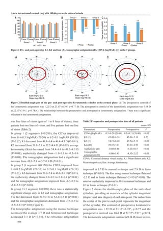
(P=0.02), K1 decreased from 48.8±6.4 to 46.4±4.3 D (P<0.01),
K2 decreased from 59.1±7.1 to 52.2±4.4 D (P=0.05), average
keratometry (Km) decreased from 54.4±6.6 to 49.3±4.3 D
(P<0.01), asphericity changed from -1.1±0.6 to -0.5±0.6
(P<0.01). The tomographic astigmatism had a significant
decrease from -10.3±2.9 to -5.7±1.8 D (P<0.01).
In group 2 (1 segment 140/150) the CDVA improved from
0.4±0.2 logMAR (20/50) to 0.2±0.1 logMAR (20/30)
(P<0.01), K2 decreased from 50.0±7.4 to 46.6±3.6 D (P=0.01),
the asphericity changed from 0.0±0.3 to 0.1±0.4 (P=0.01)
and the tomographic astigmatism improved from -6.3±2.9 to
-3.8±2.2 D (P<0.01).
In group 3 (1 segment 140/200) there was a statistically
significant change only in K2 and tomographic astigmatism.
The K2 decreased from 54.9±7.6 to 52.2±6.5 D (P<0.01)
and the tomographic astigmatism decreased from -7.5±3.9 to
-3.7±2.2 D (P<0.01; Figure 1).
The tomographic astigmatism using the manual technique
decreased the average 3.7 D and femtosecond technique
decreased 3.1 D (P>0.01). The refractive astigmatism
improved in 1.7 D in manual technique and 2.9 D in fento
technique (P>0.01). The Km using manual technique flattened
-2.5 D and in femto technique flattened -2.4 D (P>0.01). The
anterior asphericity improved in 0.4 in manual technique and
0.3 in femto technique (P>0.01).
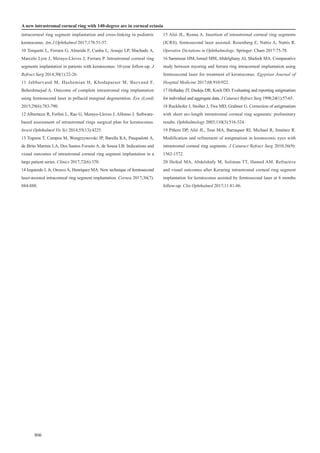
Figure 2 shows the double-angle plots of the individual
cylinders, providing an overview of the cylinder magnitude
(diopter) and axis (degree) of each data point. The radius from
the center of the plot to each point represents the magnitude
of the cylinder. The centroid of preoperative keratometric
astigmatism was 1.22 D at 23.17°±6.39°, ρ=0.77 and the
postoperative centroid was 0.68 D at 22.37°±3.91°, ρ=0.74.
The keratometric astigmatism centroid is 0.54 D closer to zero,
Table 2 Preoperative and postoperative data of all patients
mean±SD
Parameters Preoperative Postoperative P
CDVA (logMAR) 0.5±0.20 (20/60) 0.3±0.21 (20/40) <0.01
K1 (D) 45.49±6.38 45.14±5.10 0.35
K2 (D) 54.19±8.40 49.54±5.11 <0.01
Km (D) 49.87±7.01 47.34±4.90 <0.01
Asphericity (D) -0.60±0.86 -0.23±0.67 <0.01
Tomographic
astigmatism (D)
-8.00±3.45 -4.53±2.52 <0.01
CDVA: Corrected distance visual acuity; K1: Mean flattest axis; K2:
Mean steepest axis; Km: Average keratometry.
Figure 1 Pre- and post-operative K1, K2 and Km (A), tomographic astigmatism (B), CDVA (logMAR) (C) in the 3 groups.
Figure 2 Doubled-angle plot of the pre- and post-operative keratometric cylinder at the corneal plane A: The preoperative centroid of
the keratometric astigmatism was 1.22 D at 23.17°±6.39°, ρ=0.77; B: The postoperative centroid of the keratometric astigmatism was 0.68 D
at 22.37°±3.91°, ρ=0.74; C: The relationship between the preoperative and postoperative keratometric astigmatism. There was a significant
reduction in the keratometric astigmatism.
A new intrastromal corneal ring with 140-degree arc in corneal ectasia
Int J Ophthalmol, Vol. 11, No. 5, May 18, 2018 www.ijo.cn
Tel:8629-82245172 8629-82210956 Email:ijopress@163.com
805
and the standard deviation of the astigmatism was reduced by
a factor of 1.63 (6.39 D/3.91 D). The relocation of the centroid
closer to the origin and the contraction of the ellipse on the
doubled-angle plots demonstrate the amount of improvement.
DISCUSSION
The analysis of our results revealed that the implantation of
140-ICRS was efficacious and improved the visual acuity in
most patients. Postoperatively, there was a significant decrease
in Km, tomographic astigmatism and improvement of CDVA.
In group 1 (2 segments 140/200) we observed a significant
flattening effect, a marked improvement in asphericity
and a substantial reduction of tomographic astigmatism; it
seems to be a good option for cases of symmetrical cones
with hyperprolate cornea, high keratometry and very high
astigmatism. In group 2 (1 segment 140/150) and group 3
(1 segment 140/200), a significant reduction in astigmatism
was observed. There was little change in asphericity and
keratometry. This was in agreement with the effect needed
in some cases of asymmetric cones with high astigmatism,
corneal asphericity close to normal values and curvatures not
significantly steep.
A study realized by Ruckhofer et al[18]
about correction of
astigmatism with short arch segments showed a significant
correction of low to moderate compound myopic astigmatism
safely and predictably. However, the study evaluated healthy
astigmatic corneas, not eyes with keratoconus. In our study,
we analyzed corneas with ectasia and demonstrated proper
correction of tomographic astigmatism with short arc length
(140°) ICRS. These segments may provide a useful alternative
for the surgical correction of astigmatism in corneal ectasia.
Many studies have confirmed the efficacy and safety of
the ICRS in reducing sphero cylindrical error and corneal
steepening in keratoconus over the short and long term[3-4]
, but
most do not analyze astigmatism as a vector. They evaluate
changes in the magnitude of astigmatism only. However, it is
important to investigate variations in the axis of the cylinder
and to determine whether the astigmatic correction was
induced in the targeted meridian. Errors in the correction of
the astigmatic axis could induce aberrations and lead to poor
predictability of the sphero cylindrical correction.
Our study demonstrated changes in astigmatism after ICRS
implantation in patients with keratoconus using vectorial
analysis[19]
and showed that despite the reduction of the
magnitude of corneal astigmatism, not always the corrected
meridian was the planned, with a tendency for undercorrection,
especially in corneas with higher astigmatism. The importance
of the vector analysis of astigmatism is to avoid incorrect or
incomplete conclusions.
The use of femtosecond laser as a safe and accurate method
for the creation of the intrastromal tunnel was recently
proposed[14-15]
. Studies comparing the manual technique with
femtosecond technique found that there was no differences
between the improve in visual acuity and decrese in keratectomy
comparing both techniques, but the incidence of peroperative
complications is less in femtosecond technique[15]
. Thus, the
use of femtosecond laser may not provide better outcomes,
but rather an easier and more reproducible technique for the
surgeon. In this study, we did not find significant improvements
in the results when comparing the manual technique with
the femtosecond laser technique, considering an experienced
surgeon.
In conclusion, there was a significant improvement of all
parameters analyzed. The short arc intrastromal corneal ring
segments seem to be a valuable treatment, which can provide
satisfactory visual outcomes[20]
. A few potential limitations
were apparent in this study as the small sample of treated eyes
and the lack of a comparative group. Future larger, comparative
studies are needed to confirm the results found in this study.
ACKNOWLEDGEMENTS
Conflicts of Interest: Sandes J, None; Stival LRS, None;
Ávila MP, None; Ferrara P, Comercial interest in Ferrara
ICRS; Ferrara G, Comercial interest in Ferrara ICRS; Magacho
L, None; Araújo LPN, None; Torquetti L, None.
REFERENCES
1 Vega-Estrada A, Alio JL, Brenner LF, Javaloy J, Plaza Puche AB,
Barraquer RI, Teus MA, Murta J, Henriques J, Uceda-Montanes A.
Outcome analysis of intracorneal ring segments for the treatment of
keratoconus based on visual, refractive, and aberrometric impairment. Am
J Ophthalmol 2013;155(3):575-584.e1.
2 Alio JL, Vega-Estrada A, Esperanza S, Barraquer RI, Teus MA, Murta
J. Intrastromal corneal ring segments: how successful is the surgical
treatment of keratoconus? Middle East Afr J Ophthalmol 2014;21(1):3-9.
3 Vega-Estrada A, Alio JL. The use of intracorneal ring segments in
keratoconus. Eye Vis (Lond) 2016;3:8.
4 Giacomin NT, Mello GR, Medeiros CS, Kiliç A, Serpe CC, Almeida
HG, Kara-Junior N, Santhiago MR. Intracorneal ring segments
implantation for corneal ectasia. J Refract Surg 2016;32(12):829-839.
5 Kılıç A, del Barrio JLA, Estrada AV. Intracorneal ring segments: types,
indications and outcomes. In: Alió J. (eds) Keratoconus: Essentials in
Ophthalmology. Springer:Cham 2017:195-208.
6 Chan K, Hersh PS. Removal and repositioning of intracorneal ring
segments: improving corneal topography and clinical outcomes in
keratoconus and ectasia. Cornea 2017;36(2):244-248.
7 Lyra JM, Lyra D, Ribeiro G, Torquetti L, Ferrara P, Machado A.
Tomographic findings after implantation of ferrara intrastromal corneal
ring segments in keratoconus. J Refract Surg 2017;33(2):110-115.
8 Barraquer JI. Modification of refraction by means of intracorneal
inclusions. Int Ophthalmol Clin 1966;6(1):53-78.
9 Abdelmassih Y, El-Khoury S, Dirani A, Antonios R, Fadlallah A,
Cherfan CG, Chelala E, Jarade EF. Safety and efficacy of sequential
806
intracorneal ring segment implantation and cross-linking in pediatric
keratoconus. Am J Ophthalmol 2017;178:51-57.
10 Torquetti L, Ferrara G, Almeida F, Cunha L, Araujo LP, Machado A,
Marcelo Lyra J, Merayo-Lloves J, Ferrara P. Intrastromal corneal ring
segments implantation in patients with keratoconus: 10-year follow-up. J
Refract Surg 2014;30(1):22-26.
11 Jabbarvand M, Hashemian H, Khodaparast M, Bazvand F,
Beheshtnejad A. Outcome of complete intrastromal ring implantation
using femtosecond laser in pellucid marginal degeneration. Eye (Lond)
2015;29(6):783-790.
12 Albertazzi R, Ferlini L, Rao G, Merayo-Lloves J, Alfonso J. Software-
based assessment of intrastromal rings surgical plan for keratoconus.
Invest Ophthalmol Vis Sci 2014;55(13):4225.
13 Tognon T, Campos M, Wengrzynovski JP, Barella KA, Pasqualotti A,
de Brito Martins LA, Dos Santos Forseto A, de Sousa LB. Indications and
visual outcomes of intrastromal corneal ring segment implantation in a
large patient series. Clinics 2017;72(6):370.
14 Izquierdo L Jr, Orozco A, Henriquez MA. New technique of femtosecond
laser-assisted intracorneal ring segment implantation. Cornea 2017;36(7):
884-888.
15 Alió JL, Renna A. Insertion of intrastromal corneal ring segments
(ICRS), femtosecond laser assisted. Rosenberg E, Nattis A, Nattis R.
Operative Dictations in Ophthalmology. Springer: Cham 2017:75-78.
16 Sammour HM, Ismail MM, Abdelghany AI, Shafeek MA. Comparative
study between myoring and ferrara ring intracorneal implantation using
femtosecond laser for treatment of keratoconus. Egyptian Journal of
Hospital Medicine 2017;68:910-922.
17 Holladay JT, Dudeja DR, Koch DD. Evaluating and reporting astigmatism
for individual and aggregate data. J Cataract Refract Surg 1998;24(1):57-65.
18 Ruckhofer J, Stoiber J, Twa MD, Grabner G. Correction of astigmatism
with short arc-length intrastromal corneal ring segments: preliminary
results. Ophthalmology 2003;110(3):516-524.
19 Piñero DP, Alió JL, Teus MA, Barraquer RI, Michael R, Jiménez R.
Modification and refinement of astigmatism in keratoconic eyes with
intrastromal corneal ring segments. J Cataract Refract Surg 2010;36(9):
1562-1572.
20 Heikal MA, Abdelshafy M, Soliman TT, Hamed AM. Refractive
and visual outcomes after Keraring intrastromal corneal ring segment
implantation for keratoconus assisted by femtosecond laser at 6 months
follow-up. Clin Ophthalmol 2017;11:81-86.
A new intrastromal corneal ring with 140-degree arc in corneal ectasia

More Related Content

What's hot

Asphericity in ring selection
Asphericity in ring selectionAsphericity in ring selection
Asphericity in ring selection
Ferrara Ophthalmics
 
12 large case series
12 large case series12 large case series
12 large case series
Ferrara Ophthalmics
 
09 long term-follow-up
09 long term-follow-up09 long term-follow-up
09 long term-follow-up
Ferrara Ophthalmics
 
09 clinical outcomes _ 210º
09 clinical outcomes _ 210º09 clinical outcomes _ 210º
09 clinical outcomes _ 210º
Ferrara Ophthalmics
 
Ferrara ICRS in Mild Keratoconus
Ferrara ICRS in Mild KeratoconusFerrara ICRS in Mild Keratoconus
Ferrara ICRS in Mild Keratoconus
Ferrara Ophthalmics
 
12 wavefront aberrations
12 wavefront aberrations12 wavefront aberrations
12 wavefront aberrations
Ferrara Ophthalmics
 
Comparative Study of Visual Outcome between Femtosecond Lasik with Excimer La...
Comparative Study of Visual Outcome between Femtosecond Lasik with Excimer La...Comparative Study of Visual Outcome between Femtosecond Lasik with Excimer La...
Comparative Study of Visual Outcome between Femtosecond Lasik with Excimer La...
iosrjce
 
Anel de Ferrara em Crianças
Anel de Ferrara em Crianças Anel de Ferrara em Crianças
Anel de Ferrara em Crianças
Ferrara Ophthalmics
 
Multimodal evaluation of macular function in age related macular
Multimodal evaluation of macular function in age related macularMultimodal evaluation of macular function in age related macular
Multimodal evaluation of macular function in age related macular
Abdallah Ellabban
 
A DISSERTATION ON “EVALUATION OF ANTERIOR SEGMENT IMAGING TECHNIQUES IN DIAG...
A DISSERTATION ON  “EVALUATION OF ANTERIOR SEGMENT IMAGING TECHNIQUES IN DIAG...A DISSERTATION ON  “EVALUATION OF ANTERIOR SEGMENT IMAGING TECHNIQUES IN DIAG...
A DISSERTATION ON “EVALUATION OF ANTERIOR SEGMENT IMAGING TECHNIQUES IN DIAG...
Shree Bharatimaiya College of Optometry & Physiotherapy
 
DENTAL ARMAMENTARIUMS AND ITS EFFECT ON CARDIAC ARMAMENTARIUMS AND ITS FUNCTI...
DENTAL ARMAMENTARIUMS AND ITS EFFECT ON CARDIAC ARMAMENTARIUMS AND ITS FUNCTI...DENTAL ARMAMENTARIUMS AND ITS EFFECT ON CARDIAC ARMAMENTARIUMS AND ITS FUNCTI...
DENTAL ARMAMENTARIUMS AND ITS EFFECT ON CARDIAC ARMAMENTARIUMS AND ITS FUNCTI...
DrHeena tiwari
 
12 predictors of clinical outcomes
12 predictors of clinical outcomes12 predictors of clinical outcomes
12 predictors of clinical outcomes
Ferrara Ophthalmics
 
The Journal of Implant & Advanced Clinical Dentistry - Oct. 2015
The Journal of Implant & Advanced Clinical Dentistry - Oct. 2015The Journal of Implant & Advanced Clinical Dentistry - Oct. 2015
The Journal of Implant & Advanced Clinical Dentistry - Oct. 2015
Jay True
 
Evaluation Of Pachymetry,Progression Index And Back Difference Elevation Valu...
Evaluation Of Pachymetry,Progression Index And Back Difference Elevation Valu...Evaluation Of Pachymetry,Progression Index And Back Difference Elevation Valu...
Evaluation Of Pachymetry,Progression Index And Back Difference Elevation Valu...
Dr. Jagannath Boramani
 
"Crown Inclination Achieved With A Pre Adjusted Edgewise Appliance Using A Ro...
"Crown Inclination Achieved With A Pre Adjusted Edgewise Appliance Using A Ro..."Crown Inclination Achieved With A Pre Adjusted Edgewise Appliance Using A Ro...
"Crown Inclination Achieved With A Pre Adjusted Edgewise Appliance Using A Ro...
DrHeena tiwari
 
36th publication todentj - 1st name
36th publication   todentj - 1st name36th publication   todentj - 1st name
36th publication todentj - 1st name
CLOVE Dental OMNI Hospitals Andhra Hospital
 
2013 ghassemi-ohr-rekonstruktion-2
2013 ghassemi-ohr-rekonstruktion-22013 ghassemi-ohr-rekonstruktion-2
2013 ghassemi-ohr-rekonstruktion-2
Klinikum Lippe GmbH
 
12 correlation of anterior segment parameters
12 correlation of anterior segment parameters12 correlation of anterior segment parameters
12 correlation of anterior segment parameters
Ferrara Ophthalmics
 

What's hot (20)

Asphericity in ring selection
Asphericity in ring selectionAsphericity in ring selection
Asphericity in ring selection
 
12 large case series
12 large case series12 large case series
12 large case series
 
09 long term-follow-up
09 long term-follow-up09 long term-follow-up
09 long term-follow-up
 
09 clinical outcomes _ 210º
09 clinical outcomes _ 210º09 clinical outcomes _ 210º
09 clinical outcomes _ 210º
 
Ferrara ICRS in Mild Keratoconus
Ferrara ICRS in Mild KeratoconusFerrara ICRS in Mild Keratoconus
Ferrara ICRS in Mild Keratoconus
 
12 wavefront aberrations
12 wavefront aberrations12 wavefront aberrations
12 wavefront aberrations
 
Comparative Study of Visual Outcome between Femtosecond Lasik with Excimer La...
Comparative Study of Visual Outcome between Femtosecond Lasik with Excimer La...Comparative Study of Visual Outcome between Femtosecond Lasik with Excimer La...
Comparative Study of Visual Outcome between Femtosecond Lasik with Excimer La...
 
Anel de Ferrara em Crianças
Anel de Ferrara em Crianças Anel de Ferrara em Crianças
Anel de Ferrara em Crianças
 
Multimodal evaluation of macular function in age related macular
Multimodal evaluation of macular function in age related macularMultimodal evaluation of macular function in age related macular
Multimodal evaluation of macular function in age related macular
 
A DISSERTATION ON “EVALUATION OF ANTERIOR SEGMENT IMAGING TECHNIQUES IN DIAG...
A DISSERTATION ON  “EVALUATION OF ANTERIOR SEGMENT IMAGING TECHNIQUES IN DIAG...A DISSERTATION ON  “EVALUATION OF ANTERIOR SEGMENT IMAGING TECHNIQUES IN DIAG...
A DISSERTATION ON “EVALUATION OF ANTERIOR SEGMENT IMAGING TECHNIQUES IN DIAG...
 
DENTAL ARMAMENTARIUMS AND ITS EFFECT ON CARDIAC ARMAMENTARIUMS AND ITS FUNCTI...
DENTAL ARMAMENTARIUMS AND ITS EFFECT ON CARDIAC ARMAMENTARIUMS AND ITS FUNCTI...DENTAL ARMAMENTARIUMS AND ITS EFFECT ON CARDIAC ARMAMENTARIUMS AND ITS FUNCTI...
DENTAL ARMAMENTARIUMS AND ITS EFFECT ON CARDIAC ARMAMENTARIUMS AND ITS FUNCTI...
 
12 predictors of clinical outcomes
12 predictors of clinical outcomes12 predictors of clinical outcomes
12 predictors of clinical outcomes
 
The Journal of Implant & Advanced Clinical Dentistry - Oct. 2015
The Journal of Implant & Advanced Clinical Dentistry - Oct. 2015The Journal of Implant & Advanced Clinical Dentistry - Oct. 2015
The Journal of Implant & Advanced Clinical Dentistry - Oct. 2015
 
anshuj artical
anshuj articalanshuj artical
anshuj artical
 
Evaluation Of Pachymetry,Progression Index And Back Difference Elevation Valu...
Evaluation Of Pachymetry,Progression Index And Back Difference Elevation Valu...Evaluation Of Pachymetry,Progression Index And Back Difference Elevation Valu...
Evaluation Of Pachymetry,Progression Index And Back Difference Elevation Valu...
 
Scaphoid
ScaphoidScaphoid
Scaphoid
 
"Crown Inclination Achieved With A Pre Adjusted Edgewise Appliance Using A Ro...
"Crown Inclination Achieved With A Pre Adjusted Edgewise Appliance Using A Ro..."Crown Inclination Achieved With A Pre Adjusted Edgewise Appliance Using A Ro...
"Crown Inclination Achieved With A Pre Adjusted Edgewise Appliance Using A Ro...
 
36th publication todentj - 1st name
36th publication   todentj - 1st name36th publication   todentj - 1st name
36th publication todentj - 1st name
 
2013 ghassemi-ohr-rekonstruktion-2
2013 ghassemi-ohr-rekonstruktion-22013 ghassemi-ohr-rekonstruktion-2
2013 ghassemi-ohr-rekonstruktion-2
 
12 correlation of anterior segment parameters
12 correlation of anterior segment parameters12 correlation of anterior segment parameters
12 correlation of anterior segment parameters
 

Similar to Segmentos de Anel de Ferrara com arco de 140º

12 refractive tomographic biomechanical outcomes
12 refractive tomographic biomechanical outcomes12 refractive tomographic biomechanical outcomes
12 refractive tomographic biomechanical outcomes
Ferrara Ophthalmics
 
FR and the astigmatism after penetrating keratoplasty
FR and the astigmatism after penetrating keratoplastyFR and the astigmatism after penetrating keratoplasty
FR and the astigmatism after penetrating keratoplastyFerrara Ophthalmics
 
EVALUATION OF ANTERIOR SEGMENT IMAGING TECHNIQUES IN DIAGNOSIS OF KERATOCONUS
EVALUATION OF ANTERIOR SEGMENT IMAGING TECHNIQUES IN DIAGNOSIS OF KERATOCONUSEVALUATION OF ANTERIOR SEGMENT IMAGING TECHNIQUES IN DIAGNOSIS OF KERATOCONUS
EVALUATION OF ANTERIOR SEGMENT IMAGING TECHNIQUES IN DIAGNOSIS OF KERATOCONUS
Shree Bharatimaiya College of Optometry & Physiotherapy
 
Dr. Vedat Kaya (MAKALE)
Dr. Vedat Kaya (MAKALE)Dr. Vedat Kaya (MAKALE)
Dr. Vedat Kaya (MAKALE)
Lasermed Tic
 
European_Society_of_Ophthalmology_Europe.pdf
European_Society_of_Ophthalmology_Europe.pdfEuropean_Society_of_Ophthalmology_Europe.pdf
European_Society_of_Ophthalmology_Europe.pdf
gabrielladesalvo
 
A scan biometry
A scan biometryA scan biometry
A scan biometry
Mahantesh B
 
SOLIX Essential Brochure
SOLIX Essential Brochure SOLIX Essential Brochure
SOLIX Essential Brochure
Haag-Streit UK (HS-UK)
 
Pterygoid Implants
Pterygoid ImplantsPterygoid Implants
Pterygoid Implants
Bala Ganesh
 
Diagnosis of Vertical Root Fracture Using Digital Radiography, Helical Comput...
Diagnosis of Vertical Root Fracture Using Digital Radiography, Helical Comput...Diagnosis of Vertical Root Fracture Using Digital Radiography, Helical Comput...
Diagnosis of Vertical Root Fracture Using Digital Radiography, Helical Comput...
iosrjce
 
36th publication todentj - 1st name
36th publication   todentj - 1st name36th publication   todentj - 1st name
36th publication todentj - 1st name
CLOVE Dental OMNI Hospitals Andhra Hospital
 
Biometery
BiometeryBiometery
Biometery
Sheim Elteb
 
Auricular prosthesis.pptx
Auricular prosthesis.pptxAuricular prosthesis.pptx
Auricular prosthesis.pptx
Nishu Priya
 
Ferrara Ring after DALK
Ferrara Ring after DALKFerrara Ring after DALK
Ferrara Ring after DALK
Ferrara Ophthalmics
 
Trueness and precision of 5 intraoral scanners for scanning edentulous and de...
Trueness and precision of 5 intraoral scanners for scanning edentulous and de...Trueness and precision of 5 intraoral scanners for scanning edentulous and de...
Trueness and precision of 5 intraoral scanners for scanning edentulous and de...
Partha Sarathi Adhya
 
EVALUATION OF CORNEAL THICKNESS USING TWO DIFFERENT NON-CONTACT DEVICES IN AD...
EVALUATION OF CORNEAL THICKNESS USING TWO DIFFERENT NON-CONTACT DEVICES IN AD...EVALUATION OF CORNEAL THICKNESS USING TWO DIFFERENT NON-CONTACT DEVICES IN AD...
EVALUATION OF CORNEAL THICKNESS USING TWO DIFFERENT NON-CONTACT DEVICES IN AD...
Clínica Rementería
 
Influence-of-dioptric-power-on-retinal-nerve-fiber-layer-thickness-in-myopic-...
Influence-of-dioptric-power-on-retinal-nerve-fiber-layer-thickness-in-myopic-...Influence-of-dioptric-power-on-retinal-nerve-fiber-layer-thickness-in-myopic-...
Influence-of-dioptric-power-on-retinal-nerve-fiber-layer-thickness-in-myopic-...
Iqra Nehal
 
biometry for ON.ppt
biometry for ON.pptbiometry for ON.ppt
biometry for ON.ppt
mikaelgirum
 

Similar to Segmentos de Anel de Ferrara com arco de 140º (19)

12 refractive tomographic biomechanical outcomes
12 refractive tomographic biomechanical outcomes12 refractive tomographic biomechanical outcomes
12 refractive tomographic biomechanical outcomes
 
FR and the astigmatism after penetrating keratoplasty
FR and the astigmatism after penetrating keratoplastyFR and the astigmatism after penetrating keratoplasty
FR and the astigmatism after penetrating keratoplasty
 
EVALUATION OF ANTERIOR SEGMENT IMAGING TECHNIQUES IN DIAGNOSIS OF KERATOCONUS
EVALUATION OF ANTERIOR SEGMENT IMAGING TECHNIQUES IN DIAGNOSIS OF KERATOCONUSEVALUATION OF ANTERIOR SEGMENT IMAGING TECHNIQUES IN DIAGNOSIS OF KERATOCONUS
EVALUATION OF ANTERIOR SEGMENT IMAGING TECHNIQUES IN DIAGNOSIS OF KERATOCONUS
 
Dr. Vedat Kaya (MAKALE)
Dr. Vedat Kaya (MAKALE)Dr. Vedat Kaya (MAKALE)
Dr. Vedat Kaya (MAKALE)
 
European_Society_of_Ophthalmology_Europe.pdf
European_Society_of_Ophthalmology_Europe.pdfEuropean_Society_of_Ophthalmology_Europe.pdf
European_Society_of_Ophthalmology_Europe.pdf
 
A scan biometry
A scan biometryA scan biometry
A scan biometry
 
SOLIX Essential Brochure
SOLIX Essential Brochure SOLIX Essential Brochure
SOLIX Essential Brochure
 
Pterygoid Implants
Pterygoid ImplantsPterygoid Implants
Pterygoid Implants
 
Diagnosis of Vertical Root Fracture Using Digital Radiography, Helical Comput...
Diagnosis of Vertical Root Fracture Using Digital Radiography, Helical Comput...Diagnosis of Vertical Root Fracture Using Digital Radiography, Helical Comput...
Diagnosis of Vertical Root Fracture Using Digital Radiography, Helical Comput...
 
02 d003 5256 (1)
02 d003 5256 (1)02 d003 5256 (1)
02 d003 5256 (1)
 
36th publication todentj - 1st name
36th publication   todentj - 1st name36th publication   todentj - 1st name
36th publication todentj - 1st name
 
Biometery
BiometeryBiometery
Biometery
 
Auricular prosthesis.pptx
Auricular prosthesis.pptxAuricular prosthesis.pptx
Auricular prosthesis.pptx
 
Ferrara Ring after DALK
Ferrara Ring after DALKFerrara Ring after DALK
Ferrara Ring after DALK
 
Trueness and precision of 5 intraoral scanners for scanning edentulous and de...
Trueness and precision of 5 intraoral scanners for scanning edentulous and de...Trueness and precision of 5 intraoral scanners for scanning edentulous and de...
Trueness and precision of 5 intraoral scanners for scanning edentulous and de...
 
EVALUATION OF CORNEAL THICKNESS USING TWO DIFFERENT NON-CONTACT DEVICES IN AD...
EVALUATION OF CORNEAL THICKNESS USING TWO DIFFERENT NON-CONTACT DEVICES IN AD...EVALUATION OF CORNEAL THICKNESS USING TWO DIFFERENT NON-CONTACT DEVICES IN AD...
EVALUATION OF CORNEAL THICKNESS USING TWO DIFFERENT NON-CONTACT DEVICES IN AD...
 
Iej12076
Iej12076Iej12076
Iej12076
 
Influence-of-dioptric-power-on-retinal-nerve-fiber-layer-thickness-in-myopic-...
Influence-of-dioptric-power-on-retinal-nerve-fiber-layer-thickness-in-myopic-...Influence-of-dioptric-power-on-retinal-nerve-fiber-layer-thickness-in-myopic-...
Influence-of-dioptric-power-on-retinal-nerve-fiber-layer-thickness-in-myopic-...
 
biometry for ON.ppt
biometry for ON.pptbiometry for ON.ppt
biometry for ON.ppt
 

More from Ferrara Ophthalmics

Double Rings - New approachs to fine tune clinical outcomes.pdf
Double Rings - New approachs to fine tune clinical outcomes.pdfDouble Rings - New approachs to fine tune clinical outcomes.pdf
Double Rings - New approachs to fine tune clinical outcomes.pdf
Ferrara Ophthalmics
 
ICRS as a Refractive Tool.pdf
ICRS as a Refractive Tool.pdfICRS as a Refractive Tool.pdf
ICRS as a Refractive Tool.pdf
Ferrara Ophthalmics
 
Anel HM é capa da Oftalmologia em Foco.pdf
Anel HM é capa da Oftalmologia em Foco.pdfAnel HM é capa da Oftalmologia em Foco.pdf
Anel HM é capa da Oftalmologia em Foco.pdf
Ferrara Ophthalmics
 
apostila-refrativa-rio-ingles-final-pdf-revisado.pdf
apostila-refrativa-rio-ingles-final-pdf-revisado.pdfapostila-refrativa-rio-ingles-final-pdf-revisado.pdf
apostila-refrativa-rio-ingles-final-pdf-revisado.pdf
Ferrara Ophthalmics
 
apostila-refrativa-rio-final-pdf-revisado.pdf
apostila-refrativa-rio-final-pdf-revisado.pdfapostila-refrativa-rio-final-pdf-revisado.pdf
apostila-refrativa-rio-final-pdf-revisado.pdf
Ferrara Ophthalmics
 
Como planejo minha cirurgia simposio ferrara
Como planejo minha cirurgia   simposio ferraraComo planejo minha cirurgia   simposio ferrara
Como planejo minha cirurgia simposio ferrara
Ferrara Ophthalmics
 
Ferrara Ring - 30yo - 1998 - ceratocone
Ferrara Ring - 30yo - 1998 - ceratoconeFerrara Ring - 30yo - 1998 - ceratocone
Ferrara Ring - 30yo - 1998 - ceratocone
Ferrara Ophthalmics
 
O Filtro Amarelo
O Filtro AmareloO Filtro Amarelo
O Filtro Amarelo
Ferrara Ophthalmics
 
30yo Ferrara Ring
30yo Ferrara Ring30yo Ferrara Ring
30yo Ferrara Ring
Ferrara Ophthalmics
 
Ferrara ring review (2017)
Ferrara ring review (2017)Ferrara ring review (2017)
Ferrara ring review (2017)
Ferrara Ophthalmics
 
Book Chapter
Book ChapterBook Chapter
Book Chapter
Ferrara Ophthalmics
 
Mecanismo de ação e nomograma
Mecanismo de ação e nomogramaMecanismo de ação e nomograma
Mecanismo de ação e nomograma
Ferrara Ophthalmics
 
Ferrara Ring 320º
Ferrara Ring 320ºFerrara Ring 320º
Ferrara Ring 320º
Ferrara Ophthalmics
 
11 DALK
11 DALK11 DALK

More from Ferrara Ophthalmics (14)

Double Rings - New approachs to fine tune clinical outcomes.pdf
Double Rings - New approachs to fine tune clinical outcomes.pdfDouble Rings - New approachs to fine tune clinical outcomes.pdf
Double Rings - New approachs to fine tune clinical outcomes.pdf
 
ICRS as a Refractive Tool.pdf
ICRS as a Refractive Tool.pdfICRS as a Refractive Tool.pdf
ICRS as a Refractive Tool.pdf
 
Anel HM é capa da Oftalmologia em Foco.pdf
Anel HM é capa da Oftalmologia em Foco.pdfAnel HM é capa da Oftalmologia em Foco.pdf
Anel HM é capa da Oftalmologia em Foco.pdf
 
apostila-refrativa-rio-ingles-final-pdf-revisado.pdf
apostila-refrativa-rio-ingles-final-pdf-revisado.pdfapostila-refrativa-rio-ingles-final-pdf-revisado.pdf
apostila-refrativa-rio-ingles-final-pdf-revisado.pdf
 
apostila-refrativa-rio-final-pdf-revisado.pdf
apostila-refrativa-rio-final-pdf-revisado.pdfapostila-refrativa-rio-final-pdf-revisado.pdf
apostila-refrativa-rio-final-pdf-revisado.pdf
 
Como planejo minha cirurgia simposio ferrara
Como planejo minha cirurgia   simposio ferraraComo planejo minha cirurgia   simposio ferrara
Como planejo minha cirurgia simposio ferrara
 
Ferrara Ring - 30yo - 1998 - ceratocone
Ferrara Ring - 30yo - 1998 - ceratoconeFerrara Ring - 30yo - 1998 - ceratocone
Ferrara Ring - 30yo - 1998 - ceratocone
 
O Filtro Amarelo
O Filtro AmareloO Filtro Amarelo
O Filtro Amarelo
 
30yo Ferrara Ring
30yo Ferrara Ring30yo Ferrara Ring
30yo Ferrara Ring
 
Ferrara ring review (2017)
Ferrara ring review (2017)Ferrara ring review (2017)
Ferrara ring review (2017)
 
Book Chapter
Book ChapterBook Chapter
Book Chapter
 
Mecanismo de ação e nomograma
Mecanismo de ação e nomogramaMecanismo de ação e nomograma
Mecanismo de ação e nomograma
 
Ferrara Ring 320º
Ferrara Ring 320ºFerrara Ring 320º
Ferrara Ring 320º
 
11 DALK
11 DALK11 DALK
11 DALK
 

Recently uploaded

Lung Cancer: Artificial Intelligence, Synergetics, Complex System Analysis, S...
Lung Cancer: Artificial Intelligence, Synergetics, Complex System Analysis, S...Lung Cancer: Artificial Intelligence, Synergetics, Complex System Analysis, S...
Lung Cancer: Artificial Intelligence, Synergetics, Complex System Analysis, S...
Oleg Kshivets
 
ARTIFICIAL INTELLIGENCE IN HEALTHCARE.pdf
ARTIFICIAL INTELLIGENCE IN  HEALTHCARE.pdfARTIFICIAL INTELLIGENCE IN  HEALTHCARE.pdf
ARTIFICIAL INTELLIGENCE IN HEALTHCARE.pdf
Anujkumaranit
 
Physiology of Special Chemical Sensation of Taste
Physiology of Special Chemical Sensation of TastePhysiology of Special Chemical Sensation of Taste
Physiology of Special Chemical Sensation of Taste
MedicoseAcademics
 
Ocular injury ppt Upendra pal optometrist upums saifai etawah
Ocular injury  ppt  Upendra pal  optometrist upums saifai etawahOcular injury  ppt  Upendra pal  optometrist upums saifai etawah
Ocular injury ppt Upendra pal optometrist upums saifai etawah
pal078100
 
Alcohol_Dr. Jeenal Mistry MD Pharmacology.pdf
Alcohol_Dr. Jeenal Mistry MD Pharmacology.pdfAlcohol_Dr. Jeenal Mistry MD Pharmacology.pdf
Alcohol_Dr. Jeenal Mistry MD Pharmacology.pdf
Dr Jeenal Mistry
 
Hemodialysis: Chapter 3, Dialysis Water Unit - Dr.Gawad
Hemodialysis: Chapter 3, Dialysis Water Unit - Dr.GawadHemodialysis: Chapter 3, Dialysis Water Unit - Dr.Gawad
Hemodialysis: Chapter 3, Dialysis Water Unit - Dr.Gawad
NephroTube - Dr.Gawad
 
Triangles of Neck and Clinical Correlation by Dr. RIG.pptx
Triangles of Neck and Clinical Correlation by Dr. RIG.pptxTriangles of Neck and Clinical Correlation by Dr. RIG.pptx
Triangles of Neck and Clinical Correlation by Dr. RIG.pptx
Dr. Rabia Inam Gandapore
 
Report Back from SGO 2024: What’s the Latest in Cervical Cancer?
Report Back from SGO 2024: What’s the Latest in Cervical Cancer?Report Back from SGO 2024: What’s the Latest in Cervical Cancer?
Report Back from SGO 2024: What’s the Latest in Cervical Cancer?
bkling
 
TEST BANK for Operations Management, 14th Edition by William J. Stevenson, Ve...
TEST BANK for Operations Management, 14th Edition by William J. Stevenson, Ve...TEST BANK for Operations Management, 14th Edition by William J. Stevenson, Ve...
TEST BANK for Operations Management, 14th Edition by William J. Stevenson, Ve...
kevinkariuki227
 
ARTHROLOGY PPT NCISM SYLLABUS AYURVEDA STUDENTS
ARTHROLOGY PPT NCISM SYLLABUS AYURVEDA STUDENTSARTHROLOGY PPT NCISM SYLLABUS AYURVEDA STUDENTS
ARTHROLOGY PPT NCISM SYLLABUS AYURVEDA STUDENTS
Dr. Vinay Pareek
 
ANATOMY AND PHYSIOLOGY OF URINARY SYSTEM.pptx
ANATOMY AND PHYSIOLOGY OF URINARY SYSTEM.pptxANATOMY AND PHYSIOLOGY OF URINARY SYSTEM.pptx
ANATOMY AND PHYSIOLOGY OF URINARY SYSTEM.pptx
Swetaba Besh
 
Physiology of Chemical Sensation of smell.pdf
Physiology of Chemical Sensation of smell.pdfPhysiology of Chemical Sensation of smell.pdf
Physiology of Chemical Sensation of smell.pdf
MedicoseAcademics
 
Cervical & Brachial Plexus By Dr. RIG.pptx
Cervical & Brachial Plexus By Dr. RIG.pptxCervical & Brachial Plexus By Dr. RIG.pptx
Cervical & Brachial Plexus By Dr. RIG.pptx
Dr. Rabia Inam Gandapore
 
Maxilla, Mandible & Hyoid Bone & Clinical Correlations by Dr. RIG.pptx
Maxilla, Mandible & Hyoid Bone & Clinical Correlations by Dr. RIG.pptxMaxilla, Mandible & Hyoid Bone & Clinical Correlations by Dr. RIG.pptx
Maxilla, Mandible & Hyoid Bone & Clinical Correlations by Dr. RIG.pptx
Dr. Rabia Inam Gandapore
 
Surat @ℂall @Girls ꧁❤8527049040❤꧂@ℂall @Girls Service Vip Top Model Safe
Surat @ℂall @Girls ꧁❤8527049040❤꧂@ℂall @Girls Service Vip Top Model SafeSurat @ℂall @Girls ꧁❤8527049040❤꧂@ℂall @Girls Service Vip Top Model Safe
Surat @ℂall @Girls ꧁❤8527049040❤꧂@ℂall @Girls Service Vip Top Model Safe
Savita Shen $i11
 
Prix Galien International 2024 Forum Program
Prix Galien International 2024 Forum ProgramPrix Galien International 2024 Forum Program
Prix Galien International 2024 Forum Program
Levi Shapiro
 
Non-respiratory Functions of the Lungs.pdf
Non-respiratory Functions of the Lungs.pdfNon-respiratory Functions of the Lungs.pdf
Non-respiratory Functions of the Lungs.pdf
MedicoseAcademics
 
Surgical Site Infections, pathophysiology, and prevention.pptx
Surgical Site Infections, pathophysiology, and prevention.pptxSurgical Site Infections, pathophysiology, and prevention.pptx
Surgical Site Infections, pathophysiology, and prevention.pptx
jval Landero
 
KDIGO 2024 guidelines for diabetologists
KDIGO 2024 guidelines for diabetologistsKDIGO 2024 guidelines for diabetologists
KDIGO 2024 guidelines for diabetologists
د.محمود نجيب
 
Charaka Samhita Sutra sthana Chapter 15 Upakalpaniyaadhyaya
Charaka Samhita Sutra sthana Chapter 15 UpakalpaniyaadhyayaCharaka Samhita Sutra sthana Chapter 15 Upakalpaniyaadhyaya
Charaka Samhita Sutra sthana Chapter 15 Upakalpaniyaadhyaya
Dr KHALID B.M
 

Recently uploaded (20)

Lung Cancer: Artificial Intelligence, Synergetics, Complex System Analysis, S...
Lung Cancer: Artificial Intelligence, Synergetics, Complex System Analysis, S...Lung Cancer: Artificial Intelligence, Synergetics, Complex System Analysis, S...
Lung Cancer: Artificial Intelligence, Synergetics, Complex System Analysis, S...
 
ARTIFICIAL INTELLIGENCE IN HEALTHCARE.pdf
ARTIFICIAL INTELLIGENCE IN  HEALTHCARE.pdfARTIFICIAL INTELLIGENCE IN  HEALTHCARE.pdf
ARTIFICIAL INTELLIGENCE IN HEALTHCARE.pdf
 
Physiology of Special Chemical Sensation of Taste
Physiology of Special Chemical Sensation of TastePhysiology of Special Chemical Sensation of Taste
Physiology of Special Chemical Sensation of Taste
 
Ocular injury ppt Upendra pal optometrist upums saifai etawah
Ocular injury  ppt  Upendra pal  optometrist upums saifai etawahOcular injury  ppt  Upendra pal  optometrist upums saifai etawah
Ocular injury ppt Upendra pal optometrist upums saifai etawah
 
Alcohol_Dr. Jeenal Mistry MD Pharmacology.pdf
Alcohol_Dr. Jeenal Mistry MD Pharmacology.pdfAlcohol_Dr. Jeenal Mistry MD Pharmacology.pdf
Alcohol_Dr. Jeenal Mistry MD Pharmacology.pdf
 
Hemodialysis: Chapter 3, Dialysis Water Unit - Dr.Gawad
Hemodialysis: Chapter 3, Dialysis Water Unit - Dr.GawadHemodialysis: Chapter 3, Dialysis Water Unit - Dr.Gawad
Hemodialysis: Chapter 3, Dialysis Water Unit - Dr.Gawad
 
Triangles of Neck and Clinical Correlation by Dr. RIG.pptx
Triangles of Neck and Clinical Correlation by Dr. RIG.pptxTriangles of Neck and Clinical Correlation by Dr. RIG.pptx
Triangles of Neck and Clinical Correlation by Dr. RIG.pptx
 
Report Back from SGO 2024: What’s the Latest in Cervical Cancer?
Report Back from SGO 2024: What’s the Latest in Cervical Cancer?Report Back from SGO 2024: What’s the Latest in Cervical Cancer?
Report Back from SGO 2024: What’s the Latest in Cervical Cancer?
 
TEST BANK for Operations Management, 14th Edition by William J. Stevenson, Ve...
TEST BANK for Operations Management, 14th Edition by William J. Stevenson, Ve...TEST BANK for Operations Management, 14th Edition by William J. Stevenson, Ve...
TEST BANK for Operations Management, 14th Edition by William J. Stevenson, Ve...
 
ARTHROLOGY PPT NCISM SYLLABUS AYURVEDA STUDENTS
ARTHROLOGY PPT NCISM SYLLABUS AYURVEDA STUDENTSARTHROLOGY PPT NCISM SYLLABUS AYURVEDA STUDENTS
ARTHROLOGY PPT NCISM SYLLABUS AYURVEDA STUDENTS
 
ANATOMY AND PHYSIOLOGY OF URINARY SYSTEM.pptx
ANATOMY AND PHYSIOLOGY OF URINARY SYSTEM.pptxANATOMY AND PHYSIOLOGY OF URINARY SYSTEM.pptx
ANATOMY AND PHYSIOLOGY OF URINARY SYSTEM.pptx
 
Physiology of Chemical Sensation of smell.pdf
Physiology of Chemical Sensation of smell.pdfPhysiology of Chemical Sensation of smell.pdf
Physiology of Chemical Sensation of smell.pdf
 
Cervical & Brachial Plexus By Dr. RIG.pptx
Cervical & Brachial Plexus By Dr. RIG.pptxCervical & Brachial Plexus By Dr. RIG.pptx
Cervical & Brachial Plexus By Dr. RIG.pptx
 
Maxilla, Mandible & Hyoid Bone & Clinical Correlations by Dr. RIG.pptx
Maxilla, Mandible & Hyoid Bone & Clinical Correlations by Dr. RIG.pptxMaxilla, Mandible & Hyoid Bone & Clinical Correlations by Dr. RIG.pptx
Maxilla, Mandible & Hyoid Bone & Clinical Correlations by Dr. RIG.pptx
 
Surat @ℂall @Girls ꧁❤8527049040❤꧂@ℂall @Girls Service Vip Top Model Safe
Surat @ℂall @Girls ꧁❤8527049040❤꧂@ℂall @Girls Service Vip Top Model SafeSurat @ℂall @Girls ꧁❤8527049040❤꧂@ℂall @Girls Service Vip Top Model Safe
Surat @ℂall @Girls ꧁❤8527049040❤꧂@ℂall @Girls Service Vip Top Model Safe
 
Prix Galien International 2024 Forum Program
Prix Galien International 2024 Forum ProgramPrix Galien International 2024 Forum Program
Prix Galien International 2024 Forum Program
 
Non-respiratory Functions of the Lungs.pdf
Non-respiratory Functions of the Lungs.pdfNon-respiratory Functions of the Lungs.pdf
Non-respiratory Functions of the Lungs.pdf
 
Surgical Site Infections, pathophysiology, and prevention.pptx
Surgical Site Infections, pathophysiology, and prevention.pptxSurgical Site Infections, pathophysiology, and prevention.pptx
Surgical Site Infections, pathophysiology, and prevention.pptx
 
KDIGO 2024 guidelines for diabetologists
KDIGO 2024 guidelines for diabetologistsKDIGO 2024 guidelines for diabetologists
KDIGO 2024 guidelines for diabetologists
 
Charaka Samhita Sutra sthana Chapter 15 Upakalpaniyaadhyaya
Charaka Samhita Sutra sthana Chapter 15 UpakalpaniyaadhyayaCharaka Samhita Sutra sthana Chapter 15 Upakalpaniyaadhyaya
Charaka Samhita Sutra sthana Chapter 15 Upakalpaniyaadhyaya
 

Segmentos de Anel de Ferrara com arco de 140º

  • 1. 802 ·Clinical Research· Clinical outcomes after implantation of a new intrastromal corneal ring with 140-degree of arc in patients with corneal ectasia Jordana Sandes1 , Larissa R. S. Stival1 , Marcos Pereira de Ávila1 , Paulo Ferrara2 , Guilherme Ferrara2 , Leopoldo Magacho1 , Luana P. N. Araújo3 , Leonardo Torquetti4 1 Center of Reference in Ophthalmology (CEROF), Goiânia 64605-020, Brazil 2 Paulo Ferrara Eye Clinic, Belo Horizonte-MG 30110-921, Brazil 3 Fundação Altino Ventura, Recife-PE 50070-040, Brazil 4 Center of Excellence in Ophthalmology, Pará de Minas 35660-017, Brazil Correspondence to: Jordana Sandes. Street T-15, Sector Bueno, Number 715, Apartment 2601, Goiânia-GO 74230-010, Brazil. jordana.oftalmo@gmail.com Received: 2017-09-08 Accepted: 2017-11-14 Abstract ● AIM: To evaluate the clinical and tomographic outcomes after implantation of a new intrastromal corneal ring segment (ICRS) with 140-degrees of arc in eyes with corneal ectasia. ● METHODS: We evaluated patients with corneal ectasia implanted with Ferrara 140° ICRS from April 2010 to February 2015. Outcome measures included preoperative and postoperative corrected distance visual acuity (CDVA), keratometry simulated (K) reading, tomographic astigmatism and asphericity. All patients were evaluated using the Pentacam Scheimpflug system. ● RESULTS: The study evaluated 58 eyes. The mean follow- up was 16.81±10.8mo. The CDVA (logMAR) improved from 0.5±0.20 (20/60) to 0.3±0.21 (20/40) (P<0.01). The average K reduced from 49.87±7.01 to 47.34±4.90 D (P<0.01). The asphericity changed from -0.60±0.86 to -0.23±0.67 D (P<0.01). The mean preoperative tomographic astigmatism decreased from -8.0±3.45 to -4.53±2.52 D (P<0.01). ● CONCLUSION: The new ICRS model with 140-degrees of arc effectively improve the visual acuity and reduce the high astigmatism usually found in patients with corneal ectasia. ● KEYWORDS: keratoconus; intrastromal corneal ring segment; corneal ectasia DOI:10.18240/ijo.2018.05.14 Citation: Sandes J, Stival LRS, Ávila MP, Ferrara P, Ferrara G, Magacho L, Araújo LPN, Torquetti L. Clinical outcomes after implantation of a new intrastromal corneal ring with 140-degree of arc in patients with corneal ectasia. Int J Ophthalmol 2018;11(5):802-806 INTRODUCTION Intrastromal corneal ring segments (ICRS) are an efficacious alternative in patients with clear corneas who have unsatisfactory corrected visual acuity with glasses or contact lens and contact lens intolerance[1-5] . It acts according to Barraquer’s postulate which states that tissue addition in corneal periphery leads to corneal flattening. The ring diameter (optical zone) determines how much the cornea will be flattened. The thicker and smaller the optical zone of the implanted segment, the more flattening effect and myopic reduction is achieved[6-8] . The main advantages of this procedure are its safety, reversibility, and stability, as well as the fact that the segments do not affect the visual axis[9-10] . There are different models of ICRS with varying sizes and arc thicknesses. Theoretically, a shorter segment induces a greater astigmatic correction and induces lesser asphericity change, comparing with long arch segments. The new model presents a short arc length of 140 degrees (140-ICRS) with the main advantage of astigmatism reduction. The primary indication is in keratoconus patients with high astigmatism[11-12] . The intrastromal tunnel for ring implantation was initially manually constructed; however, complications such as epithelial defects, depth asymmetry and perforation, were reported[13] . Femtosecond laser has recently been used to create the tunnel for ring implantation. This technique reportedly creates a tunnel with precise depth, width and location leading to faster visual recovery and less incidence of surgical complications[14-15] . The main purpose of this study is to evaluate the clinical results of the implantation of 140-ICRS regarding its efficacy and capacity of corneal astigmatism reduction. Moreover we compared the clinical outcomes of patients implanted with the Ferrara 140-ICRS using the manual and femtosecond laser assisted technique. SUBJECTS AND METHODS This retrospective study included keratoconus eyes with high astigmatism that were intolerant to contact lens or disease progression and presented visual acuity worse than 0.2 logMAR (20/30). An informed consent was given to all eligible patients prior to data collection, requesting permission A new intrastromal corneal ring with 140-degree arc in corneal ectasia
  • 2. Int J Ophthalmol, Vol. 11, No. 5, May 18, 2018 www.ijo.cn Tel:8629-82245172 8629-82210956 Email:ijopress@163.com 803 for the research and use of data from their medical records relating to the pre and postoperative periods. All bioethical principles were considered in accordance with the Declaration of Helsinki and Brazilian regulations. The same surgeon (Paulo Ferrara) performed all surgical procedures using the manual or femtosecond laser-assisted technique for ICRS implantation. Both techniques have been widely described in the literature[2,5,13-15] . Patients were randomized to receive a manual or laser-assisted surgical technique. The corneal depth of ICRS were 80% for all cases (manual and laser). In the manual technique, the surgery was performed under topical anesthesia, and the visual axis is marked; a 5-mm optical zone and incision side were aligned with the desired axis in which the incision would be made, in the steepest axis. The diamond blade was set at 80% of corneal depth at the incision site. After the incison, a stromal spreader creates a pocket on each side of the incision. Two semi-circular dissecting spatulas were consecutively inserted through the incision and gently pushed with some quick rotary back and forth movements. Following channel creation, the ring segments were implanted. Using the femtosecond laser, LDV Z4 (Ziemer, Switzerland), tunnel depth is set at 80% of the thinnest corneal thickness within the probable ring track. The channel’s inner diameter is set to 4.4 mm, the outer diameter 5.6 mm and the entry cut thickness was 1 μm (at the steepest topographic axis). The ring energy used for channel creation is 1.3 mJ. The femtosecond laser takes approximately 15s to create the channel. The segments are implanted immediately after channel creation before the disappearing of the bubbles, which reveals the exact tunnel location. The segments were placed in the final position with a Sinskey hook through a dialing hole at both ends of the segment[14-16] . According to the Ferrara Ring nomogram, for 140-ICRS, the selection of the thickness of the segment to be implanted varies with the preoperative tomographic astigmatism. For asymmetric keratoconus a single segment was implanted and for symmetrical keratoconus 2 segments were implanted. Asymmetry means that, by dividing the cornea into two halves from the more curved meridian considering the anterior sagittal map of the pentacam, asymmetric corneas are those that the halves are unequal, and symmetrical corneas are those that the halves are very similar. Descriptive analysis such as age, sex, technique and follow- up was collected for all patients. Statistical analysis included preoperative and postoperative, corrected distance visual acuity (CDVA), refractive astigmatism, tomographic astigmatism, keratometry simulated (K) readings, mean flattest axis (K1), mean steepest axis (K2) and asphericity. Corneal tomography and pachymetry were obtained using the software included within the Pentacam rotation Scheimpflug camera (Oculus Pentacam, Wetzlar, Germany). Statistical analysis was carried out using the Minitab software (Minitab Inc., Chicago, USA). Analysis of Astigmatism The astigmatism results were analyzed arithmetically (nonvector analysis) and using vector analysis when concerning the cylindrical axis. Although observed changes in cylinders were commonly reported, they do not accurately reflect the actual nature of the change in the cylinder. The magnitude and axis of the cylinder are related to the spherical power. The vector analysis used for calculating surgically induced astigmatism change was a Doubled-Angle polar plot. Due to astigmatism traverses an entire cycle in 180 degrees, the doubled-angle polar plot was described as the most appropriate plot for aggregating astigmatism data. In this method, the centroid is the mean of a set of x and y values. The standard deviation can be represented in a graphic by an area surrounding the centroid. The shape of this area will vary depending on the length of the major and minor axis. The shape factor (ρ) has been used to describe this relationship[17] . Statistical Analysis Normality of data was evaluated with the Kolmogorov-Smirnov test. The analysis of primary outcome measures was based on a normal distribution of the data. Student’s t-test for paired variables was used to compare pre and postoperative data considering a significance level of P<0.05. Graphic analysis was made using the Microsoft Excel 2007 (USA) and SPSS Sigma Plot 12.0 (USA). RESULTS Fifty eight eyes/patients were evaluated. The average follow- up was 16.81±10.8mo. The mean age was 33.3±13.2y. Forty six patients (79.3%) were male, and twelve patients (20.7%) were female. Considering the analysis by groups, in group 1 (17 eyes) 2 segments of 200 μm (140/200) were implanted; in group 2 (30 eyes) a single 150 μm segment (140/150) was implanted and in group 3 (11 eyes) a single 200 μm segment (140/200) was implanted (Table 1). Last follow up, 12.2% of patients maintained the same CDVA, 13.7% of patients had the CDVA worsened, and 74.1% of patients had improvement in CDVA. The improved average Table 1 Segment thickness according to preoperative tomographic astigmatism Tomographic astigmatism (D) Segment thickness Asymmetric keratoconus <4.00 1 segment 150 μm >4.00 and <8.00 1 segment 200 μm >8.00 1 segment 250 μm Symmetric keratoconus <6.00 2 segments 150 μm >6.00 and <10.00 2 segments 200 μm >10.00 2 segments 250 μm
  • 3. 804 was four lines of vision (gain of 1 to 9 lines of vision), three patients lost two lines of vision, and five patients lost one line of vision (Table 2). In group 1 (2 segments 140/200), the CDVA improved from 0.4±0.3 logMAR (20/50) to 0.2±0.1 logMAR (20/30) (P=0.02), K1 decreased from 48.8±6.4 to 46.4±4.3 D (P<0.01), K2 decreased from 59.1±7.1 to 52.2±4.4 D (P=0.05), average keratometry (Km) decreased from 54.4±6.6 to 49.3±4.3 D (P<0.01), asphericity changed from -1.1±0.6 to -0.5±0.6 (P<0.01). The tomographic astigmatism had a significant decrease from -10.3±2.9 to -5.7±1.8 D (P<0.01). In group 2 (1 segment 140/150) the CDVA improved from 0.4±0.2 logMAR (20/50) to 0.2±0.1 logMAR (20/30) (P<0.01), K2 decreased from 50.0±7.4 to 46.6±3.6 D (P=0.01), the asphericity changed from 0.0±0.3 to 0.1±0.4 (P=0.01) and the tomographic astigmatism improved from -6.3±2.9 to -3.8±2.2 D (P<0.01). In group 3 (1 segment 140/200) there was a statistically significant change only in K2 and tomographic astigmatism. The K2 decreased from 54.9±7.6 to 52.2±6.5 D (P<0.01) and the tomographic astigmatism decreased from -7.5±3.9 to -3.7±2.2 D (P<0.01; Figure 1). The tomographic astigmatism using the manual technique decreased the average 3.7 D and femtosecond technique decreased 3.1 D (P>0.01). The refractive astigmatism improved in 1.7 D in manual technique and 2.9 D in fento technique (P>0.01). The Km using manual technique flattened -2.5 D and in femto technique flattened -2.4 D (P>0.01). The anterior asphericity improved in 0.4 in manual technique and 0.3 in femto technique (P>0.01). Figure 2 shows the double-angle plots of the individual cylinders, providing an overview of the cylinder magnitude (diopter) and axis (degree) of each data point. The radius from the center of the plot to each point represents the magnitude of the cylinder. The centroid of preoperative keratometric astigmatism was 1.22 D at 23.17°±6.39°, ρ=0.77 and the postoperative centroid was 0.68 D at 22.37°±3.91°, ρ=0.74. The keratometric astigmatism centroid is 0.54 D closer to zero, Table 2 Preoperative and postoperative data of all patients mean±SD Parameters Preoperative Postoperative P CDVA (logMAR) 0.5±0.20 (20/60) 0.3±0.21 (20/40) <0.01 K1 (D) 45.49±6.38 45.14±5.10 0.35 K2 (D) 54.19±8.40 49.54±5.11 <0.01 Km (D) 49.87±7.01 47.34±4.90 <0.01 Asphericity (D) -0.60±0.86 -0.23±0.67 <0.01 Tomographic astigmatism (D) -8.00±3.45 -4.53±2.52 <0.01 CDVA: Corrected distance visual acuity; K1: Mean flattest axis; K2: Mean steepest axis; Km: Average keratometry. Figure 1 Pre- and post-operative K1, K2 and Km (A), tomographic astigmatism (B), CDVA (logMAR) (C) in the 3 groups. Figure 2 Doubled-angle plot of the pre- and post-operative keratometric cylinder at the corneal plane A: The preoperative centroid of the keratometric astigmatism was 1.22 D at 23.17°±6.39°, ρ=0.77; B: The postoperative centroid of the keratometric astigmatism was 0.68 D at 22.37°±3.91°, ρ=0.74; C: The relationship between the preoperative and postoperative keratometric astigmatism. There was a significant reduction in the keratometric astigmatism. A new intrastromal corneal ring with 140-degree arc in corneal ectasia
  • 4. Int J Ophthalmol, Vol. 11, No. 5, May 18, 2018 www.ijo.cn Tel:8629-82245172 8629-82210956 Email:ijopress@163.com 805 and the standard deviation of the astigmatism was reduced by a factor of 1.63 (6.39 D/3.91 D). The relocation of the centroid closer to the origin and the contraction of the ellipse on the doubled-angle plots demonstrate the amount of improvement. DISCUSSION The analysis of our results revealed that the implantation of 140-ICRS was efficacious and improved the visual acuity in most patients. Postoperatively, there was a significant decrease in Km, tomographic astigmatism and improvement of CDVA. In group 1 (2 segments 140/200) we observed a significant flattening effect, a marked improvement in asphericity and a substantial reduction of tomographic astigmatism; it seems to be a good option for cases of symmetrical cones with hyperprolate cornea, high keratometry and very high astigmatism. In group 2 (1 segment 140/150) and group 3 (1 segment 140/200), a significant reduction in astigmatism was observed. There was little change in asphericity and keratometry. This was in agreement with the effect needed in some cases of asymmetric cones with high astigmatism, corneal asphericity close to normal values and curvatures not significantly steep. A study realized by Ruckhofer et al[18] about correction of astigmatism with short arch segments showed a significant correction of low to moderate compound myopic astigmatism safely and predictably. However, the study evaluated healthy astigmatic corneas, not eyes with keratoconus. In our study, we analyzed corneas with ectasia and demonstrated proper correction of tomographic astigmatism with short arc length (140°) ICRS. These segments may provide a useful alternative for the surgical correction of astigmatism in corneal ectasia. Many studies have confirmed the efficacy and safety of the ICRS in reducing sphero cylindrical error and corneal steepening in keratoconus over the short and long term[3-4] , but most do not analyze astigmatism as a vector. They evaluate changes in the magnitude of astigmatism only. However, it is important to investigate variations in the axis of the cylinder and to determine whether the astigmatic correction was induced in the targeted meridian. Errors in the correction of the astigmatic axis could induce aberrations and lead to poor predictability of the sphero cylindrical correction. Our study demonstrated changes in astigmatism after ICRS implantation in patients with keratoconus using vectorial analysis[19] and showed that despite the reduction of the magnitude of corneal astigmatism, not always the corrected meridian was the planned, with a tendency for undercorrection, especially in corneas with higher astigmatism. The importance of the vector analysis of astigmatism is to avoid incorrect or incomplete conclusions. The use of femtosecond laser as a safe and accurate method for the creation of the intrastromal tunnel was recently proposed[14-15] . Studies comparing the manual technique with femtosecond technique found that there was no differences between the improve in visual acuity and decrese in keratectomy comparing both techniques, but the incidence of peroperative complications is less in femtosecond technique[15] . Thus, the use of femtosecond laser may not provide better outcomes, but rather an easier and more reproducible technique for the surgeon. In this study, we did not find significant improvements in the results when comparing the manual technique with the femtosecond laser technique, considering an experienced surgeon. In conclusion, there was a significant improvement of all parameters analyzed. The short arc intrastromal corneal ring segments seem to be a valuable treatment, which can provide satisfactory visual outcomes[20] . A few potential limitations were apparent in this study as the small sample of treated eyes and the lack of a comparative group. Future larger, comparative studies are needed to confirm the results found in this study. ACKNOWLEDGEMENTS Conflicts of Interest: Sandes J, None; Stival LRS, None; Ávila MP, None; Ferrara P, Comercial interest in Ferrara ICRS; Ferrara G, Comercial interest in Ferrara ICRS; Magacho L, None; Araújo LPN, None; Torquetti L, None. REFERENCES 1 Vega-Estrada A, Alio JL, Brenner LF, Javaloy J, Plaza Puche AB, Barraquer RI, Teus MA, Murta J, Henriques J, Uceda-Montanes A. Outcome analysis of intracorneal ring segments for the treatment of keratoconus based on visual, refractive, and aberrometric impairment. Am J Ophthalmol 2013;155(3):575-584.e1. 2 Alio JL, Vega-Estrada A, Esperanza S, Barraquer RI, Teus MA, Murta J. Intrastromal corneal ring segments: how successful is the surgical treatment of keratoconus? Middle East Afr J Ophthalmol 2014;21(1):3-9. 3 Vega-Estrada A, Alio JL. The use of intracorneal ring segments in keratoconus. Eye Vis (Lond) 2016;3:8. 4 Giacomin NT, Mello GR, Medeiros CS, Kiliç A, Serpe CC, Almeida HG, Kara-Junior N, Santhiago MR. Intracorneal ring segments implantation for corneal ectasia. J Refract Surg 2016;32(12):829-839. 5 Kılıç A, del Barrio JLA, Estrada AV. Intracorneal ring segments: types, indications and outcomes. In: Alió J. (eds) Keratoconus: Essentials in Ophthalmology. Springer:Cham 2017:195-208. 6 Chan K, Hersh PS. Removal and repositioning of intracorneal ring segments: improving corneal topography and clinical outcomes in keratoconus and ectasia. Cornea 2017;36(2):244-248. 7 Lyra JM, Lyra D, Ribeiro G, Torquetti L, Ferrara P, Machado A. Tomographic findings after implantation of ferrara intrastromal corneal ring segments in keratoconus. J Refract Surg 2017;33(2):110-115. 8 Barraquer JI. Modification of refraction by means of intracorneal inclusions. Int Ophthalmol Clin 1966;6(1):53-78. 9 Abdelmassih Y, El-Khoury S, Dirani A, Antonios R, Fadlallah A, Cherfan CG, Chelala E, Jarade EF. Safety and efficacy of sequential
  • 5. 806 intracorneal ring segment implantation and cross-linking in pediatric keratoconus. Am J Ophthalmol 2017;178:51-57. 10 Torquetti L, Ferrara G, Almeida F, Cunha L, Araujo LP, Machado A, Marcelo Lyra J, Merayo-Lloves J, Ferrara P. Intrastromal corneal ring segments implantation in patients with keratoconus: 10-year follow-up. J Refract Surg 2014;30(1):22-26. 11 Jabbarvand M, Hashemian H, Khodaparast M, Bazvand F, Beheshtnejad A. Outcome of complete intrastromal ring implantation using femtosecond laser in pellucid marginal degeneration. Eye (Lond) 2015;29(6):783-790. 12 Albertazzi R, Ferlini L, Rao G, Merayo-Lloves J, Alfonso J. Software- based assessment of intrastromal rings surgical plan for keratoconus. Invest Ophthalmol Vis Sci 2014;55(13):4225. 13 Tognon T, Campos M, Wengrzynovski JP, Barella KA, Pasqualotti A, de Brito Martins LA, Dos Santos Forseto A, de Sousa LB. Indications and visual outcomes of intrastromal corneal ring segment implantation in a large patient series. Clinics 2017;72(6):370. 14 Izquierdo L Jr, Orozco A, Henriquez MA. New technique of femtosecond laser-assisted intracorneal ring segment implantation. Cornea 2017;36(7): 884-888. 15 Alió JL, Renna A. Insertion of intrastromal corneal ring segments (ICRS), femtosecond laser assisted. Rosenberg E, Nattis A, Nattis R. Operative Dictations in Ophthalmology. Springer: Cham 2017:75-78. 16 Sammour HM, Ismail MM, Abdelghany AI, Shafeek MA. Comparative study between myoring and ferrara ring intracorneal implantation using femtosecond laser for treatment of keratoconus. Egyptian Journal of Hospital Medicine 2017;68:910-922. 17 Holladay JT, Dudeja DR, Koch DD. Evaluating and reporting astigmatism for individual and aggregate data. J Cataract Refract Surg 1998;24(1):57-65. 18 Ruckhofer J, Stoiber J, Twa MD, Grabner G. Correction of astigmatism with short arc-length intrastromal corneal ring segments: preliminary results. Ophthalmology 2003;110(3):516-524. 19 Piñero DP, Alió JL, Teus MA, Barraquer RI, Michael R, Jiménez R. Modification and refinement of astigmatism in keratoconic eyes with intrastromal corneal ring segments. J Cataract Refract Surg 2010;36(9): 1562-1572. 20 Heikal MA, Abdelshafy M, Soliman TT, Hamed AM. Refractive and visual outcomes after Keraring intrastromal corneal ring segment implantation for keratoconus assisted by femtosecond laser at 6 months follow-up. Clin Ophthalmol 2017;11:81-86. A new intrastromal corneal ring with 140-degree arc in corneal ectasia