SlideShare a Scribd company logo
International Journal of Keratoconus and Ectatic Corneal Diseases, July-December 2017;6(2):1-4 1
IJKECD
Intrastromal Corneal Ring Segments in Children with Keratoconus
ABSTRACT
Purpose: To evaluate the long-term follow-up of Ferrara intra-
stromal corneal ring segments (ICRSs) (Ferrara Ophthalmics,
Belo Horizonte, Brazil) implantation for the management of
keratoconus in children.
Study design: Dr Paulo Ferrara Eye Clinic, Belo Horizonte,
MG, Brazil.
Materials and methods: A total of 58 eyes of 37 children with
keratoconus were included. One or two ring segments were
inserted into the cornea, embracing the keratoconus area.
Statistical analysis included preoperative and postoperative
uncorrected distance visual acuity (UDVA), corrected distance
visual acuity (CDVA), asphericity, pachymetry, and keratometry.
Results: Ferrara ICRS implantation significantly improved the
mean UDVA and CDVA. Corneal tomography (Pentacam®
)
showed corneal flattening in all eyes implanted with the Ferrara
ring. The mean K decreased, and the corneal asphericity and
pachymetry increased in all cases.
Conclusion: The Ferrara ICRS improved all parameters after
2 years of implantation in children with keratoconus. There
was significant corneal flattening after ring implantation with
improvement of the UDVA and the CDVA. All studied param-
eters remained stable over time.
Keywords: Children, Ferrara ring, Intrastromal corneal ring
segments, Keratoconus.
How to cite this article: Ferrara G, Ferrara P, Torquetti L.
Intrastromal Corneal Ring Segments in Children with Kerato-
conus. Int J Kerat Ect Cor Dis 2017;6(2):1-4.
Source of support: Nil
Conflict of interest: None
INTRODUCTION
Keratoconus is a corneal ectatic disease characterized
by noninflammatory progressive thinning of unknown
cause in which the cornea assumes a conical shape.
Intrastromal corneal ring segments have been used
to correct ectatic corneal diseases in order to reduce the
corneal steepening, reduce the irregular astigmatism, and
improve the visual acuity (VA).1-7
Besides, the segments
may be a surgical alternative to at least delay, if not elimi-
nate, the need of lamellar or penetrating keratoplasty.
The Ferrara ICRSs are made of PMMA Perspex CQ
acrylic. They vary in thickness, and are available in 0.15,
0.20, 0.25, 0.30, and 0.35 mm. The segment cross-section is
triangular, and the base for every thickness and diameter
is 0.60 mm. The segments have 90°, 120°, 160°, or 210° of arc.
Many studies have demonstrated the efficacy of intra-
stromal rings to treat many corneal conditions, such as
keratoconus,1-7
postlaser-assisted in situ keratomileusis
corneal ectasia,8
postradial keratotomy ectasia,9
astigma-
tism,10
and myopia.11-14
The changes in corneal structure
induced by additive technologies can be roughly pre-
dicted by Barraquer’s thickness law, i.e., when material is
added to the periphery of the cornea or an equal amount
of material is removed from the central area, a flattening
effect is achieved. The corrective result varies in direct
proportion to the thickness of the implant and in inverse
proportion to its diameter. The thicker and smaller the
diameter of the device, the higher the corrective result.15
In order to investigate the long-term VA and mechani-
cal stability after Ferrara ICRS implantation in children
with keratoconus, we conducted the current retrospec-
tive study.
MATERIALS AND METHODS
We retrospectively reviewed patient records of 58 eyes
of 37 children with keratoconus, which were followed
for a period of at least 6 months. The main indication for
ICRS implantation was contact lens intolerance and/or
progression of the ectasia. The progression of the disease
was defined by worsening of UDVA, CDVA (loss of at
least 1 line of VA), progressive intolerance to contact lens
wear, and progressive corneal steepening documented by
corneal topography. Patients were excluded if any of the
following criteria applied after preoperative examination:
Advanced keratoconus with significant apical opacity and
scarring, hydrops, thin corneas, with thickness below
300 µm in the ring track, intense atopia, and any ongoing
infectious process, local or systemic.
Patients were separated into two groups, initial and
advanced, according to Amsler–Krumeich classification.
Statistical analysis included preoperative and post-
operative UDVA, CDVA, keratometry, pachymetry at the
Intrastromal Corneal Ring Segments in Children
with Keratoconus
1
Guilherme Ferrara, 2
Paulo Ferrara, 3
Leonardo Torquetti
IJKECD
Original Paper
10.5005/jp-journals-10025-1140
1-3
Private Practitioner
1,2
Paulo Ferrara Eye Clinic, Belo Horizonte, Minas Gerais, Brazil
3
Centro de Excelência em Oftalmologia, Pará de Minas, Brazil
CorrespondingAuthor: Guilherme Ferrara,Av. Contorno, 4747
– Suite 601, Belo Horizonte-30110921, Minas Gerais, Brazil
2
Guilherme Ferrara et al
thinnest point of the cornea, and corneal asphericity. The
anterior segment parameters were obtained from Penta-
cam (Oculus Pentacam®, Germany). Statistical analysis
was carried out using the MINITAB software (version
3.3.1). Student’s t-test for paired data was used to compare
preoperative and postoperative data.
All surgeries were performed by the same surgeon
(P.F.) using the manual technique for ICRS implanta-
tion, as previously described.1-5
The segments were
implanted according to a previously described Ferrara
nomogram.16,17
After surgery Ketorolac drops were used every
15 minutes for 3 hours, and a combination of 0.1% dexa-
methasone and 0.3% moxifloxacin or ciprofloxacin drops
was used every 4 hours for 7 days, as well as hypromellose
(Alcon) every 6 hours for 30 days.
RESULTS
Fifty-eight eyes of 37 patients were studied. Thirteen
eyes remained untreated and three eyes underwent
lamellar keratoplasty due to advanced keratoconus. The
mean age of patients was 13 ± 2.1 years (8–16 years). All
patients completed at least 6 months of follow-up (average
20 months, 6–81). No preoperative or postoperative com-
plications occurred.
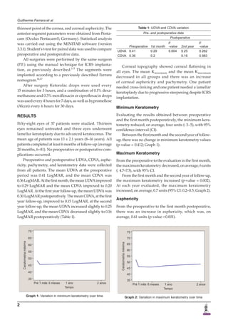
Preoperative and postoperative UDVA, CDVA, asphe-
ricity, pachymetry, and keratometry data were collected
from all patients. The mean UDVA at the preoperative
period was 0.41 LogMAR, and the mean CDVA was
0.36LogMAR.Atthefirstmonth,themeanUDVAimproved
to 0.29 LogMAR and the mean CDVA improved to 0.20
LogMAR. At the first year follow-up, the mean UDVA was
0.30 LogMAR postoperatively. The mean CDVA, at the first
year follow-up, improved to 0.15 LogMAR, at the second
year follow-up, the mean UDVA increased slightly to 0.25
LogMAR, and the mean CDVA decreased slightly to 0.16
LogMAR postoperatively (Table 1).
Corneal topography showed corneal flattening in
all eyes. The mean Kminimum and the mean Kmaximum
decreased in all groups and there was an increase
of corneal asphericity and pachymetry. One patient
needed cross-linking and one patient needed a lamellar
keratoplasty due to progressive steepening despite ICRS
implantation.
Minimum Keratometry
Evaluating the results obtained between preoperative
and the first month postoperatively, the minimum kera-
tometry reduced, on average, four units (: 3–5), with 95%
confidence interval (CI).
Between the first month and the second year of follow-
up, there was no change in minimum keratometry values
(p-value = 0.412; Graph 1).
Maximum Keratometry
From the preoperative to the evaluation in the first month,
the maximum keratometry decreased, on average, 6 units
(: 4.7–7.3), with 95% CI.
From the first month and the second year of follow-up,
the maximum keratometry increased (p-value = 0.002).
At each year evaluated, the maximum keratometry
increased, on average, 0.7 units (95% CI: 0.2–0.5; Graph 2).
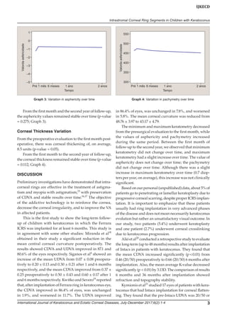
Asphericity
From the preoperative to the first month postoperative,
there was an increase in asphericity, which was, on
average, 0.61 units (p-value < 0.001).
Table 1: UDVA and CDVA variation
Pre- and postoperative data
Preoperative
Postoperative
1st month
p
-value 2nd year
p
-value
UDVA 0.41 0.29 0.004 0.25 0.262
CDVA 0.36 0.2 0.16 0.983
Graph 1: Variation in minimum keratometry over time Graph 2: Variation in maximum keratometry over time
International Journal of Keratoconus and Ectatic Corneal Diseases, July-December 2017;6(2):1-4 3
IJKECD
Intrastromal Corneal Ring Segments in Children with Keratoconus
From the first month and the second year of follow-up,
the asphericity values remained stable over time (p-value
= 0.275; Graph 3).
Corneal Thickness Variation
From the preoperative evaluation to the first month post-
operative, there was corneal thickening of, on average,
8.5 units (p-value = 0.05).
From the first month to the second year of follow-up,
the corneal thickness remained stable over time (p-value
= 0.112; Graph 4).
DISCUSSION
Preliminary investigations have demonstrated that intra-
corneal rings are effective in the treatment of astigma-
tism and myopia with astigmatism,15
with preservation
of CDVA and stable results over time.16,17
The objective
of the addictive technology is to reinforce the cornea,
decrease the corneal irregularity, and to improve the VA
in affected patients.
This is the first study to show the long-term follow-
up of children with keratoconus in which the Ferrara
ICRS was implanted for at least 6 months. This study is
in agreement with some other studies: Miranda et al18
obtained in their study a significant reduction in the
mean central corneal curvature postoperatively. The
results showed CDVA and UDVA improved in 87.1 and
80.6% of the eyes respectively. Siganos et al4
showed an
increase of the mean UDVA from 0.07 ± 0.08 preopera-
tively to 0.20 ± 0.13 and 0.30 ± 0.21 after 1 and 6 months
respectively, and the mean CDVA improved from 0.37 ±
0.25 preoperatively to 0.50 ± 0.43 and 0.60 ± 0.17 after 1
and 6 months respectively. Kwitko and Severo19
reported
that, after implantation of Ferrara ring in keratoconus eye,
the CDVA improved in 86.4% of eyes, was unchanged
in 1.9%, and worsened in 11.7%. The UDVA improved
in 86.4% of eyes, was unchanged in 7.8%, and worsened
in 5.8%. The mean corneal curvature was reduced from
48.76 ± 3.97 to 43.17 ± 4.79.
The minimum and maximum keratometry decreased
from the presurgical evaluation to the first month, while
the values of asphericity and pachymetry increased
during the same period. Between the first month of
follow-up to the second year, we observed that minimum
keratometry did not change over time, and maximum
keratometry had a slight increase over time. The value of
asphericity does not change over time; the pachymetry
did not change over time. Although there was a slight
increase in maximum keratometry over time (0.7 diop-
ters per year, on average), this increase was not clinically
significant.
Based on our personal (unpublished) data, about 5% of
patients go to penetrating or lamellar keratoplasty due to
progressive corneal scarring, despite proper ICRS implan-
tation. It is important to emphasize that these patients
usually had ring implantation in very advanced phases
of the disease and does not mean necessarily keratoconus
evolution but rather an unsatisfactory visual outcome. In
our study, two patients (5.4%) underwent keratoplasty
and one patient (2.7%) underwent corneal crosslinking
due to keratoconus progression.
Alió et al20
conducted a retrospective study to evaluate
the long-term (up to 48 months) results after implantation
of Intacs in patients with keratoconus. They found that
the mean CDVA increased significantly (p < 0.01) from
0.46 (20/50) preoperatively to 0.66 (20/30) 6 months after
implantation. Also, the mean average K-value decreased
significantly (p < 0.01) by 3.13D. The comparison of results
6 months and 36 months after implantation showed
refraction and topography stability.
Kymionis et al21
studied 17 eyes of patients with kera-
toconus that had Intacs implantation for corneal flatten-
ing. They found that the pre-Intacs UDVA was 20/50 or
Graph 3: Variation in asphericity over time Graph 4: Variation in pachymetry over time
4
Guilherme Ferrara et al
worse in all eyes, whereas, at the last follow-up examina-
tion, 59% had UDVA of 20/50 or better. Most of the eyes
(59%) experienced a gain of one up to eight lines of VA.
Previous studies showed that the intrastromal ring
flattens the cornea and keeps this effect for a long period
of time. There is no significant resteepening of the cornea
over time in adults. We found that the preoperative kera-
tometry values were higher in our study when compared
with similar studies.20-22
Moreover, the preoperative
UDVA and CDVA were worse in our study. This can be
explained by the behavior of keratoconus in some chil-
dren, which can rapidly evolve, and when the surgery is
indicated the disease is more advanced.
There was improvement of all parameters in our
study. The pattern of reduction of keratometry parameters
was similar to other studies. Despite that, at 5 years of
follow-up we found corneal resteepening on maximum
keratometry, which was not found in other studies when
compared with our results. Regarding the VA, there was
improvement in UDVA and CDVA, but not as much as
in adults.
The present study showed that the Ferrara ring,
despite the small sample of patients, could be a valuable
tool to provide topographic and visual improvement in
children with keratoconus. Corneal re-steepening can
occur after ICRS implantation. It plays an important role
in delaying the progression of keratoconus and postpones
a corneal grafting surgery. Further studies with larger
samples and longer follow-up periods must be warranted
to confirm the presented results.
REFERENCES
	 1.	 Colin J, Velou S. Implantation of Intacs and a refractive intra-
ocular lens to correct keratoconus. J Cataract Refract Surg
2003 Apr;29(4):832-834.
	 2.	 Asbell PA, Uçakhan OO. Long-term follow-up of Intacs from a
single center. J Cataract Refract Surg 2001 Sep;27(9):1456-1468.
	 3.	 Colin J, Cochener B, Savary G, Malet F. Correcting kerato-
conus with intracorneal rings. J Cataract Refract Surg 2000
Aug;26(8):1117-1122.
	 4.	 SiganosD,FerraraP,ChatzinikolasK,BessisN,PapastergiouG.
Ferrara intrastromal corneal rings for the correction of kerato-
conus. J Cataract Refract Surg 2002 Nov;28(11):1947-1951.
	 5.	 Colin J, Cochener B, Savary G, Malet F, Holmes-Higgin D.
INTACS inserts for treating keratoconus; one-year results.
Ophthalmology 2001 Aug;108(8):1409-1414.
	 6.	 Siganos CS, Kymionis GD, Kartakis N, Theodorakis MA,
Astyrakakis N, Pallikaris IG. Management of keratoconus
with Intacs. Am J Ophthalmol 2003 Jan;135(1):64-70.
	 7.	 Assil KK, Barrett AM, Fouraker BD, Schanzlin DJ. One-year
results of the intrastromal corneal ring in nonfunctional
human eyes. Intrastromal Corneal Ring Study Group. Arch
Ophthalmol 1995 Feb;113(2):159-167.
	 8.	 Siganos CS, Kymionis GD, Astyrakakis N, Pallikaris IG. Man-
agement of corneal ectasia after laser in situ keratomileusis
with INTACS. J Refract Surg 2002 Jan-Feb;18(1):43-46.
	 9.	 Silva FBD, Alves EAF, Cunha PFA. Utilização do Anel de
Ferrara na estabilização e correção da ectasia corneana pós
PRK. Arq Bras Oftalmol 2000 Jun;63(3):215-218.
	 10.	 Ruckhofer J, Stoiber J, Twa MD, Grabner G. Correction of
astigmatism with short arc-length intrastromal corneal
ring segments: preliminary results. Ophthalmology 2003
Mar;110(3):516-524.
	 11.	 Nosé W, Neves RA, Burris TE, Schanzlin DJ, Belfort Júnior R.
Intrastromal corneal ring: 12-month sighted myopic eyes. J
Refract Surg 1996 Jan;12(1):20-28.
	 12.	 Schanzlin DJ, Asbell PA, Burris TE, Durrie DS. The intra-
stromal ring segments. Phase II results for the correction of
myopia. Ophthalmology 1997 Jul;104(7):1067-1078.
	 13.	 Holmes-Higgin DK, Burris TE, Lapidus JA, Greenlick MR.
Risk factors for self-reported visual symptoms with Intacs
inserts for myopia. Ophthalmology 2002 Jan;109(1):46-56.
	 14.	 Asbell PA, Uçakhan OO, Abbott RL, Assil KA, Burris TE,
Durrie DS, Lindstrom RL, Schanzlin DJ, Verity SM, Waring
GO 3rd. Intrastromal corneal ring segments: reversibility of
refractive effect. J Refract Surg 2001 Jan-Feb;17(1):25-31.
	 15.	 Barraquer JI. Modification of refraction by means of intracor-
neal inclusions. Int Ophthalmol Clin 1966 Spring;6(1):53-78.
	 16.	 Ferrara de A, Cunha P. Técnica cirúrgica para correção de
miopia. Anel corneano intra-estromal. Rev Bras Oftalmol
1995;54(8):577-588.
	 17.	 Bock RH, Maumenee AE. Corneal fluid metabolism:
experiments and observations. Arch Ophthalmol 1953
Sep;50(3):282-285.
	 18.	 Miranda D, Sartori M, Francesconi C, Allemann N, Ferrara P,
Campos M. Ferrara intrastromal corneal ring segments for
severe keratoconus. J Refract Surg 2003 Nov-Dec;19(6):645-653.
	 19.	 Kwitko S, Severo N. Ferrara intracorneal ring segments for
keratoconus. J Cataract Refract Surg 2004 Apr;30(4):812-820.
	 20.	 Alió JI, Shabayek MH, Artola A. Intracorneal ring segments
for keratoconus correction: long-term follow-up. J Cataract
Refract Surg 2006 Jun;32(6):978-985.
	 21.	 Kymionis GD, Siganos CS, Tsiklis NS, Anastasakis A, Yoo SH,
Pallikaris AI, Astyrakakis N, Pallikaris IG. Long-term
follow-up of Intacs in Keratoconus. Am J Ophthalmol 2007
Feb;143(2):236-244.
	 22.	 Colin J, Malet FJ. Intacs for the correction of keratoconus: two-
year follow-up. J Cataract Refract Surg 2007 Jan;33(1):69-74.

More Related Content

What's hot

Dr. Vedat Kaya (MAKALE)
Dr. Vedat Kaya (MAKALE)Dr. Vedat Kaya (MAKALE)
Dr. Vedat Kaya (MAKALE)
Lasermed Tic
 
Asphericity in ring selection
Asphericity in ring selectionAsphericity in ring selection
Asphericity in ring selection
Ferrara Ophthalmics
 
Resultados preliminares do implante de um novo anel associado ao PRK para pre...
Resultados preliminares do implante de um novo anel associado ao PRK para pre...Resultados preliminares do implante de um novo anel associado ao PRK para pre...
Resultados preliminares do implante de um novo anel associado ao PRK para pre...
Ferrara Ophthalmics
 
Análise da tomografia corneana pós Anel de Ferrara
Análise da tomografia corneana pós Anel de FerraraAnálise da tomografia corneana pós Anel de Ferrara
Análise da tomografia corneana pós Anel de Ferrara
Ferrara Ophthalmics
 
FR and the astigmatism after penetrating keratoplasty
FR and the astigmatism after penetrating keratoplastyFR and the astigmatism after penetrating keratoplasty
FR and the astigmatism after penetrating keratoplastyFerrara Ophthalmics
 
Asphericity changes after Ferrara Ring
Asphericity changes after Ferrara RingAsphericity changes after Ferrara Ring
Asphericity changes after Ferrara RingFerrara Ophthalmics
 
Ferrara ICRS in Mild Keratoconus
Ferrara ICRS in Mild KeratoconusFerrara ICRS in Mild Keratoconus
Ferrara ICRS in Mild Keratoconus
Ferrara Ophthalmics
 
12 wavefront aberrations
12 wavefront aberrations12 wavefront aberrations
12 wavefront aberrations
Ferrara Ophthalmics
 
Ferrara Ring after DALK
Ferrara Ring after DALKFerrara Ring after DALK
Ferrara Ring after DALK
Ferrara Ophthalmics
 
12 correlation of anterior segment parameters
12 correlation of anterior segment parameters12 correlation of anterior segment parameters
12 correlation of anterior segment parameters
Ferrara Ophthalmics
 
Comparative Study of Visual Outcome between Femtosecond Lasik with Excimer La...
Comparative Study of Visual Outcome between Femtosecond Lasik with Excimer La...Comparative Study of Visual Outcome between Femtosecond Lasik with Excimer La...
Comparative Study of Visual Outcome between Femtosecond Lasik with Excimer La...
iosrjce
 
MyoRing in pellucid marginal degeneration
MyoRing in pellucid marginal degenerationMyoRing in pellucid marginal degeneration
MyoRing in pellucid marginal degeneration
Sahar Mojaled Nobari
 
Tessuti per DSAEK: le aspettative del chirurgo
Tessuti per DSAEK: le aspettative del chirurgoTessuti per DSAEK: le aspettative del chirurgo
Tessuti per DSAEK: le aspettative del chirurgo
Luca Avoni
 
Segmentos de Anel de Ferrara com arco de 140º
Segmentos de Anel de Ferrara com arco de 140ºSegmentos de Anel de Ferrara com arco de 140º
Segmentos de Anel de Ferrara com arco de 140º
Ferrara Ophthalmics
 
12 predictors of clinical outcomes
12 predictors of clinical outcomes12 predictors of clinical outcomes
12 predictors of clinical outcomes
Ferrara Ophthalmics
 
Otoplasty: New Modification of the Mustardé technique
Otoplasty: New Modification of the Mustardé techniqueOtoplasty: New Modification of the Mustardé technique
Otoplasty: New Modification of the Mustardé technique
Dr. Mohamed El-Rouby دكتور محمد الروبي
 
A Tomografia e o Implante de Anel de Ferrara
A Tomografia e o Implante de Anel de FerraraA Tomografia e o Implante de Anel de Ferrara
A Tomografia e o Implante de Anel de Ferrara
Ferrara Ophthalmics
 
Stability and safety of MyoRing implantation in keratoconic eyes
Stability and safety of MyoRing implantation in keratoconic eyesStability and safety of MyoRing implantation in keratoconic eyes
Stability and safety of MyoRing implantation in keratoconic eyes
Sahar Mojaled Nobari
 
Torica precizon webinar Newtech 30 giugno2020 drCanali
Torica precizon webinar Newtech 30 giugno2020 drCanaliTorica precizon webinar Newtech 30 giugno2020 drCanali
Torica precizon webinar Newtech 30 giugno2020 drCanali
Nicola Canali
 

What's hot (20)

Dr. Vedat Kaya (MAKALE)
Dr. Vedat Kaya (MAKALE)Dr. Vedat Kaya (MAKALE)
Dr. Vedat Kaya (MAKALE)
 
Asphericity in ring selection
Asphericity in ring selectionAsphericity in ring selection
Asphericity in ring selection
 
Resultados preliminares do implante de um novo anel associado ao PRK para pre...
Resultados preliminares do implante de um novo anel associado ao PRK para pre...Resultados preliminares do implante de um novo anel associado ao PRK para pre...
Resultados preliminares do implante de um novo anel associado ao PRK para pre...
 
Análise da tomografia corneana pós Anel de Ferrara
Análise da tomografia corneana pós Anel de FerraraAnálise da tomografia corneana pós Anel de Ferrara
Análise da tomografia corneana pós Anel de Ferrara
 
FR and the astigmatism after penetrating keratoplasty
FR and the astigmatism after penetrating keratoplastyFR and the astigmatism after penetrating keratoplasty
FR and the astigmatism after penetrating keratoplasty
 
Asphericity changes after Ferrara Ring
Asphericity changes after Ferrara RingAsphericity changes after Ferrara Ring
Asphericity changes after Ferrara Ring
 
Ferrara ICRS in Mild Keratoconus
Ferrara ICRS in Mild KeratoconusFerrara ICRS in Mild Keratoconus
Ferrara ICRS in Mild Keratoconus
 
12 wavefront aberrations
12 wavefront aberrations12 wavefront aberrations
12 wavefront aberrations
 
Ferrara Ring after DALK
Ferrara Ring after DALKFerrara Ring after DALK
Ferrara Ring after DALK
 
12 correlation of anterior segment parameters
12 correlation of anterior segment parameters12 correlation of anterior segment parameters
12 correlation of anterior segment parameters
 
Comparative Study of Visual Outcome between Femtosecond Lasik with Excimer La...
Comparative Study of Visual Outcome between Femtosecond Lasik with Excimer La...Comparative Study of Visual Outcome between Femtosecond Lasik with Excimer La...
Comparative Study of Visual Outcome between Femtosecond Lasik with Excimer La...
 
MyoRing in pellucid marginal degeneration
MyoRing in pellucid marginal degenerationMyoRing in pellucid marginal degeneration
MyoRing in pellucid marginal degeneration
 
Tessuti per DSAEK: le aspettative del chirurgo
Tessuti per DSAEK: le aspettative del chirurgoTessuti per DSAEK: le aspettative del chirurgo
Tessuti per DSAEK: le aspettative del chirurgo
 
Segmentos de Anel de Ferrara com arco de 140º
Segmentos de Anel de Ferrara com arco de 140ºSegmentos de Anel de Ferrara com arco de 140º
Segmentos de Anel de Ferrara com arco de 140º
 
12 predictors of clinical outcomes
12 predictors of clinical outcomes12 predictors of clinical outcomes
12 predictors of clinical outcomes
 
Otoplasty: New Modification of the Mustardé technique
Otoplasty: New Modification of the Mustardé techniqueOtoplasty: New Modification of the Mustardé technique
Otoplasty: New Modification of the Mustardé technique
 
A Tomografia e o Implante de Anel de Ferrara
A Tomografia e o Implante de Anel de FerraraA Tomografia e o Implante de Anel de Ferrara
A Tomografia e o Implante de Anel de Ferrara
 
Stability and safety of MyoRing implantation in keratoconic eyes
Stability and safety of MyoRing implantation in keratoconic eyesStability and safety of MyoRing implantation in keratoconic eyes
Stability and safety of MyoRing implantation in keratoconic eyes
 
Aaberg jr en bloc emm 2014
Aaberg jr en bloc emm 2014Aaberg jr en bloc emm 2014
Aaberg jr en bloc emm 2014
 
Torica precizon webinar Newtech 30 giugno2020 drCanali
Torica precizon webinar Newtech 30 giugno2020 drCanaliTorica precizon webinar Newtech 30 giugno2020 drCanali
Torica precizon webinar Newtech 30 giugno2020 drCanali
 

Similar to Anel de Ferrara em Crianças

12 refractive tomographic biomechanical outcomes
12 refractive tomographic biomechanical outcomes12 refractive tomographic biomechanical outcomes
12 refractive tomographic biomechanical outcomes
Ferrara Ophthalmics
 
EVALUATION OF ANTERIOR SEGMENT IMAGING TECHNIQUES IN DIAGNOSIS OF KERATOCONUS
EVALUATION OF ANTERIOR SEGMENT IMAGING TECHNIQUES IN DIAGNOSIS OF KERATOCONUSEVALUATION OF ANTERIOR SEGMENT IMAGING TECHNIQUES IN DIAGNOSIS OF KERATOCONUS
EVALUATION OF ANTERIOR SEGMENT IMAGING TECHNIQUES IN DIAGNOSIS OF KERATOCONUS
Shree Bharatimaiya College of Optometry & Physiotherapy
 
Ferrara ring review (2017)
Ferrara ring review (2017)Ferrara ring review (2017)
Ferrara ring review (2017)
Ferrara Ophthalmics
 
Topical dorzolamide for macular edema in the early phase after vitrectomy and...
Topical dorzolamide for macular edema in the early phase after vitrectomy and...Topical dorzolamide for macular edema in the early phase after vitrectomy and...
Topical dorzolamide for macular edema in the early phase after vitrectomy and...
Avaleks-Kiev
 
10 Ferrara Ring for ectasia after refractive surgery
10 Ferrara Ring for ectasia after refractive surgery10 Ferrara Ring for ectasia after refractive surgery
10 Ferrara Ring for ectasia after refractive surgery
Ferrara Ophthalmics
 
10 corneal endothelial profile
10 corneal endothelial profile10 corneal endothelial profile
10 corneal endothelial profile
Ferrara Ophthalmics
 
Influence-of-dioptric-power-on-retinal-nerve-fiber-layer-thickness-in-myopic-...
Influence-of-dioptric-power-on-retinal-nerve-fiber-layer-thickness-in-myopic-...Influence-of-dioptric-power-on-retinal-nerve-fiber-layer-thickness-in-myopic-...
Influence-of-dioptric-power-on-retinal-nerve-fiber-layer-thickness-in-myopic-...
Iqra Nehal
 
European_Society_of_Ophthalmology_Europe.pdf
European_Society_of_Ophthalmology_Europe.pdfEuropean_Society_of_Ophthalmology_Europe.pdf
European_Society_of_Ophthalmology_Europe.pdf
gabrielladesalvo
 
Corneal pachymetry by ben okeeffe
Corneal pachymetry by ben okeeffeCorneal pachymetry by ben okeeffe
Corneal pachymetry by ben okeeffe
OphthalmicDocs Chiong
 
1 s2.0-s0886335012010061-main
1 s2.0-s0886335012010061-main1 s2.0-s0886335012010061-main
1 s2.0-s0886335012010061-mainAmr Elmarasy
 
A DISSERTATION ON “EVALUATION OF ANTERIOR SEGMENT IMAGING TECHNIQUES IN DIAG...
A DISSERTATION ON  “EVALUATION OF ANTERIOR SEGMENT IMAGING TECHNIQUES IN DIAG...A DISSERTATION ON  “EVALUATION OF ANTERIOR SEGMENT IMAGING TECHNIQUES IN DIAG...
A DISSERTATION ON “EVALUATION OF ANTERIOR SEGMENT IMAGING TECHNIQUES IN DIAG...
Shree Bharatimaiya College of Optometry & Physiotherapy
 
Pterygium Excision with Free Conjunctival Limbal Autograft
Pterygium Excision with Free Conjunctival Limbal AutograftPterygium Excision with Free Conjunctival Limbal Autograft
Pterygium Excision with Free Conjunctival Limbal Autograft
iosrjce
 
An Optometrist S Personal Perspective Of Aspects Of Refractive Surgery
An Optometrist S Personal Perspective Of Aspects Of Refractive SurgeryAn Optometrist S Personal Perspective Of Aspects Of Refractive Surgery
An Optometrist S Personal Perspective Of Aspects Of Refractive Surgery
Erin Taylor
 
Macular Abnormality after Successful Surgery for Idiopathic Macular Hole Asse...
Macular Abnormality after Successful Surgery for Idiopathic Macular Hole Asse...Macular Abnormality after Successful Surgery for Idiopathic Macular Hole Asse...
Macular Abnormality after Successful Surgery for Idiopathic Macular Hole Asse...
CrimsonpublishersMSOR
 

Similar to Anel de Ferrara em Crianças (16)

12 refractive tomographic biomechanical outcomes
12 refractive tomographic biomechanical outcomes12 refractive tomographic biomechanical outcomes
12 refractive tomographic biomechanical outcomes
 
EVALUATION OF ANTERIOR SEGMENT IMAGING TECHNIQUES IN DIAGNOSIS OF KERATOCONUS
EVALUATION OF ANTERIOR SEGMENT IMAGING TECHNIQUES IN DIAGNOSIS OF KERATOCONUSEVALUATION OF ANTERIOR SEGMENT IMAGING TECHNIQUES IN DIAGNOSIS OF KERATOCONUS
EVALUATION OF ANTERIOR SEGMENT IMAGING TECHNIQUES IN DIAGNOSIS OF KERATOCONUS
 
Ferrara Ring l An Overview
Ferrara Ring l An OverviewFerrara Ring l An Overview
Ferrara Ring l An Overview
 
Ferrara ring review (2017)
Ferrara ring review (2017)Ferrara ring review (2017)
Ferrara ring review (2017)
 
Topical dorzolamide for macular edema in the early phase after vitrectomy and...
Topical dorzolamide for macular edema in the early phase after vitrectomy and...Topical dorzolamide for macular edema in the early phase after vitrectomy and...
Topical dorzolamide for macular edema in the early phase after vitrectomy and...
 
10 Ferrara Ring for ectasia after refractive surgery
10 Ferrara Ring for ectasia after refractive surgery10 Ferrara Ring for ectasia after refractive surgery
10 Ferrara Ring for ectasia after refractive surgery
 
10 corneal endothelial profile
10 corneal endothelial profile10 corneal endothelial profile
10 corneal endothelial profile
 
poster final
poster finalposter final
poster final
 
Influence-of-dioptric-power-on-retinal-nerve-fiber-layer-thickness-in-myopic-...
Influence-of-dioptric-power-on-retinal-nerve-fiber-layer-thickness-in-myopic-...Influence-of-dioptric-power-on-retinal-nerve-fiber-layer-thickness-in-myopic-...
Influence-of-dioptric-power-on-retinal-nerve-fiber-layer-thickness-in-myopic-...
 
European_Society_of_Ophthalmology_Europe.pdf
European_Society_of_Ophthalmology_Europe.pdfEuropean_Society_of_Ophthalmology_Europe.pdf
European_Society_of_Ophthalmology_Europe.pdf
 
Corneal pachymetry by ben okeeffe
Corneal pachymetry by ben okeeffeCorneal pachymetry by ben okeeffe
Corneal pachymetry by ben okeeffe
 
1 s2.0-s0886335012010061-main
1 s2.0-s0886335012010061-main1 s2.0-s0886335012010061-main
1 s2.0-s0886335012010061-main
 
A DISSERTATION ON “EVALUATION OF ANTERIOR SEGMENT IMAGING TECHNIQUES IN DIAG...
A DISSERTATION ON  “EVALUATION OF ANTERIOR SEGMENT IMAGING TECHNIQUES IN DIAG...A DISSERTATION ON  “EVALUATION OF ANTERIOR SEGMENT IMAGING TECHNIQUES IN DIAG...
A DISSERTATION ON “EVALUATION OF ANTERIOR SEGMENT IMAGING TECHNIQUES IN DIAG...
 
Pterygium Excision with Free Conjunctival Limbal Autograft
Pterygium Excision with Free Conjunctival Limbal AutograftPterygium Excision with Free Conjunctival Limbal Autograft
Pterygium Excision with Free Conjunctival Limbal Autograft
 
An Optometrist S Personal Perspective Of Aspects Of Refractive Surgery
An Optometrist S Personal Perspective Of Aspects Of Refractive SurgeryAn Optometrist S Personal Perspective Of Aspects Of Refractive Surgery
An Optometrist S Personal Perspective Of Aspects Of Refractive Surgery
 
Macular Abnormality after Successful Surgery for Idiopathic Macular Hole Asse...
Macular Abnormality after Successful Surgery for Idiopathic Macular Hole Asse...Macular Abnormality after Successful Surgery for Idiopathic Macular Hole Asse...
Macular Abnormality after Successful Surgery for Idiopathic Macular Hole Asse...
 

More from Ferrara Ophthalmics

Double Rings - New approachs to fine tune clinical outcomes.pdf
Double Rings - New approachs to fine tune clinical outcomes.pdfDouble Rings - New approachs to fine tune clinical outcomes.pdf
Double Rings - New approachs to fine tune clinical outcomes.pdf
Ferrara Ophthalmics
 
ICRS as a Refractive Tool.pdf
ICRS as a Refractive Tool.pdfICRS as a Refractive Tool.pdf
ICRS as a Refractive Tool.pdf
Ferrara Ophthalmics
 
Anel HM é capa da Oftalmologia em Foco.pdf
Anel HM é capa da Oftalmologia em Foco.pdfAnel HM é capa da Oftalmologia em Foco.pdf
Anel HM é capa da Oftalmologia em Foco.pdf
Ferrara Ophthalmics
 
apostila-refrativa-rio-ingles-final-pdf-revisado.pdf
apostila-refrativa-rio-ingles-final-pdf-revisado.pdfapostila-refrativa-rio-ingles-final-pdf-revisado.pdf
apostila-refrativa-rio-ingles-final-pdf-revisado.pdf
Ferrara Ophthalmics
 
apostila-refrativa-rio-final-pdf-revisado.pdf
apostila-refrativa-rio-final-pdf-revisado.pdfapostila-refrativa-rio-final-pdf-revisado.pdf
apostila-refrativa-rio-final-pdf-revisado.pdf
Ferrara Ophthalmics
 
Como planejo minha cirurgia simposio ferrara
Como planejo minha cirurgia   simposio ferraraComo planejo minha cirurgia   simposio ferrara
Como planejo minha cirurgia simposio ferrara
Ferrara Ophthalmics
 
Ferrara Ring - 30yo - 1998 - ceratocone
Ferrara Ring - 30yo - 1998 - ceratoconeFerrara Ring - 30yo - 1998 - ceratocone
Ferrara Ring - 30yo - 1998 - ceratocone
Ferrara Ophthalmics
 
O Filtro Amarelo
O Filtro AmareloO Filtro Amarelo
O Filtro Amarelo
Ferrara Ophthalmics
 
30yo Ferrara Ring
30yo Ferrara Ring30yo Ferrara Ring
30yo Ferrara Ring
Ferrara Ophthalmics
 
Book Chapter
Book ChapterBook Chapter
Book Chapter
Ferrara Ophthalmics
 
Mecanismo de ação e nomograma
Mecanismo de ação e nomogramaMecanismo de ação e nomograma
Mecanismo de ação e nomograma
Ferrara Ophthalmics
 
Ferrara Ring 320º
Ferrara Ring 320ºFerrara Ring 320º
Ferrara Ring 320º
Ferrara Ophthalmics
 
11 DALK
11 DALK11 DALK

More from Ferrara Ophthalmics (13)

Double Rings - New approachs to fine tune clinical outcomes.pdf
Double Rings - New approachs to fine tune clinical outcomes.pdfDouble Rings - New approachs to fine tune clinical outcomes.pdf
Double Rings - New approachs to fine tune clinical outcomes.pdf
 
ICRS as a Refractive Tool.pdf
ICRS as a Refractive Tool.pdfICRS as a Refractive Tool.pdf
ICRS as a Refractive Tool.pdf
 
Anel HM é capa da Oftalmologia em Foco.pdf
Anel HM é capa da Oftalmologia em Foco.pdfAnel HM é capa da Oftalmologia em Foco.pdf
Anel HM é capa da Oftalmologia em Foco.pdf
 
apostila-refrativa-rio-ingles-final-pdf-revisado.pdf
apostila-refrativa-rio-ingles-final-pdf-revisado.pdfapostila-refrativa-rio-ingles-final-pdf-revisado.pdf
apostila-refrativa-rio-ingles-final-pdf-revisado.pdf
 
apostila-refrativa-rio-final-pdf-revisado.pdf
apostila-refrativa-rio-final-pdf-revisado.pdfapostila-refrativa-rio-final-pdf-revisado.pdf
apostila-refrativa-rio-final-pdf-revisado.pdf
 
Como planejo minha cirurgia simposio ferrara
Como planejo minha cirurgia   simposio ferraraComo planejo minha cirurgia   simposio ferrara
Como planejo minha cirurgia simposio ferrara
 
Ferrara Ring - 30yo - 1998 - ceratocone
Ferrara Ring - 30yo - 1998 - ceratoconeFerrara Ring - 30yo - 1998 - ceratocone
Ferrara Ring - 30yo - 1998 - ceratocone
 
O Filtro Amarelo
O Filtro AmareloO Filtro Amarelo
O Filtro Amarelo
 
30yo Ferrara Ring
30yo Ferrara Ring30yo Ferrara Ring
30yo Ferrara Ring
 
Book Chapter
Book ChapterBook Chapter
Book Chapter
 
Mecanismo de ação e nomograma
Mecanismo de ação e nomogramaMecanismo de ação e nomograma
Mecanismo de ação e nomograma
 
Ferrara Ring 320º
Ferrara Ring 320ºFerrara Ring 320º
Ferrara Ring 320º
 
11 DALK
11 DALK11 DALK
11 DALK
 

Recently uploaded

Top-Vitamin-Supplement-Brands-in-India List
Top-Vitamin-Supplement-Brands-in-India ListTop-Vitamin-Supplement-Brands-in-India List
Top-Vitamin-Supplement-Brands-in-India List
SwisschemDerma
 
Triangles of Neck and Clinical Correlation by Dr. RIG.pptx
Triangles of Neck and Clinical Correlation by Dr. RIG.pptxTriangles of Neck and Clinical Correlation by Dr. RIG.pptx
Triangles of Neck and Clinical Correlation by Dr. RIG.pptx
Dr. Rabia Inam Gandapore
 
Thyroid Gland- Gross Anatomy by Dr. Rabia Inam Gandapore.pptx
Thyroid Gland- Gross Anatomy by Dr. Rabia Inam Gandapore.pptxThyroid Gland- Gross Anatomy by Dr. Rabia Inam Gandapore.pptx
Thyroid Gland- Gross Anatomy by Dr. Rabia Inam Gandapore.pptx
Dr. Rabia Inam Gandapore
 
Top 10 Best Ayurvedic Kidney Stone Syrups in India
Top 10 Best Ayurvedic Kidney Stone Syrups in IndiaTop 10 Best Ayurvedic Kidney Stone Syrups in India
Top 10 Best Ayurvedic Kidney Stone Syrups in India
Swastik Ayurveda
 
BRACHYTHERAPY OVERVIEW AND APPLICATORS
BRACHYTHERAPY OVERVIEW  AND  APPLICATORSBRACHYTHERAPY OVERVIEW  AND  APPLICATORS
BRACHYTHERAPY OVERVIEW AND APPLICATORS
Krishan Murari
 
The Electrocardiogram - Physiologic Principles
The Electrocardiogram - Physiologic PrinciplesThe Electrocardiogram - Physiologic Principles
The Electrocardiogram - Physiologic Principles
MedicoseAcademics
 
Hemodialysis: Chapter 4, Dialysate Circuit - Dr.Gawad
Hemodialysis: Chapter 4, Dialysate Circuit - Dr.GawadHemodialysis: Chapter 4, Dialysate Circuit - Dr.Gawad
Hemodialysis: Chapter 4, Dialysate Circuit - Dr.Gawad
NephroTube - Dr.Gawad
 
Identification and nursing management of congenital malformations .pptx
Identification and nursing management of congenital malformations .pptxIdentification and nursing management of congenital malformations .pptx
Identification and nursing management of congenital malformations .pptx
MGM SCHOOL/COLLEGE OF NURSING
 
Physiology of Special Chemical Sensation of Taste
Physiology of Special Chemical Sensation of TastePhysiology of Special Chemical Sensation of Taste
Physiology of Special Chemical Sensation of Taste
MedicoseAcademics
 
Novas diretrizes da OMS para os cuidados perinatais de mais qualidade
Novas diretrizes da OMS para os cuidados perinatais de mais qualidadeNovas diretrizes da OMS para os cuidados perinatais de mais qualidade
Novas diretrizes da OMS para os cuidados perinatais de mais qualidade
Prof. Marcus Renato de Carvalho
 
SURGICAL ANATOMY OF THE RETROPERITONEUM, ADRENALS, KIDNEYS AND URETERS.pptx
SURGICAL ANATOMY OF THE RETROPERITONEUM, ADRENALS, KIDNEYS AND URETERS.pptxSURGICAL ANATOMY OF THE RETROPERITONEUM, ADRENALS, KIDNEYS AND URETERS.pptx
SURGICAL ANATOMY OF THE RETROPERITONEUM, ADRENALS, KIDNEYS AND URETERS.pptx
Bright Chipili
 
Colonic and anorectal physiology with surgical implications
Colonic and anorectal physiology with surgical implicationsColonic and anorectal physiology with surgical implications
Colonic and anorectal physiology with surgical implications
Dr Maria Tamanna
 
Physiology of Chemical Sensation of smell.pdf
Physiology of Chemical Sensation of smell.pdfPhysiology of Chemical Sensation of smell.pdf
Physiology of Chemical Sensation of smell.pdf
MedicoseAcademics
 
ABDOMINAL TRAUMA in pediatrics part one.
ABDOMINAL TRAUMA in pediatrics part one.ABDOMINAL TRAUMA in pediatrics part one.
ABDOMINAL TRAUMA in pediatrics part one.
drhasanrajab
 
Pictures of Superficial & Deep Fascia.ppt.pdf
Pictures of Superficial & Deep Fascia.ppt.pdfPictures of Superficial & Deep Fascia.ppt.pdf
Pictures of Superficial & Deep Fascia.ppt.pdf
Dr. Rabia Inam Gandapore
 
Best Ayurvedic medicine for Gas and Indigestion
Best Ayurvedic medicine for Gas and IndigestionBest Ayurvedic medicine for Gas and Indigestion
Best Ayurvedic medicine for Gas and Indigestion
Swastik Ayurveda
 
Superficial & Deep Fascia of the NECK.pptx
Superficial & Deep Fascia of the NECK.pptxSuperficial & Deep Fascia of the NECK.pptx
Superficial & Deep Fascia of the NECK.pptx
Dr. Rabia Inam Gandapore
 
A Classical Text Review on Basavarajeeyam
A Classical Text Review on BasavarajeeyamA Classical Text Review on Basavarajeeyam
A Classical Text Review on Basavarajeeyam
Dr. Jyothirmai Paindla
 
Cervical & Brachial Plexus By Dr. RIG.pptx
Cervical & Brachial Plexus By Dr. RIG.pptxCervical & Brachial Plexus By Dr. RIG.pptx
Cervical & Brachial Plexus By Dr. RIG.pptx
Dr. Rabia Inam Gandapore
 
NVBDCP.pptx Nation vector borne disease control program
NVBDCP.pptx Nation vector borne disease control programNVBDCP.pptx Nation vector borne disease control program
NVBDCP.pptx Nation vector borne disease control program
Sapna Thakur
 

Recently uploaded (20)

Top-Vitamin-Supplement-Brands-in-India List
Top-Vitamin-Supplement-Brands-in-India ListTop-Vitamin-Supplement-Brands-in-India List
Top-Vitamin-Supplement-Brands-in-India List
 
Triangles of Neck and Clinical Correlation by Dr. RIG.pptx
Triangles of Neck and Clinical Correlation by Dr. RIG.pptxTriangles of Neck and Clinical Correlation by Dr. RIG.pptx
Triangles of Neck and Clinical Correlation by Dr. RIG.pptx
 
Thyroid Gland- Gross Anatomy by Dr. Rabia Inam Gandapore.pptx
Thyroid Gland- Gross Anatomy by Dr. Rabia Inam Gandapore.pptxThyroid Gland- Gross Anatomy by Dr. Rabia Inam Gandapore.pptx
Thyroid Gland- Gross Anatomy by Dr. Rabia Inam Gandapore.pptx
 
Top 10 Best Ayurvedic Kidney Stone Syrups in India
Top 10 Best Ayurvedic Kidney Stone Syrups in IndiaTop 10 Best Ayurvedic Kidney Stone Syrups in India
Top 10 Best Ayurvedic Kidney Stone Syrups in India
 
BRACHYTHERAPY OVERVIEW AND APPLICATORS
BRACHYTHERAPY OVERVIEW  AND  APPLICATORSBRACHYTHERAPY OVERVIEW  AND  APPLICATORS
BRACHYTHERAPY OVERVIEW AND APPLICATORS
 
The Electrocardiogram - Physiologic Principles
The Electrocardiogram - Physiologic PrinciplesThe Electrocardiogram - Physiologic Principles
The Electrocardiogram - Physiologic Principles
 
Hemodialysis: Chapter 4, Dialysate Circuit - Dr.Gawad
Hemodialysis: Chapter 4, Dialysate Circuit - Dr.GawadHemodialysis: Chapter 4, Dialysate Circuit - Dr.Gawad
Hemodialysis: Chapter 4, Dialysate Circuit - Dr.Gawad
 
Identification and nursing management of congenital malformations .pptx
Identification and nursing management of congenital malformations .pptxIdentification and nursing management of congenital malformations .pptx
Identification and nursing management of congenital malformations .pptx
 
Physiology of Special Chemical Sensation of Taste
Physiology of Special Chemical Sensation of TastePhysiology of Special Chemical Sensation of Taste
Physiology of Special Chemical Sensation of Taste
 
Novas diretrizes da OMS para os cuidados perinatais de mais qualidade
Novas diretrizes da OMS para os cuidados perinatais de mais qualidadeNovas diretrizes da OMS para os cuidados perinatais de mais qualidade
Novas diretrizes da OMS para os cuidados perinatais de mais qualidade
 
SURGICAL ANATOMY OF THE RETROPERITONEUM, ADRENALS, KIDNEYS AND URETERS.pptx
SURGICAL ANATOMY OF THE RETROPERITONEUM, ADRENALS, KIDNEYS AND URETERS.pptxSURGICAL ANATOMY OF THE RETROPERITONEUM, ADRENALS, KIDNEYS AND URETERS.pptx
SURGICAL ANATOMY OF THE RETROPERITONEUM, ADRENALS, KIDNEYS AND URETERS.pptx
 
Colonic and anorectal physiology with surgical implications
Colonic and anorectal physiology with surgical implicationsColonic and anorectal physiology with surgical implications
Colonic and anorectal physiology with surgical implications
 
Physiology of Chemical Sensation of smell.pdf
Physiology of Chemical Sensation of smell.pdfPhysiology of Chemical Sensation of smell.pdf
Physiology of Chemical Sensation of smell.pdf
 
ABDOMINAL TRAUMA in pediatrics part one.
ABDOMINAL TRAUMA in pediatrics part one.ABDOMINAL TRAUMA in pediatrics part one.
ABDOMINAL TRAUMA in pediatrics part one.
 
Pictures of Superficial & Deep Fascia.ppt.pdf
Pictures of Superficial & Deep Fascia.ppt.pdfPictures of Superficial & Deep Fascia.ppt.pdf
Pictures of Superficial & Deep Fascia.ppt.pdf
 
Best Ayurvedic medicine for Gas and Indigestion
Best Ayurvedic medicine for Gas and IndigestionBest Ayurvedic medicine for Gas and Indigestion
Best Ayurvedic medicine for Gas and Indigestion
 
Superficial & Deep Fascia of the NECK.pptx
Superficial & Deep Fascia of the NECK.pptxSuperficial & Deep Fascia of the NECK.pptx
Superficial & Deep Fascia of the NECK.pptx
 
A Classical Text Review on Basavarajeeyam
A Classical Text Review on BasavarajeeyamA Classical Text Review on Basavarajeeyam
A Classical Text Review on Basavarajeeyam
 
Cervical & Brachial Plexus By Dr. RIG.pptx
Cervical & Brachial Plexus By Dr. RIG.pptxCervical & Brachial Plexus By Dr. RIG.pptx
Cervical & Brachial Plexus By Dr. RIG.pptx
 
NVBDCP.pptx Nation vector borne disease control program
NVBDCP.pptx Nation vector borne disease control programNVBDCP.pptx Nation vector borne disease control program
NVBDCP.pptx Nation vector borne disease control program
 

Anel de Ferrara em Crianças

  • 1. International Journal of Keratoconus and Ectatic Corneal Diseases, July-December 2017;6(2):1-4 1 IJKECD Intrastromal Corneal Ring Segments in Children with Keratoconus ABSTRACT Purpose: To evaluate the long-term follow-up of Ferrara intra- stromal corneal ring segments (ICRSs) (Ferrara Ophthalmics, Belo Horizonte, Brazil) implantation for the management of keratoconus in children. Study design: Dr Paulo Ferrara Eye Clinic, Belo Horizonte, MG, Brazil. Materials and methods: A total of 58 eyes of 37 children with keratoconus were included. One or two ring segments were inserted into the cornea, embracing the keratoconus area. Statistical analysis included preoperative and postoperative uncorrected distance visual acuity (UDVA), corrected distance visual acuity (CDVA), asphericity, pachymetry, and keratometry. Results: Ferrara ICRS implantation significantly improved the mean UDVA and CDVA. Corneal tomography (Pentacam® ) showed corneal flattening in all eyes implanted with the Ferrara ring. The mean K decreased, and the corneal asphericity and pachymetry increased in all cases. Conclusion: The Ferrara ICRS improved all parameters after 2 years of implantation in children with keratoconus. There was significant corneal flattening after ring implantation with improvement of the UDVA and the CDVA. All studied param- eters remained stable over time. Keywords: Children, Ferrara ring, Intrastromal corneal ring segments, Keratoconus. How to cite this article: Ferrara G, Ferrara P, Torquetti L. Intrastromal Corneal Ring Segments in Children with Kerato- conus. Int J Kerat Ect Cor Dis 2017;6(2):1-4. Source of support: Nil Conflict of interest: None INTRODUCTION Keratoconus is a corneal ectatic disease characterized by noninflammatory progressive thinning of unknown cause in which the cornea assumes a conical shape. Intrastromal corneal ring segments have been used to correct ectatic corneal diseases in order to reduce the corneal steepening, reduce the irregular astigmatism, and improve the visual acuity (VA).1-7 Besides, the segments may be a surgical alternative to at least delay, if not elimi- nate, the need of lamellar or penetrating keratoplasty. The Ferrara ICRSs are made of PMMA Perspex CQ acrylic. They vary in thickness, and are available in 0.15, 0.20, 0.25, 0.30, and 0.35 mm. The segment cross-section is triangular, and the base for every thickness and diameter is 0.60 mm. The segments have 90°, 120°, 160°, or 210° of arc. Many studies have demonstrated the efficacy of intra- stromal rings to treat many corneal conditions, such as keratoconus,1-7 postlaser-assisted in situ keratomileusis corneal ectasia,8 postradial keratotomy ectasia,9 astigma- tism,10 and myopia.11-14 The changes in corneal structure induced by additive technologies can be roughly pre- dicted by Barraquer’s thickness law, i.e., when material is added to the periphery of the cornea or an equal amount of material is removed from the central area, a flattening effect is achieved. The corrective result varies in direct proportion to the thickness of the implant and in inverse proportion to its diameter. The thicker and smaller the diameter of the device, the higher the corrective result.15 In order to investigate the long-term VA and mechani- cal stability after Ferrara ICRS implantation in children with keratoconus, we conducted the current retrospec- tive study. MATERIALS AND METHODS We retrospectively reviewed patient records of 58 eyes of 37 children with keratoconus, which were followed for a period of at least 6 months. The main indication for ICRS implantation was contact lens intolerance and/or progression of the ectasia. The progression of the disease was defined by worsening of UDVA, CDVA (loss of at least 1 line of VA), progressive intolerance to contact lens wear, and progressive corneal steepening documented by corneal topography. Patients were excluded if any of the following criteria applied after preoperative examination: Advanced keratoconus with significant apical opacity and scarring, hydrops, thin corneas, with thickness below 300 µm in the ring track, intense atopia, and any ongoing infectious process, local or systemic. Patients were separated into two groups, initial and advanced, according to Amsler–Krumeich classification. Statistical analysis included preoperative and post- operative UDVA, CDVA, keratometry, pachymetry at the Intrastromal Corneal Ring Segments in Children with Keratoconus 1 Guilherme Ferrara, 2 Paulo Ferrara, 3 Leonardo Torquetti IJKECD Original Paper 10.5005/jp-journals-10025-1140 1-3 Private Practitioner 1,2 Paulo Ferrara Eye Clinic, Belo Horizonte, Minas Gerais, Brazil 3 Centro de Excelência em Oftalmologia, Pará de Minas, Brazil CorrespondingAuthor: Guilherme Ferrara,Av. Contorno, 4747 – Suite 601, Belo Horizonte-30110921, Minas Gerais, Brazil
  • 2. 2 Guilherme Ferrara et al thinnest point of the cornea, and corneal asphericity. The anterior segment parameters were obtained from Penta- cam (Oculus Pentacam®, Germany). Statistical analysis was carried out using the MINITAB software (version 3.3.1). Student’s t-test for paired data was used to compare preoperative and postoperative data. All surgeries were performed by the same surgeon (P.F.) using the manual technique for ICRS implanta- tion, as previously described.1-5 The segments were implanted according to a previously described Ferrara nomogram.16,17 After surgery Ketorolac drops were used every 15 minutes for 3 hours, and a combination of 0.1% dexa- methasone and 0.3% moxifloxacin or ciprofloxacin drops was used every 4 hours for 7 days, as well as hypromellose (Alcon) every 6 hours for 30 days. RESULTS Fifty-eight eyes of 37 patients were studied. Thirteen eyes remained untreated and three eyes underwent lamellar keratoplasty due to advanced keratoconus. The mean age of patients was 13 ± 2.1 years (8–16 years). All patients completed at least 6 months of follow-up (average 20 months, 6–81). No preoperative or postoperative com- plications occurred. Preoperative and postoperative UDVA, CDVA, asphe- ricity, pachymetry, and keratometry data were collected from all patients. The mean UDVA at the preoperative period was 0.41 LogMAR, and the mean CDVA was 0.36LogMAR.Atthefirstmonth,themeanUDVAimproved to 0.29 LogMAR and the mean CDVA improved to 0.20 LogMAR. At the first year follow-up, the mean UDVA was 0.30 LogMAR postoperatively. The mean CDVA, at the first year follow-up, improved to 0.15 LogMAR, at the second year follow-up, the mean UDVA increased slightly to 0.25 LogMAR, and the mean CDVA decreased slightly to 0.16 LogMAR postoperatively (Table 1). Corneal topography showed corneal flattening in all eyes. The mean Kminimum and the mean Kmaximum decreased in all groups and there was an increase of corneal asphericity and pachymetry. One patient needed cross-linking and one patient needed a lamellar keratoplasty due to progressive steepening despite ICRS implantation. Minimum Keratometry Evaluating the results obtained between preoperative and the first month postoperatively, the minimum kera- tometry reduced, on average, four units (: 3–5), with 95% confidence interval (CI). Between the first month and the second year of follow- up, there was no change in minimum keratometry values (p-value = 0.412; Graph 1). Maximum Keratometry From the preoperative to the evaluation in the first month, the maximum keratometry decreased, on average, 6 units (: 4.7–7.3), with 95% CI. From the first month and the second year of follow-up, the maximum keratometry increased (p-value = 0.002). At each year evaluated, the maximum keratometry increased, on average, 0.7 units (95% CI: 0.2–0.5; Graph 2). Asphericity From the preoperative to the first month postoperative, there was an increase in asphericity, which was, on average, 0.61 units (p-value < 0.001). Table 1: UDVA and CDVA variation Pre- and postoperative data Preoperative Postoperative 1st month p -value 2nd year p -value UDVA 0.41 0.29 0.004 0.25 0.262 CDVA 0.36 0.2 0.16 0.983 Graph 1: Variation in minimum keratometry over time Graph 2: Variation in maximum keratometry over time
  • 3. International Journal of Keratoconus and Ectatic Corneal Diseases, July-December 2017;6(2):1-4 3 IJKECD Intrastromal Corneal Ring Segments in Children with Keratoconus From the first month and the second year of follow-up, the asphericity values remained stable over time (p-value = 0.275; Graph 3). Corneal Thickness Variation From the preoperative evaluation to the first month post- operative, there was corneal thickening of, on average, 8.5 units (p-value = 0.05). From the first month to the second year of follow-up, the corneal thickness remained stable over time (p-value = 0.112; Graph 4). DISCUSSION Preliminary investigations have demonstrated that intra- corneal rings are effective in the treatment of astigma- tism and myopia with astigmatism,15 with preservation of CDVA and stable results over time.16,17 The objective of the addictive technology is to reinforce the cornea, decrease the corneal irregularity, and to improve the VA in affected patients. This is the first study to show the long-term follow- up of children with keratoconus in which the Ferrara ICRS was implanted for at least 6 months. This study is in agreement with some other studies: Miranda et al18 obtained in their study a significant reduction in the mean central corneal curvature postoperatively. The results showed CDVA and UDVA improved in 87.1 and 80.6% of the eyes respectively. Siganos et al4 showed an increase of the mean UDVA from 0.07 ± 0.08 preopera- tively to 0.20 ± 0.13 and 0.30 ± 0.21 after 1 and 6 months respectively, and the mean CDVA improved from 0.37 ± 0.25 preoperatively to 0.50 ± 0.43 and 0.60 ± 0.17 after 1 and 6 months respectively. Kwitko and Severo19 reported that, after implantation of Ferrara ring in keratoconus eye, the CDVA improved in 86.4% of eyes, was unchanged in 1.9%, and worsened in 11.7%. The UDVA improved in 86.4% of eyes, was unchanged in 7.8%, and worsened in 5.8%. The mean corneal curvature was reduced from 48.76 ± 3.97 to 43.17 ± 4.79. The minimum and maximum keratometry decreased from the presurgical evaluation to the first month, while the values of asphericity and pachymetry increased during the same period. Between the first month of follow-up to the second year, we observed that minimum keratometry did not change over time, and maximum keratometry had a slight increase over time. The value of asphericity does not change over time; the pachymetry did not change over time. Although there was a slight increase in maximum keratometry over time (0.7 diop- ters per year, on average), this increase was not clinically significant. Based on our personal (unpublished) data, about 5% of patients go to penetrating or lamellar keratoplasty due to progressive corneal scarring, despite proper ICRS implan- tation. It is important to emphasize that these patients usually had ring implantation in very advanced phases of the disease and does not mean necessarily keratoconus evolution but rather an unsatisfactory visual outcome. In our study, two patients (5.4%) underwent keratoplasty and one patient (2.7%) underwent corneal crosslinking due to keratoconus progression. Alió et al20 conducted a retrospective study to evaluate the long-term (up to 48 months) results after implantation of Intacs in patients with keratoconus. They found that the mean CDVA increased significantly (p < 0.01) from 0.46 (20/50) preoperatively to 0.66 (20/30) 6 months after implantation. Also, the mean average K-value decreased significantly (p < 0.01) by 3.13D. The comparison of results 6 months and 36 months after implantation showed refraction and topography stability. Kymionis et al21 studied 17 eyes of patients with kera- toconus that had Intacs implantation for corneal flatten- ing. They found that the pre-Intacs UDVA was 20/50 or Graph 3: Variation in asphericity over time Graph 4: Variation in pachymetry over time
  • 4. 4 Guilherme Ferrara et al worse in all eyes, whereas, at the last follow-up examina- tion, 59% had UDVA of 20/50 or better. Most of the eyes (59%) experienced a gain of one up to eight lines of VA. Previous studies showed that the intrastromal ring flattens the cornea and keeps this effect for a long period of time. There is no significant resteepening of the cornea over time in adults. We found that the preoperative kera- tometry values were higher in our study when compared with similar studies.20-22 Moreover, the preoperative UDVA and CDVA were worse in our study. This can be explained by the behavior of keratoconus in some chil- dren, which can rapidly evolve, and when the surgery is indicated the disease is more advanced. There was improvement of all parameters in our study. The pattern of reduction of keratometry parameters was similar to other studies. Despite that, at 5 years of follow-up we found corneal resteepening on maximum keratometry, which was not found in other studies when compared with our results. Regarding the VA, there was improvement in UDVA and CDVA, but not as much as in adults. The present study showed that the Ferrara ring, despite the small sample of patients, could be a valuable tool to provide topographic and visual improvement in children with keratoconus. Corneal re-steepening can occur after ICRS implantation. It plays an important role in delaying the progression of keratoconus and postpones a corneal grafting surgery. Further studies with larger samples and longer follow-up periods must be warranted to confirm the presented results. REFERENCES 1. Colin J, Velou S. Implantation of Intacs and a refractive intra- ocular lens to correct keratoconus. J Cataract Refract Surg 2003 Apr;29(4):832-834. 2. Asbell PA, Uçakhan OO. Long-term follow-up of Intacs from a single center. J Cataract Refract Surg 2001 Sep;27(9):1456-1468. 3. Colin J, Cochener B, Savary G, Malet F. Correcting kerato- conus with intracorneal rings. J Cataract Refract Surg 2000 Aug;26(8):1117-1122. 4. SiganosD,FerraraP,ChatzinikolasK,BessisN,PapastergiouG. Ferrara intrastromal corneal rings for the correction of kerato- conus. J Cataract Refract Surg 2002 Nov;28(11):1947-1951. 5. Colin J, Cochener B, Savary G, Malet F, Holmes-Higgin D. INTACS inserts for treating keratoconus; one-year results. Ophthalmology 2001 Aug;108(8):1409-1414. 6. Siganos CS, Kymionis GD, Kartakis N, Theodorakis MA, Astyrakakis N, Pallikaris IG. Management of keratoconus with Intacs. Am J Ophthalmol 2003 Jan;135(1):64-70. 7. Assil KK, Barrett AM, Fouraker BD, Schanzlin DJ. One-year results of the intrastromal corneal ring in nonfunctional human eyes. Intrastromal Corneal Ring Study Group. Arch Ophthalmol 1995 Feb;113(2):159-167. 8. Siganos CS, Kymionis GD, Astyrakakis N, Pallikaris IG. Man- agement of corneal ectasia after laser in situ keratomileusis with INTACS. J Refract Surg 2002 Jan-Feb;18(1):43-46. 9. Silva FBD, Alves EAF, Cunha PFA. Utilização do Anel de Ferrara na estabilização e correção da ectasia corneana pós PRK. Arq Bras Oftalmol 2000 Jun;63(3):215-218. 10. Ruckhofer J, Stoiber J, Twa MD, Grabner G. Correction of astigmatism with short arc-length intrastromal corneal ring segments: preliminary results. Ophthalmology 2003 Mar;110(3):516-524. 11. Nosé W, Neves RA, Burris TE, Schanzlin DJ, Belfort Júnior R. Intrastromal corneal ring: 12-month sighted myopic eyes. J Refract Surg 1996 Jan;12(1):20-28. 12. Schanzlin DJ, Asbell PA, Burris TE, Durrie DS. The intra- stromal ring segments. Phase II results for the correction of myopia. Ophthalmology 1997 Jul;104(7):1067-1078. 13. Holmes-Higgin DK, Burris TE, Lapidus JA, Greenlick MR. Risk factors for self-reported visual symptoms with Intacs inserts for myopia. Ophthalmology 2002 Jan;109(1):46-56. 14. Asbell PA, Uçakhan OO, Abbott RL, Assil KA, Burris TE, Durrie DS, Lindstrom RL, Schanzlin DJ, Verity SM, Waring GO 3rd. Intrastromal corneal ring segments: reversibility of refractive effect. J Refract Surg 2001 Jan-Feb;17(1):25-31. 15. Barraquer JI. Modification of refraction by means of intracor- neal inclusions. Int Ophthalmol Clin 1966 Spring;6(1):53-78. 16. Ferrara de A, Cunha P. Técnica cirúrgica para correção de miopia. Anel corneano intra-estromal. Rev Bras Oftalmol 1995;54(8):577-588. 17. Bock RH, Maumenee AE. Corneal fluid metabolism: experiments and observations. Arch Ophthalmol 1953 Sep;50(3):282-285. 18. Miranda D, Sartori M, Francesconi C, Allemann N, Ferrara P, Campos M. Ferrara intrastromal corneal ring segments for severe keratoconus. J Refract Surg 2003 Nov-Dec;19(6):645-653. 19. Kwitko S, Severo N. Ferrara intracorneal ring segments for keratoconus. J Cataract Refract Surg 2004 Apr;30(4):812-820. 20. Alió JI, Shabayek MH, Artola A. Intracorneal ring segments for keratoconus correction: long-term follow-up. J Cataract Refract Surg 2006 Jun;32(6):978-985. 21. Kymionis GD, Siganos CS, Tsiklis NS, Anastasakis A, Yoo SH, Pallikaris AI, Astyrakakis N, Pallikaris IG. Long-term follow-up of Intacs in Keratoconus. Am J Ophthalmol 2007 Feb;143(2):236-244. 22. Colin J, Malet FJ. Intacs for the correction of keratoconus: two- year follow-up. J Cataract Refract Surg 2007 Jan;33(1):69-74.