The document discusses components of a complete blood count (CBC) including:
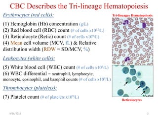
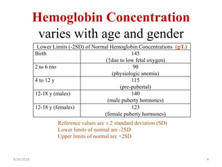
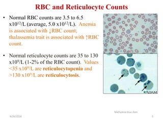
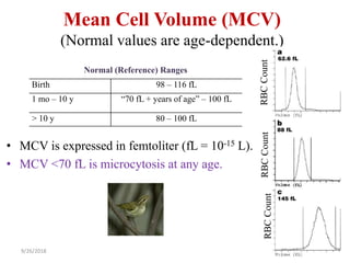
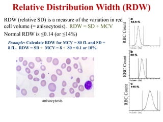
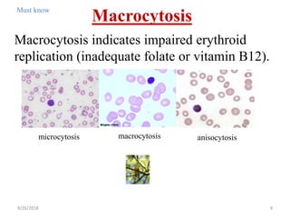
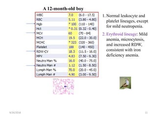
1. Erythrocytes (red blood cells) characterized by hemoglobin concentration, red blood cell count, mean cell volume, and other measures.
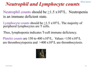
2. Leukocytes (white blood cells) characterized by white blood cell count and differential.
3. Thrombocytes (platelets) characterized by platelet count.
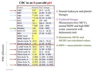
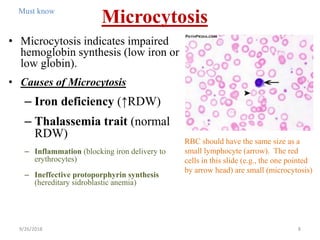
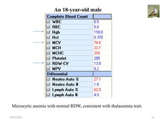
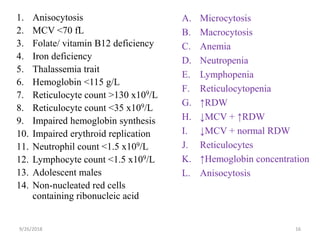
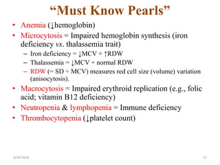
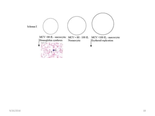
The CBC describes the tri-lineage of hematopoiesis including the erythroid, leukocyte, and thrombocyte lineages. Key information and interpretations of CBC results are provided such as distinguishing between microcytosis caused by iron deficiency versus thalassemia trait.


![PERI-PROSTHETIC FRACTURE NAIL-PLATE CONSTRUCT [NPC].pptx](https://cdn.slidesharecdn.com/ss_thumbnails/drarunkumardrmohamedashrafperiprostheticfrasturenail-plateconstructnpc-260209164459-7e9d15a1-thumbnail.jpg?width=640&height=640&fit=bounds)


