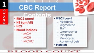
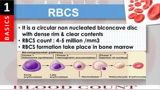
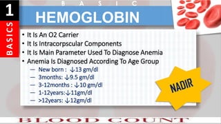
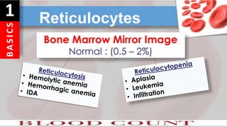
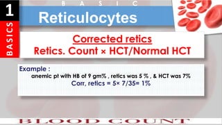
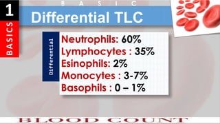
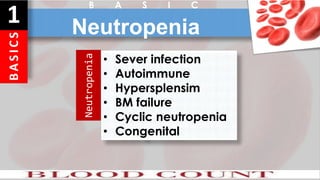
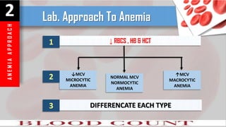
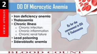
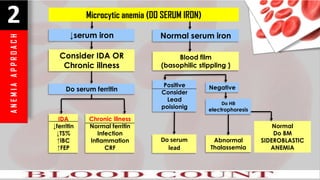
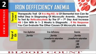
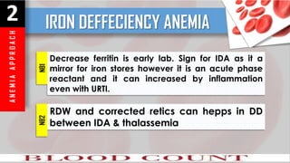
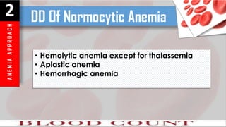
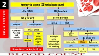
DISCOVER
PROF / ABDUL HAMID ABDUL MONEM , MD
108 takes
AHMED
SOLIMAN
MD
1
B A S I C S
B
A
S
I
C
S
1 CBC Report
Contents
• RBCS count
• HB (gm/dl)
• HCT
• Blood indices
⁻ MCV
⁻ MCH
⁻ MCHC
• WBCS count
₋ Netrophils
₋ Segmented
₋ Staff
₋ Lymphocytes
₋ Esinophils
₋ Monocytes
₋ Abnormal cells
• Platelets
B A S I C
B
A
S
I
C
S
1 RBCS
• It is a circular non nucleated biconcave disc
with dense rim & clear contents
• RBCS count : 4-5 million /mm3
• RBCS formation take place in bone marrow
B A S I C
B
A
S
I
C
S
1
• It Is An O2 Carrier
• It Is Intracorpscular Components
• It Is Main Parameter Used To Diagnose Anemia
• Anemia Is Diagnosed According To Age Group
― New born : ↓13 gm/dl
― 3months: ↓9.5 gm/dl
― 3-12months : ↓10 gm/dl
― 1-12years:↓11gm/dl
― >12years: ↓12gm/dl
HEMOGLOBIN
B A S I C
B
A
S
I
C
S
1
• It is volume of RBCS as regard to the whole blood
• Average in newborn : 50%.
• Infant & child : 35% - 40% .
• Decrease HB & HCT : Anemia
• Increase HB & HCT : Polycythemia
HEMATOCRIT
(HCT)
B
A
S
I
C
S
1
• MCV = HCT % × 10 / RBCS
• ↓MCV : Microcytosis
• ↑MCV: Macrocytosis
• Normal MCV : Normocytosis
• Average : 90 + 9 FL
• Lower Limit = 70 Age/Year
• Upper Limit
(↓10 Years →90 Fl , ↑10 Years →95 Fl)
MEAN CURPSCULAR VOLUME
(MCV)
B
A
S
I
C
S
1
MCH = Average quantity of hemoglobin in RBCS
MCH = HB (gm%)×10 / RBCS = 32+ 2 pgm
It ↓ in iron deficiency & thalassemia
B A S I C
MEAN CORPUSCULAR
HAEMOGLOBIN
B
A
S
I
C
S
1 RDW
• RDW : red cell distribution width
• It denotes size variability of RBCS
• It is quantification of ansiocytosis
• Normal : 10 – 15%
• Used in DD between IDA & Thalassemia
• It is markedly increased in IDA up to 30 – 40%
B
A
S
I
C
S
1 Reticulocytes
Bone Marrow Mirror Image
Normal : (0.5 – 2%)
B
A
S
I
C
S
1 Reticulocytes
B A S I C
Example :
anemic pt with HB of 9 gm% , retics was 5 % , & HCT was 7%
Corr, retics = 5× 7/35= 1%
Corrected retics
Retics. Count × HCT/Normal HCT
B
A
S
I
C
S
1 WBCS
B A S I C
TLC below 10 years : 6000 – 14000
TLC above 10 years : 4000 - 12000
Normal
• Infection
• Inflammation
• Malignancy
Leukocytosis
• Aplastic anemia
• Malignancy
• Infiltration
Leukopenia
B
A
S
I
C
S
1 Differential TLC
B A S I C
Neutrophils: 60%
Lymphocytes : 35%
Esinophils: 2%
Monocytes : 3-7%
Basophils : 0 – 1%
Differential
B
A
S
I
C
S
1 Neutropenia
B A S I C
• Sever infection
• Autoimmune
• Hypersplensim
• BM failure
• Cyclic neutropenia
• Congenital
Neutropenia
B
A
S
I
C
S
1 Eosinophilia
B A S I C
Eosinophilia
Asthma
Eczema
Parasitic
IBD(UC)
Malignancy
B
A
S
I
C
S
1 Lymphocytosis
B A S I C
Lymphocytosis
Viral infection
TB
ALL
Whooping
cough
B
A
S
I
C
S
1 Abnormal cells
B A S I C
Band cells
N: less than 20% of total neutrophils
Bandemia :↑ band cells (bacterial infection).
Metamylocytes & myleocytes
in prepheripheral blood is abnormal & denotes
bacterial infection .
B
A
S
I
C
S
1 Platelets
B A S I C
Platelets are produced by bone marrow
Normal count : (150,000 – 450,000)
Function
Hemostasis
• Platelets aggregation
• Platelets adhesion
• Clot formation
Disorders
• Thrombocytopenia
• Thromboasthenia
• Thrombocytosis
2
ANEMIA APPROACH
A
N
E
M
I
A
A
P
P
R
O
A
C
H
2 Lab. Approach To Anemia
↓ RBCS , HB & HCT
↓MCV
MICROCYTIC
ANEMIA
NORMAL MCV
NORMOCYTIC
ANEMIA
↑MCV
MACROCYTIC
ANEMIA
DIFFERENCATE EACH TYPE
1
2
3
A
N
E
M
I
A
A
P
P
R
O
A
C
H
2 DD Of Microcytic Anemia
• Iron deficiency anemia
• Thalassemia
• Chronic illness
o Chronic infection
o Chronic inflammation
o Chronic renal failure
• Lead poisoning
• Sideroblastic anemia
A
N
E
M
I
A
A
P
P
R
O
A
C
H
2 Microcytic anemia (DO SERUM IRON)
↓serum iron
Consider IDA OR
Chronic illness
Do serum ferritin
IDA
↓ferritin
↓TS%
↑IBC
↑FEP
Chronic illness
Normal ferritin
Infection
Inflammation
CRF
Normal serum iron
Blood film
(basophilic stippling )
Positive
Consider
Lead
poisionig
Do serum
lead
Negative
Do HB
electrophoresis
Abnormal
Thalassemia
Normal
Do BM
SIDEROBLASTIC
ANEMIA
A
N
E
M
I
A
A
P
P
R
O
A
C
H
2 IRON DEFFECIENCY ANEMIA
Therapeutic Trail Of 4-6 Mg/KG /d Of Elemental Iron Can Be
Initial Step In Diagnosing Of Microcytic Anemia , Response
To Test By Reticulocytosis By The 5th – 7th Day And Increase
HB I Gm With In 1- 2 Weeks Is Diagnostic , If No Response
You Can Evaluate The Other Causes Of Microcytic Anemia .
Iron depletion Iron deficiency Anemia
↓ferritin ↓ferritin
↓transferrin saturation
↑EEP
↓ferritin
↓transferrin saturation
↑EEP
↓HB
↓MCV
Therapeuti
c
test
Stage
of
IDA
A
N
E
M
I
A
A
P
P
R
O
A
C
H
2 IRON DEFFECIENCY ANEMIA
Decrease ferritin is early lab. Sign for IDA as it a
mirror for iron stores however it is an acute phase
reactant and it can increased by inflammation
even with URTI.
NB1
RDW and corrected retics can hepps in DD
between IDA & thalassemia
NB2
A
N
E
M
I
A
A
P
P
R
O
A
C
H
2 DD Of Normocytic Anemia
• Hemolytic anemia except for thalassemia
• Aplastic anemia
• Hemorrhagic anemia
A
N
E
M
I
A
A
P
P
R
O
A
C
H
2 Normocytic anemia (DO reticulocyte count)
Low retics
PLT & WBCS
Normal
Consider
Pure red
cell
anemia
BLAST CELLS
High retics
Serum bilirubin
Normal
History of
acute
hemorrhage
& investigate
for bleeding
High
comb's test
Positive
AHA
Negative
Pancytopenia
YES NO
LEUKEMIA
Aplastic
anemia
A leukemic
leukemia
BM
infiltration
Bone Marrow Aspiration
Do blood film
Heinz bodies Sickle Sphrocyte
SCA
HB
electro
Spherocytosis
OFT
G6PD
ENZYME ESSAY
A
N
E
M
I
A
A
P
P
R
O
A
C
H
2 Normocytic anemia
At what time the retics count is normal or
decreased in hemolytic anemia ?
• Aplastic crisis : parvo virus infection
• Decrsed erythropoietin : CKD
• Decreased micronutrient for erythropoiesis : decrees iron &
folate
• Autoimmune hemolytic anemia when the autoantibodies
reacts with antigens that are presently on reticulocytes which
increase clearance of the cells
A
N
E
M
I
A
A
P
P
R
O
A
C
H
2 Normocytic anemia
• Direct : coombs reagent + patient RBCS if agglutination
(positive)
• Indirect : coombs reagent + patient serum + known
RBCS if agglutination (positive)
• Coombs test is negative in about 10% of patients with
AHA the patient should be treated for AIHA if the
disease is strongly suspected even direct coombs is
negative
Coombs
test
A
N
E
M
I
A
A
P
P
R
O
A
C
H
2 Normocytic anemia
Amount Of Enzyme Activity Depend On The Age Of RBCS
G6PD
• It is difficult to be diagnosed by HB electrophoresis
• It should requested for HbH & hb Bart , Hb HPLC ,high performance liquid
chromatography
May needed in diagnosis of anemia with bone
marrow failure in cases of osteoporosis
Alpha
thalassemia
Skeletal
survey
A
N
E
M
I
A
A
P
P
R
O
A
C
H
2 DD Of Macrocytic Anemia
• Pernicious anemia (do serum B12)
• Folic acid deficiency ( Do serum folic acid)
• Aplastic anemia (bone marrow )
• WBCS ,platelets may also decreased in
megaloblastic anemia (pancytopenia)
• Hypersegmented neutrophils are characterstic >3
lobes
• Schilling test : help to diagnose the the cause of B12
deffeciency
3
BLOOD FILM
B
L
O
O
D
F
I
L
M
3
B
L
O
O
D
F
I
L
M
3
B
L
O
O
D
F
I
L
M
3
B
L
O
O
D
F
I
L
M
3
B
L
O
O
D
F
I
L
M
3
B
L
O
O
D
F
I
L
M
3
B
L
O
O
D
F
I
L
M
3
CREDIT
AHMAD SOLIMMAN ADUL HALIM
AHMAD MOHMOD ATTIA

Discover CBC_BY AHMED SOLIMAN MD

  • 1.
    DISCOVER PROF / ABDULHAMID ABDUL MONEM , MD 108 takes AHMED SOLIMAN MD
  • 2.
    1 B A SI C S
  • 3.
    B A S I C S 1 CBC Report Contents •RBCS count • HB (gm/dl) • HCT • Blood indices ⁻ MCV ⁻ MCH ⁻ MCHC • WBCS count ₋ Netrophils ₋ Segmented ₋ Staff ₋ Lymphocytes ₋ Esinophils ₋ Monocytes ₋ Abnormal cells • Platelets B A S I C
  • 4.
    B A S I C S 1 RBCS • Itis a circular non nucleated biconcave disc with dense rim & clear contents • RBCS count : 4-5 million /mm3 • RBCS formation take place in bone marrow B A S I C
  • 5.
    B A S I C S 1 • It IsAn O2 Carrier • It Is Intracorpscular Components • It Is Main Parameter Used To Diagnose Anemia • Anemia Is Diagnosed According To Age Group ― New born : ↓13 gm/dl ― 3months: ↓9.5 gm/dl ― 3-12months : ↓10 gm/dl ― 1-12years:↓11gm/dl ― >12years: ↓12gm/dl HEMOGLOBIN B A S I C
  • 6.
    B A S I C S 1 • It isvolume of RBCS as regard to the whole blood • Average in newborn : 50%. • Infant & child : 35% - 40% . • Decrease HB & HCT : Anemia • Increase HB & HCT : Polycythemia HEMATOCRIT (HCT)
  • 7.
    B A S I C S 1 • MCV =HCT % × 10 / RBCS • ↓MCV : Microcytosis • ↑MCV: Macrocytosis • Normal MCV : Normocytosis • Average : 90 + 9 FL • Lower Limit = 70 Age/Year • Upper Limit (↓10 Years →90 Fl , ↑10 Years →95 Fl) MEAN CURPSCULAR VOLUME (MCV)
  • 8.
    B A S I C S 1 MCH = Averagequantity of hemoglobin in RBCS MCH = HB (gm%)×10 / RBCS = 32+ 2 pgm It ↓ in iron deficiency & thalassemia B A S I C MEAN CORPUSCULAR HAEMOGLOBIN
  • 9.
    B A S I C S 1 RDW • RDW: red cell distribution width • It denotes size variability of RBCS • It is quantification of ansiocytosis • Normal : 10 – 15% • Used in DD between IDA & Thalassemia • It is markedly increased in IDA up to 30 – 40%
  • 10.
    B A S I C S 1 Reticulocytes Bone MarrowMirror Image Normal : (0.5 – 2%)
  • 11.
    B A S I C S 1 Reticulocytes B AS I C Example : anemic pt with HB of 9 gm% , retics was 5 % , & HCT was 7% Corr, retics = 5× 7/35= 1% Corrected retics Retics. Count × HCT/Normal HCT
  • 12.
    B A S I C S 1 WBCS B AS I C TLC below 10 years : 6000 – 14000 TLC above 10 years : 4000 - 12000 Normal • Infection • Inflammation • Malignancy Leukocytosis • Aplastic anemia • Malignancy • Infiltration Leukopenia
  • 13.
    B A S I C S 1 Differential TLC BA S I C Neutrophils: 60% Lymphocytes : 35% Esinophils: 2% Monocytes : 3-7% Basophils : 0 – 1% Differential
  • 14.
    B A S I C S 1 Neutropenia B AS I C • Sever infection • Autoimmune • Hypersplensim • BM failure • Cyclic neutropenia • Congenital Neutropenia
  • 15.
    B A S I C S 1 Eosinophilia B AS I C Eosinophilia Asthma Eczema Parasitic IBD(UC) Malignancy
  • 16.
    B A S I C S 1 Lymphocytosis B AS I C Lymphocytosis Viral infection TB ALL Whooping cough
  • 17.
    B A S I C S 1 Abnormal cells BA S I C Band cells N: less than 20% of total neutrophils Bandemia :↑ band cells (bacterial infection). Metamylocytes & myleocytes in prepheripheral blood is abnormal & denotes bacterial infection .
  • 18.
    B A S I C S 1 Platelets B AS I C Platelets are produced by bone marrow Normal count : (150,000 – 450,000) Function Hemostasis • Platelets aggregation • Platelets adhesion • Clot formation Disorders • Thrombocytopenia • Thromboasthenia • Thrombocytosis
  • 19.
  • 20.
    A N E M I A A P P R O A C H 2 Lab. ApproachTo Anemia ↓ RBCS , HB & HCT ↓MCV MICROCYTIC ANEMIA NORMAL MCV NORMOCYTIC ANEMIA ↑MCV MACROCYTIC ANEMIA DIFFERENCATE EACH TYPE 1 2 3
  • 21.
    A N E M I A A P P R O A C H 2 DD OfMicrocytic Anemia • Iron deficiency anemia • Thalassemia • Chronic illness o Chronic infection o Chronic inflammation o Chronic renal failure • Lead poisoning • Sideroblastic anemia
  • 22.
    A N E M I A A P P R O A C H 2 Microcytic anemia(DO SERUM IRON) ↓serum iron Consider IDA OR Chronic illness Do serum ferritin IDA ↓ferritin ↓TS% ↑IBC ↑FEP Chronic illness Normal ferritin Infection Inflammation CRF Normal serum iron Blood film (basophilic stippling ) Positive Consider Lead poisionig Do serum lead Negative Do HB electrophoresis Abnormal Thalassemia Normal Do BM SIDEROBLASTIC ANEMIA
  • 23.
    A N E M I A A P P R O A C H 2 IRON DEFFECIENCYANEMIA Therapeutic Trail Of 4-6 Mg/KG /d Of Elemental Iron Can Be Initial Step In Diagnosing Of Microcytic Anemia , Response To Test By Reticulocytosis By The 5th – 7th Day And Increase HB I Gm With In 1- 2 Weeks Is Diagnostic , If No Response You Can Evaluate The Other Causes Of Microcytic Anemia . Iron depletion Iron deficiency Anemia ↓ferritin ↓ferritin ↓transferrin saturation ↑EEP ↓ferritin ↓transferrin saturation ↑EEP ↓HB ↓MCV Therapeuti c test Stage of IDA
  • 24.
    A N E M I A A P P R O A C H 2 IRON DEFFECIENCYANEMIA Decrease ferritin is early lab. Sign for IDA as it a mirror for iron stores however it is an acute phase reactant and it can increased by inflammation even with URTI. NB1 RDW and corrected retics can hepps in DD between IDA & thalassemia NB2
  • 25.
    A N E M I A A P P R O A C H 2 DD OfNormocytic Anemia • Hemolytic anemia except for thalassemia • Aplastic anemia • Hemorrhagic anemia
  • 26.
    A N E M I A A P P R O A C H 2 Normocytic anemia(DO reticulocyte count) Low retics PLT & WBCS Normal Consider Pure red cell anemia BLAST CELLS High retics Serum bilirubin Normal History of acute hemorrhage & investigate for bleeding High comb's test Positive AHA Negative Pancytopenia YES NO LEUKEMIA Aplastic anemia A leukemic leukemia BM infiltration Bone Marrow Aspiration Do blood film Heinz bodies Sickle Sphrocyte SCA HB electro Spherocytosis OFT G6PD ENZYME ESSAY
  • 27.
    A N E M I A A P P R O A C H 2 Normocytic anemia Atwhat time the retics count is normal or decreased in hemolytic anemia ? • Aplastic crisis : parvo virus infection • Decrsed erythropoietin : CKD • Decreased micronutrient for erythropoiesis : decrees iron & folate • Autoimmune hemolytic anemia when the autoantibodies reacts with antigens that are presently on reticulocytes which increase clearance of the cells
  • 28.
    A N E M I A A P P R O A C H 2 Normocytic anemia •Direct : coombs reagent + patient RBCS if agglutination (positive) • Indirect : coombs reagent + patient serum + known RBCS if agglutination (positive) • Coombs test is negative in about 10% of patients with AHA the patient should be treated for AIHA if the disease is strongly suspected even direct coombs is negative Coombs test
  • 29.
    A N E M I A A P P R O A C H 2 Normocytic anemia AmountOf Enzyme Activity Depend On The Age Of RBCS G6PD • It is difficult to be diagnosed by HB electrophoresis • It should requested for HbH & hb Bart , Hb HPLC ,high performance liquid chromatography May needed in diagnosis of anemia with bone marrow failure in cases of osteoporosis Alpha thalassemia Skeletal survey
  • 30.
    A N E M I A A P P R O A C H 2 DD OfMacrocytic Anemia • Pernicious anemia (do serum B12) • Folic acid deficiency ( Do serum folic acid) • Aplastic anemia (bone marrow ) • WBCS ,platelets may also decreased in megaloblastic anemia (pancytopenia) • Hypersegmented neutrophils are characterstic >3 lobes • Schilling test : help to diagnose the the cause of B12 deffeciency
  • 31.
  • 32.
  • 33.
  • 34.
  • 35.
  • 36.
  • 37.
  • 38.
  • 39.
    CREDIT AHMAD SOLIMMAN ADULHALIM AHMAD MOHMOD ATTIA