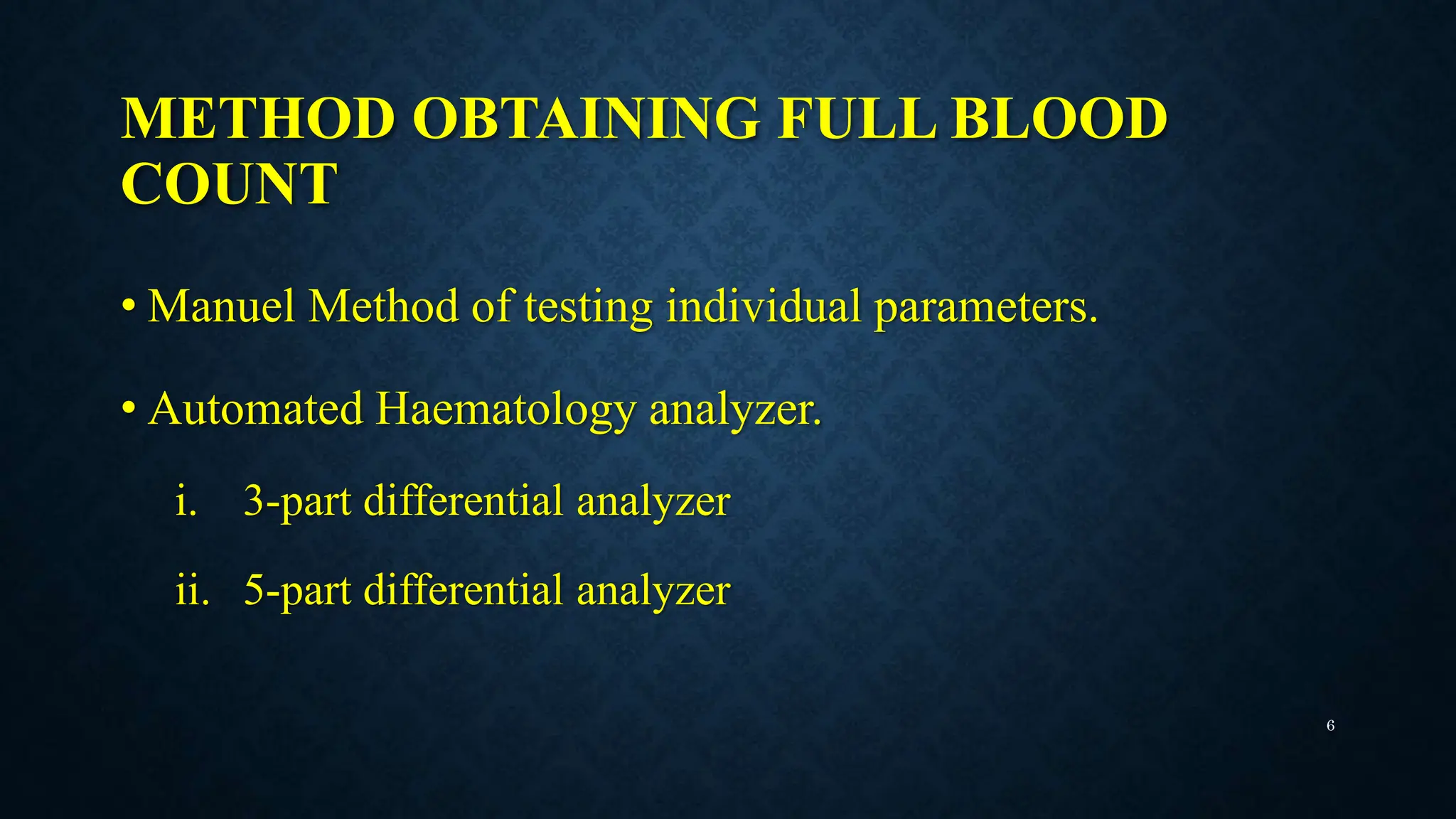
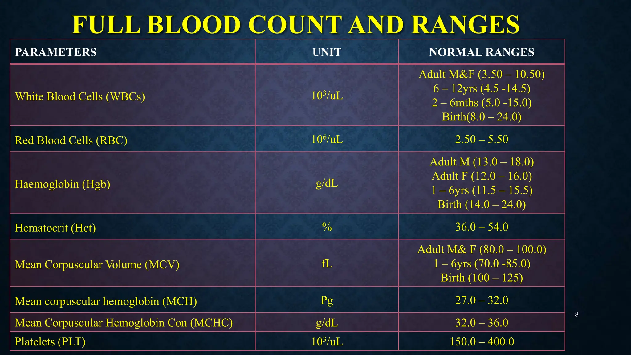
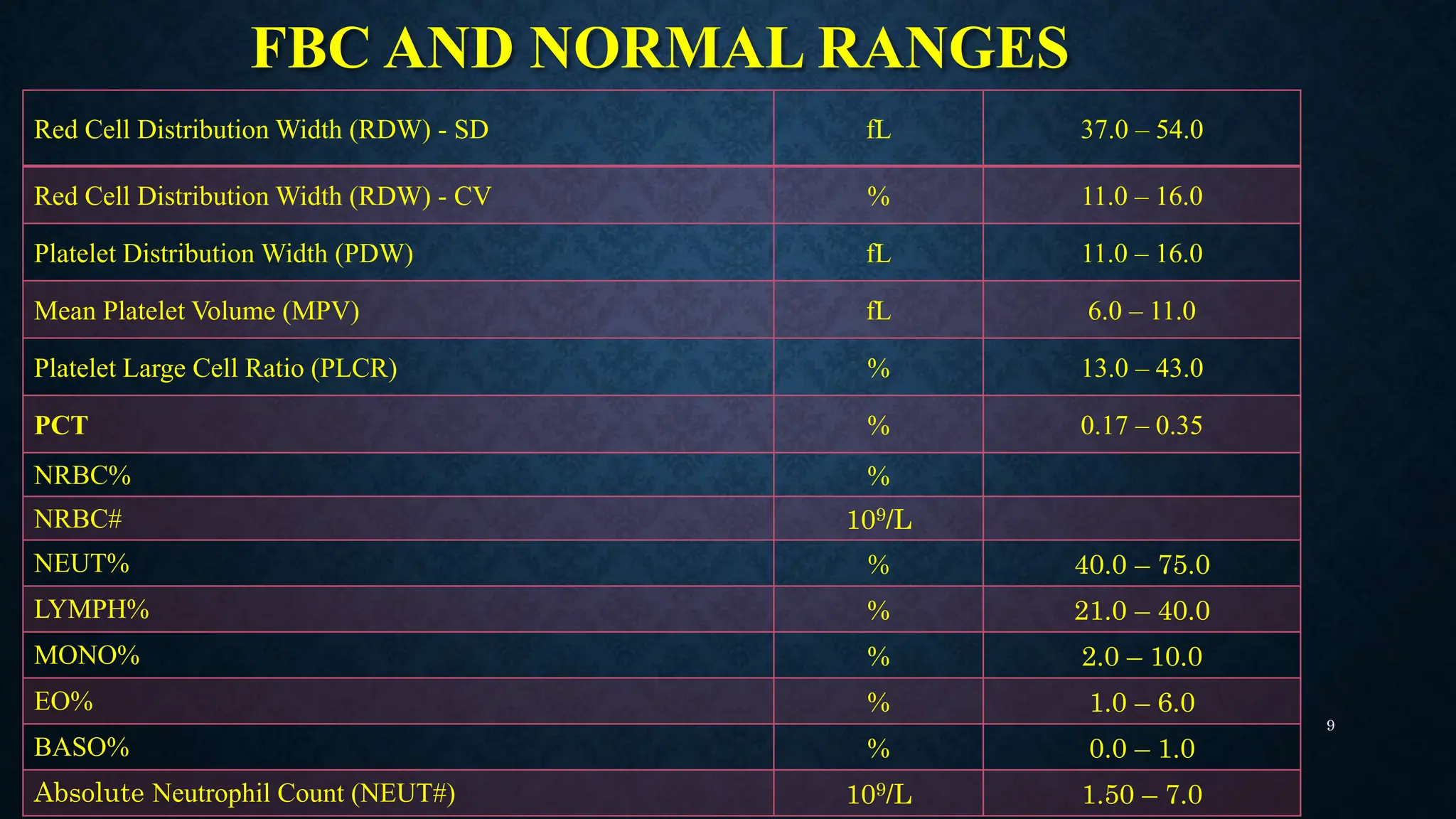
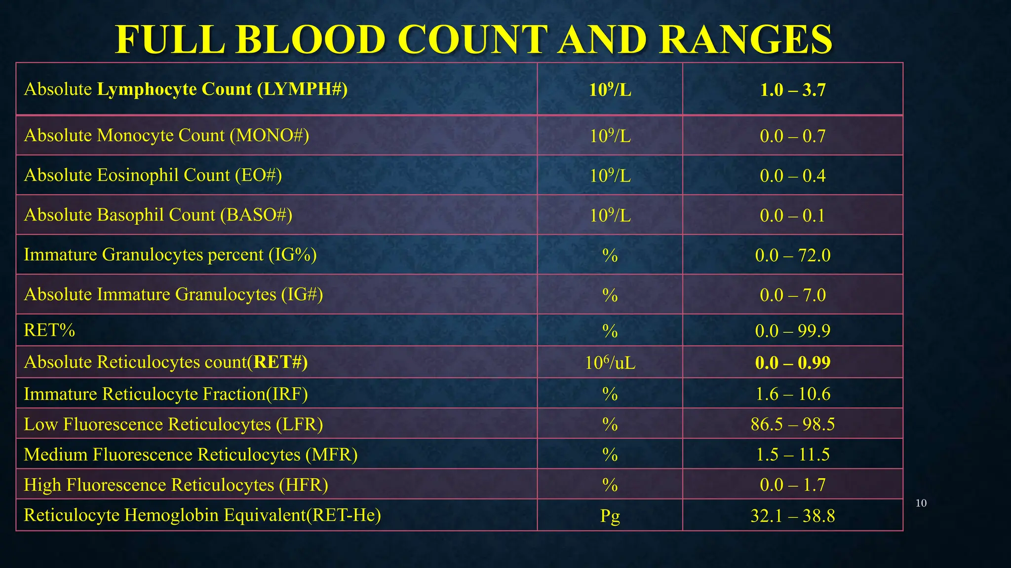
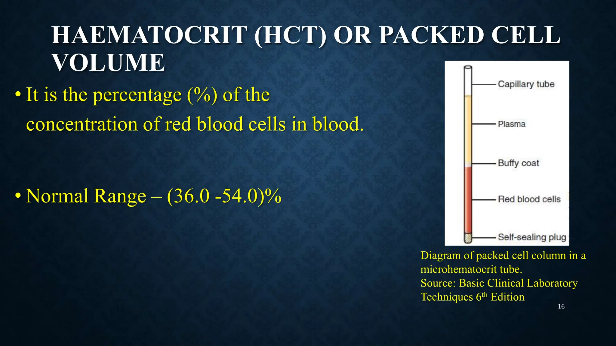
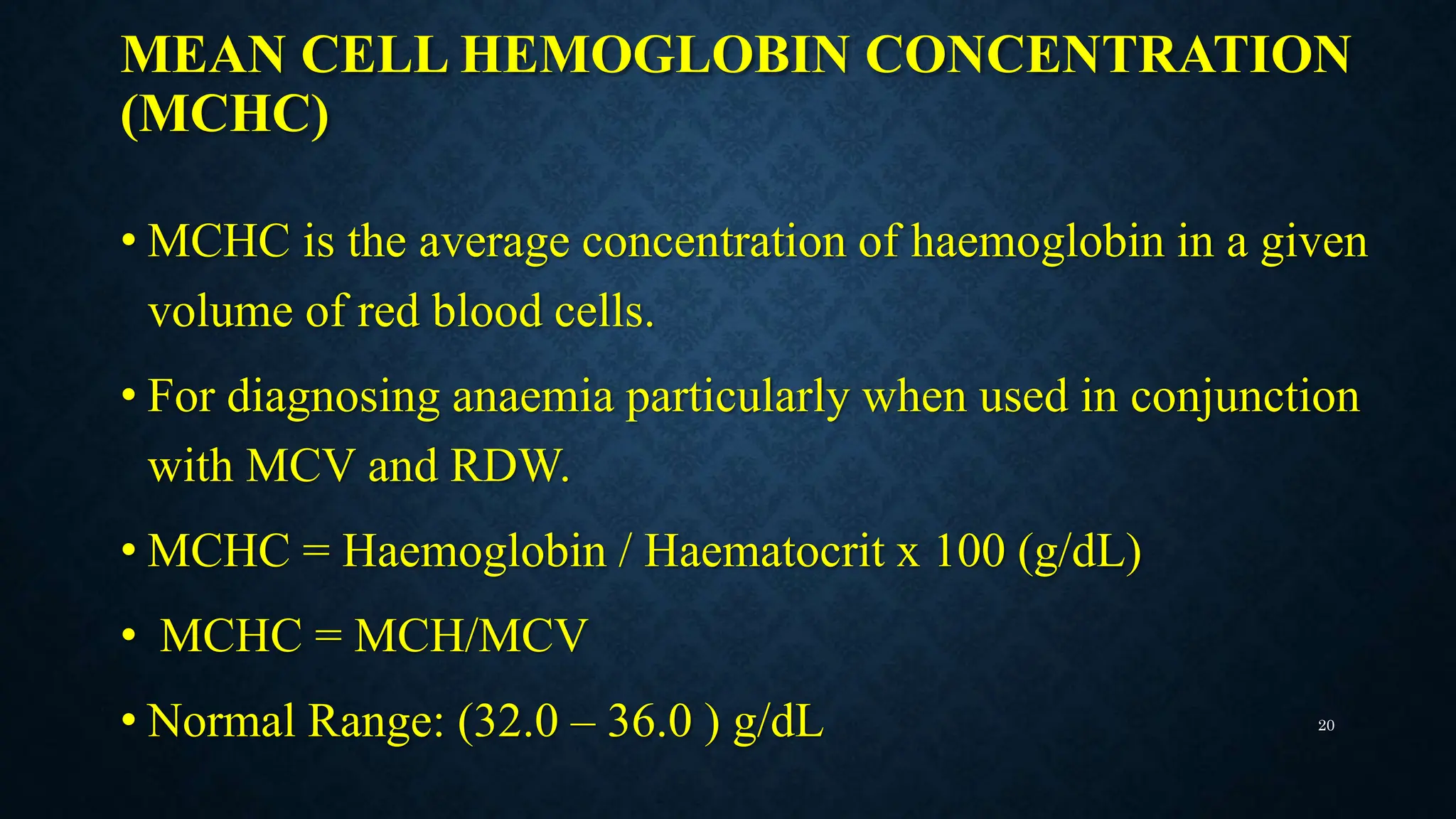
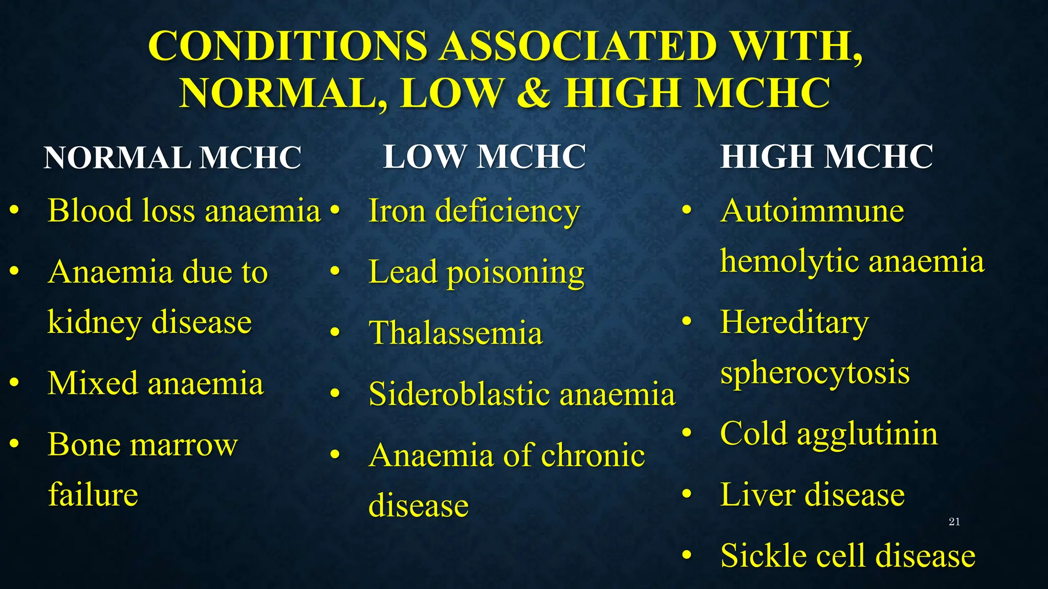
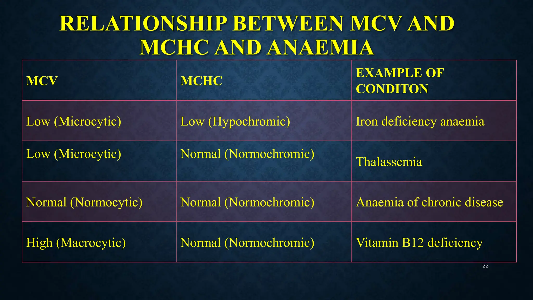
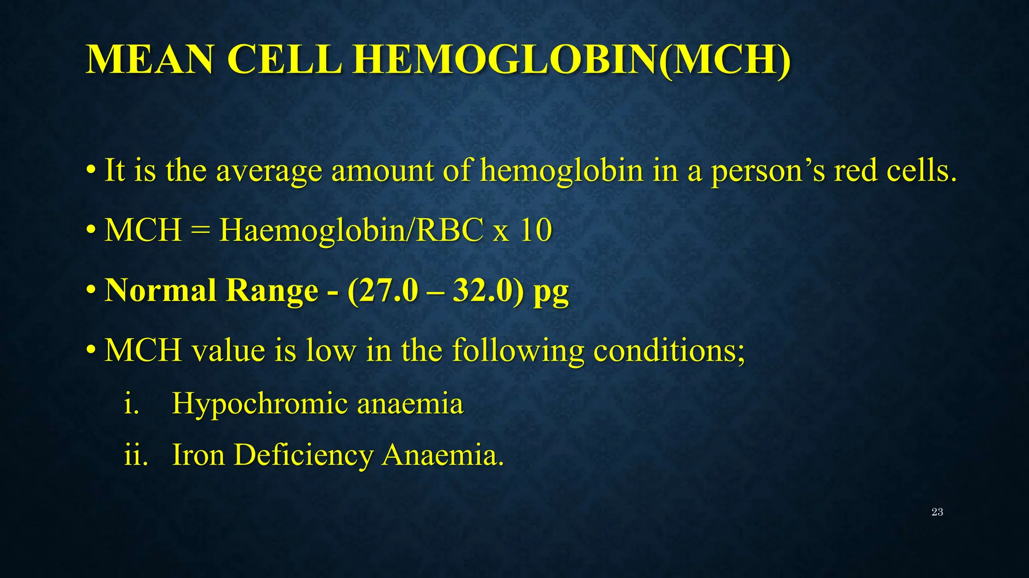
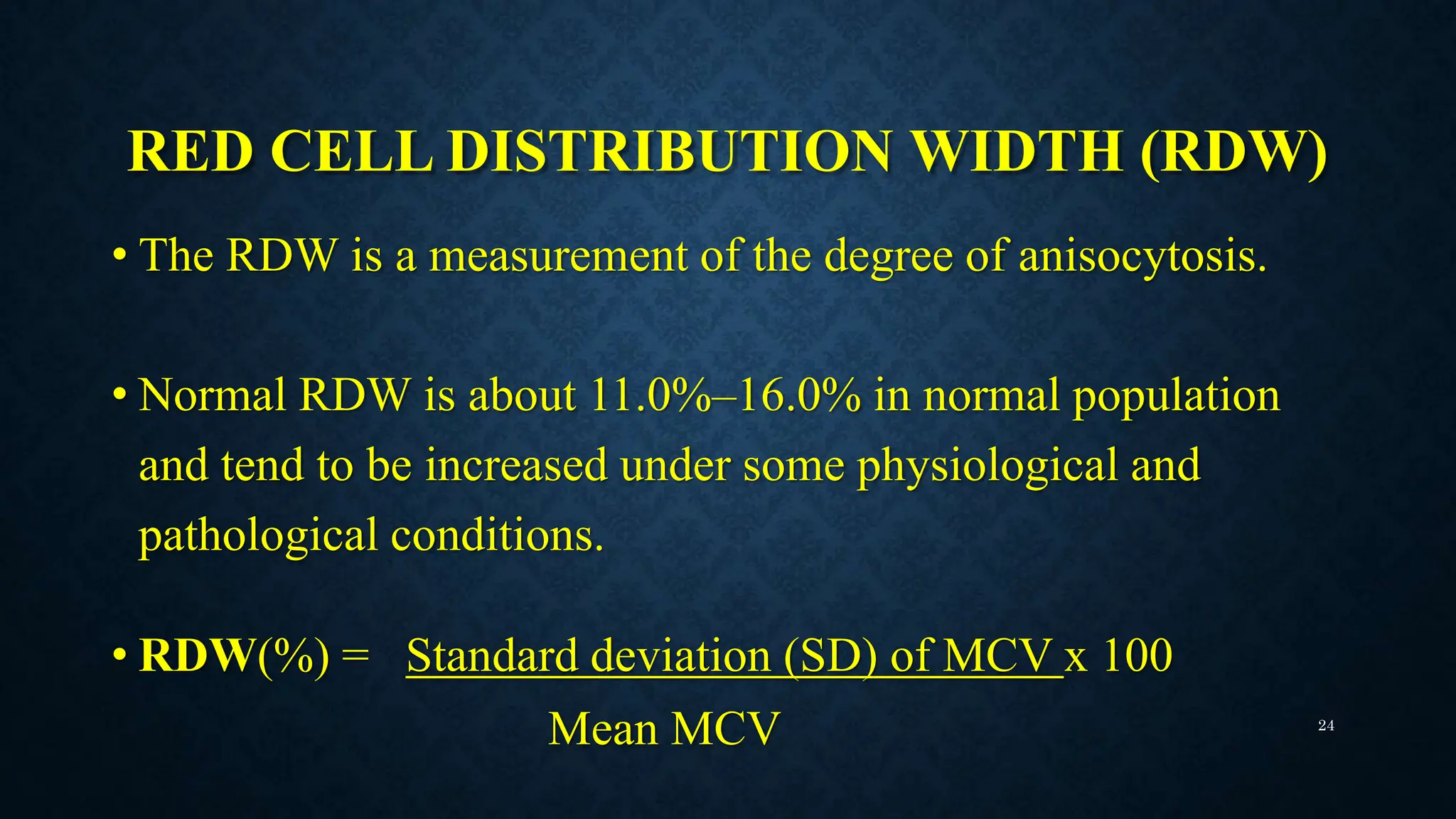
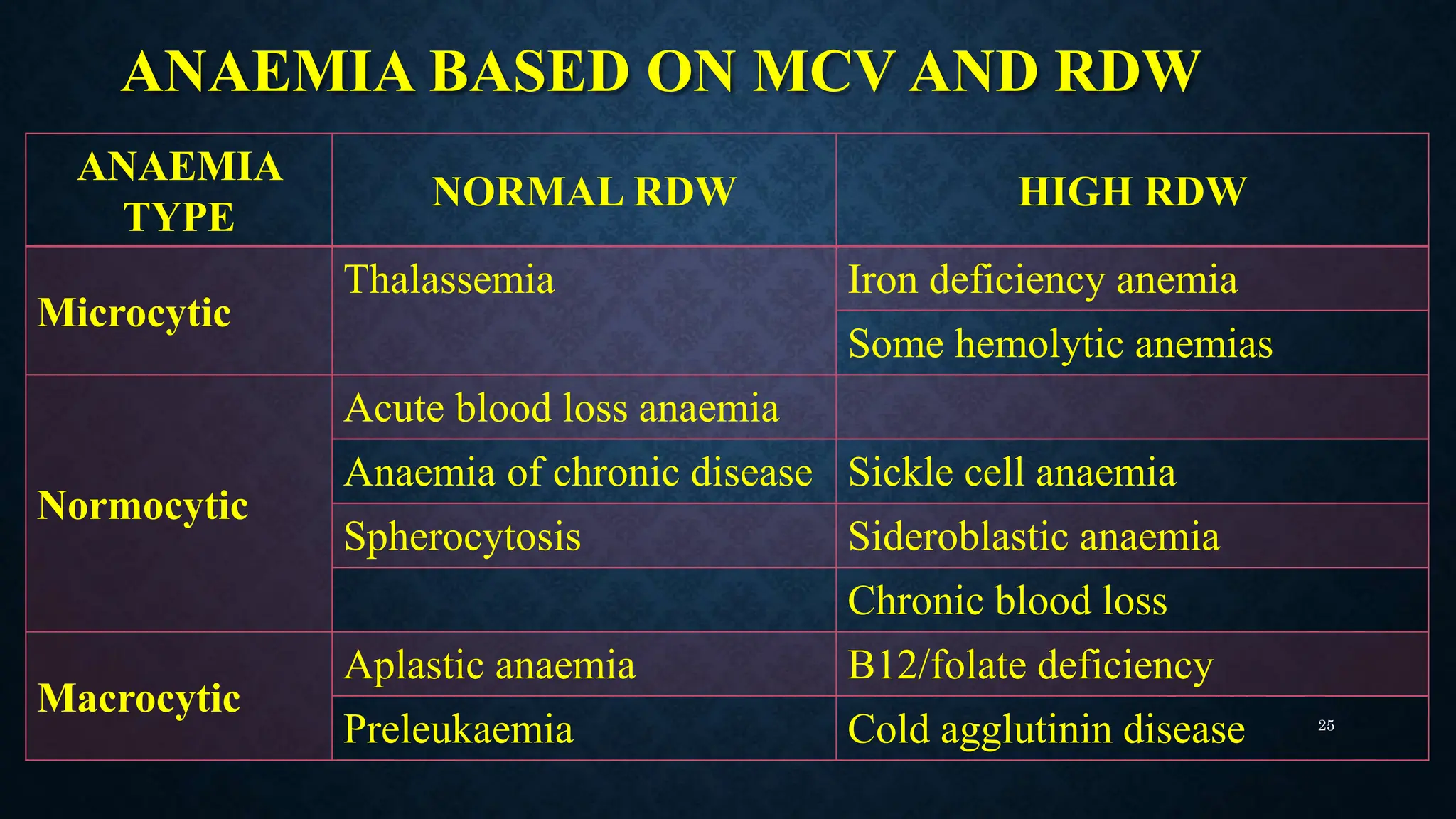
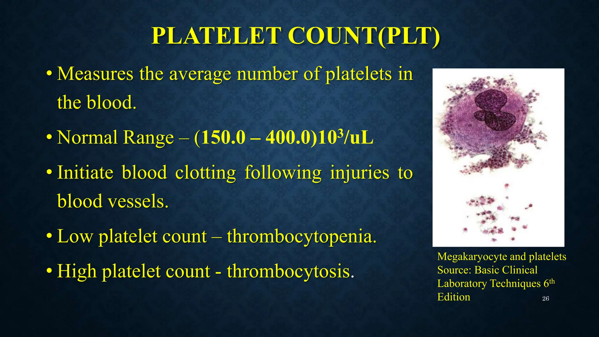
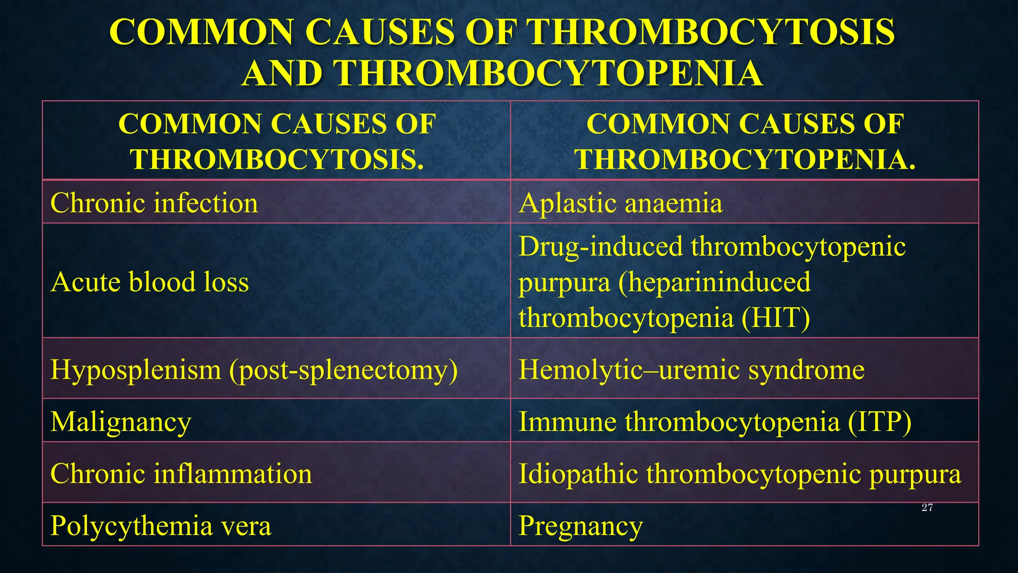
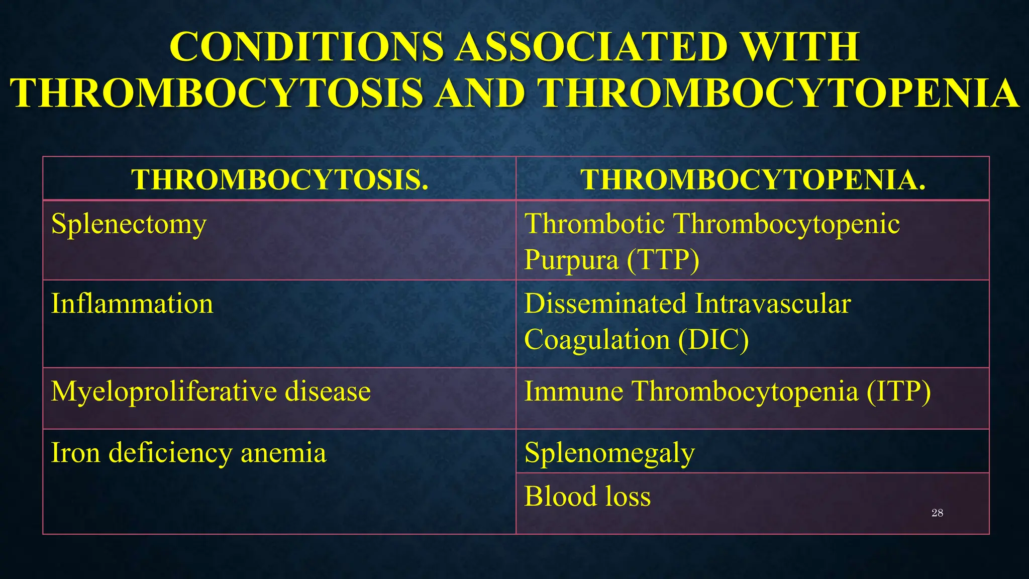
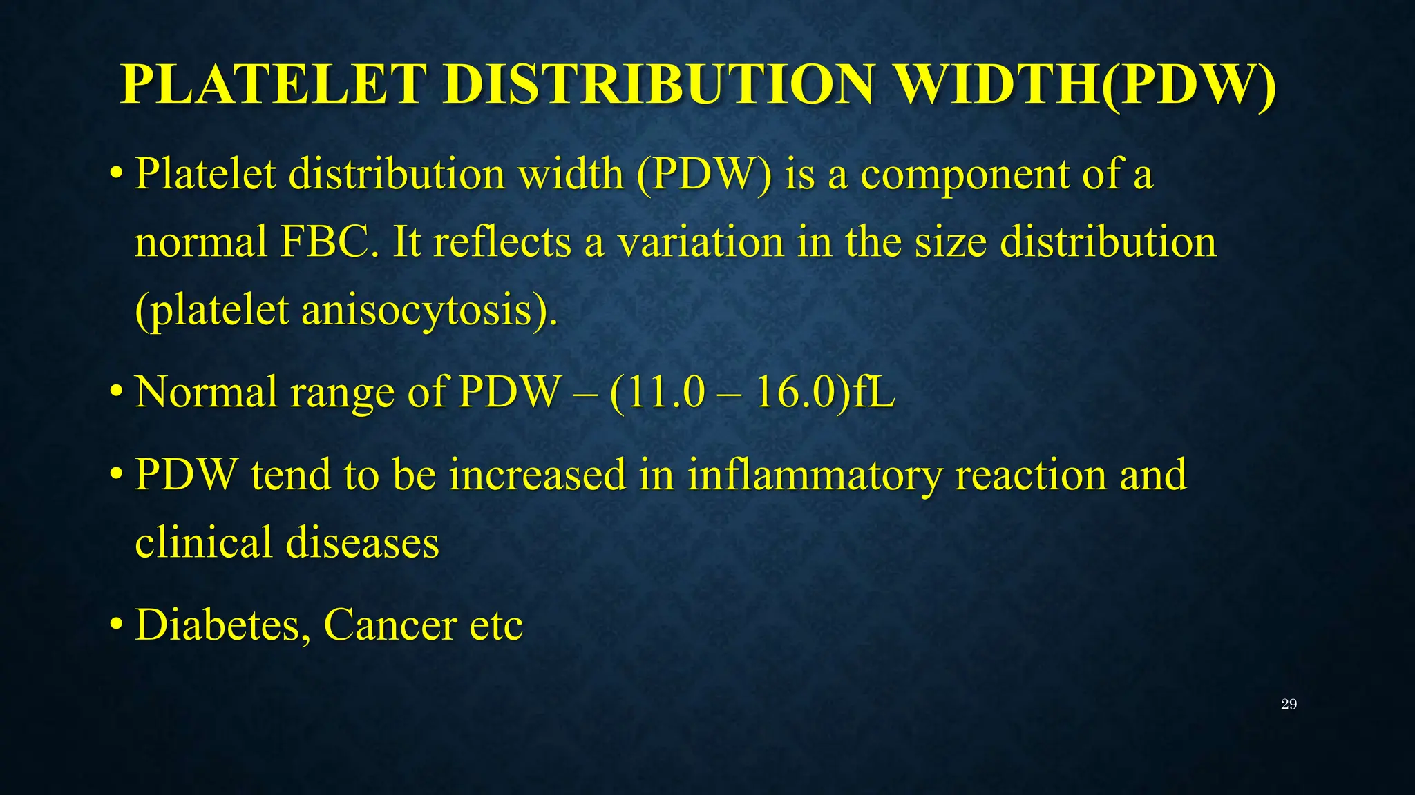
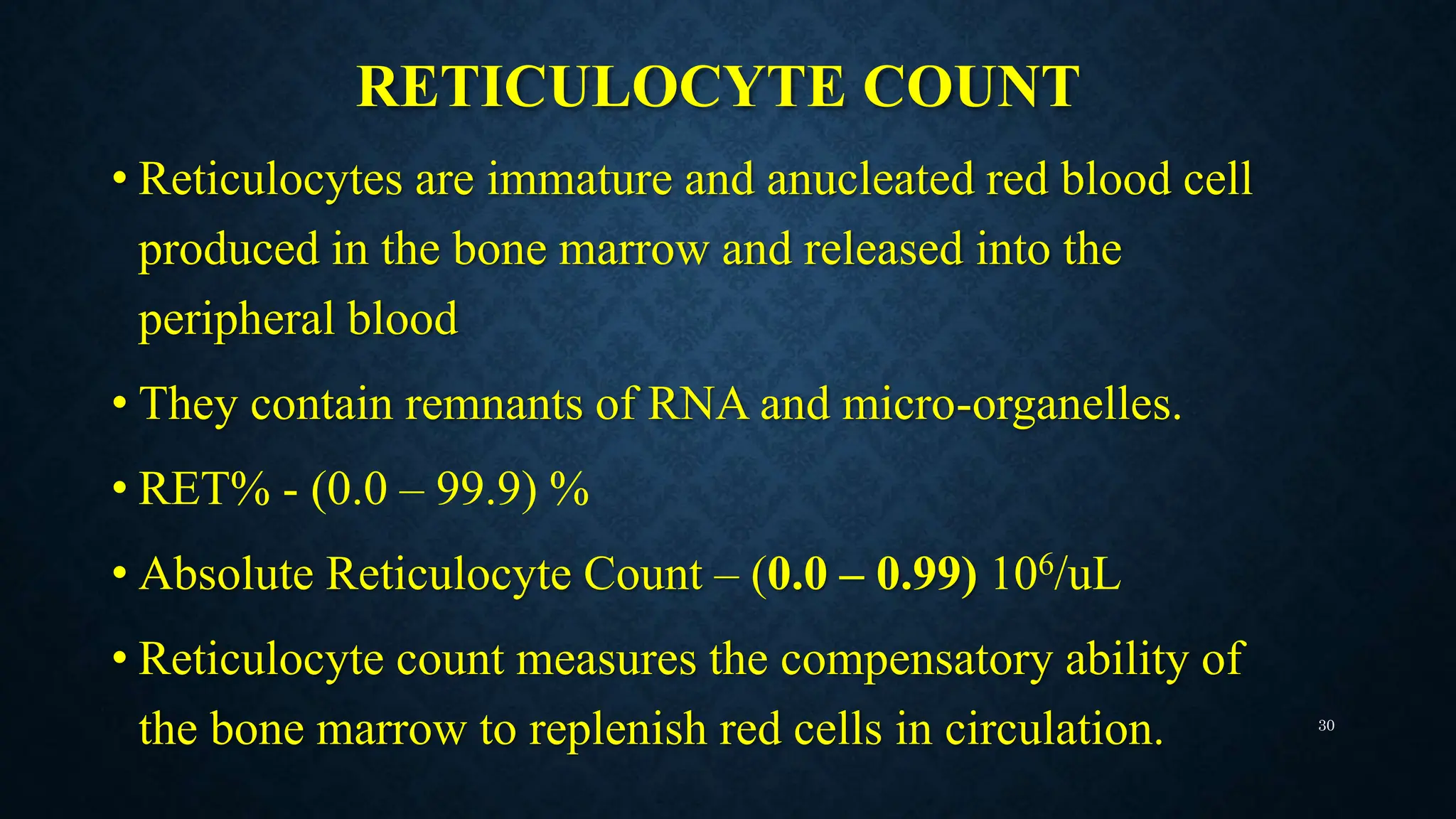
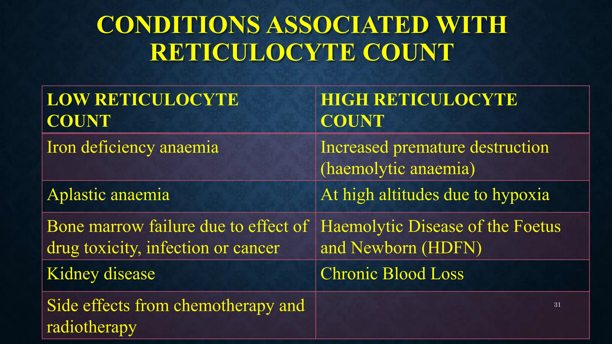
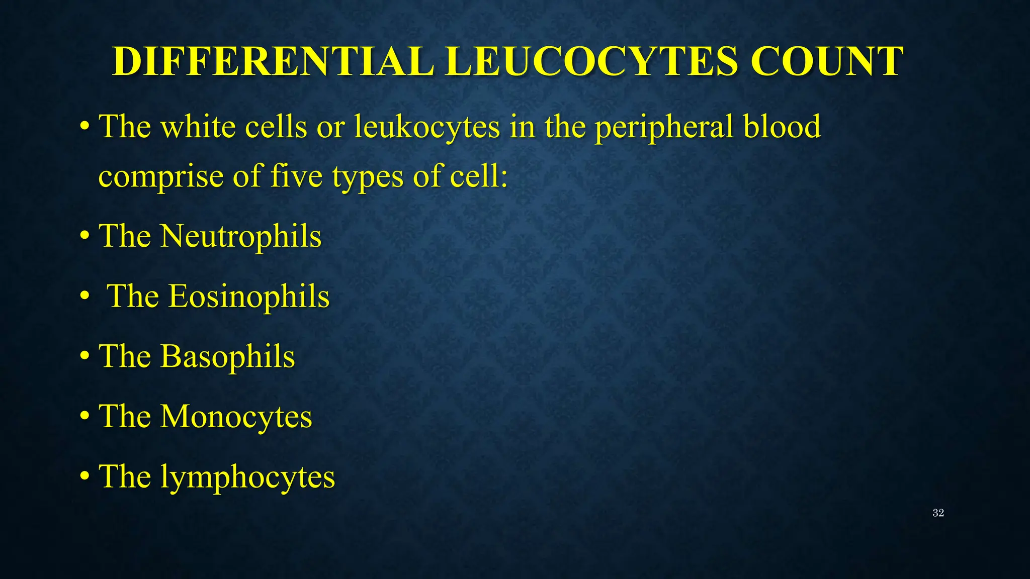
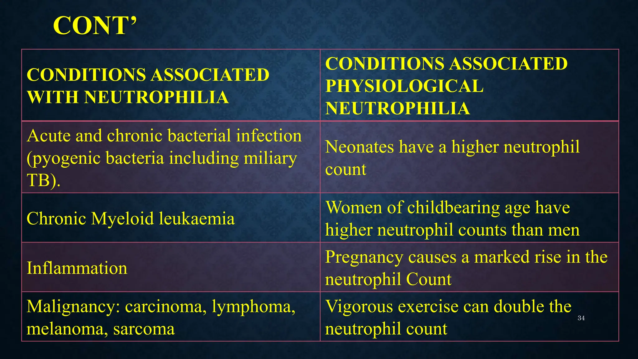
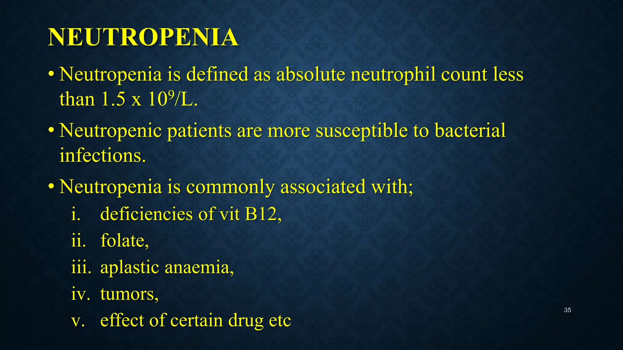
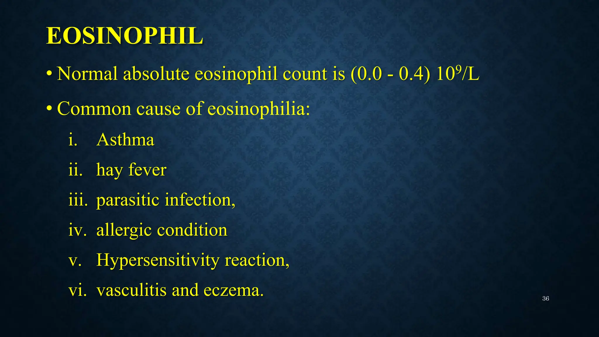
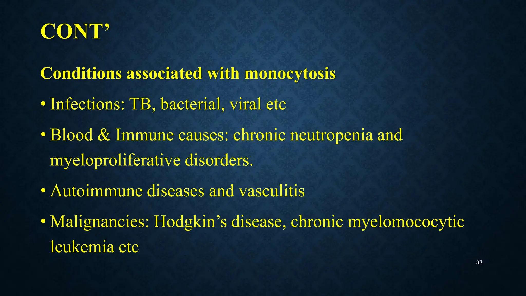
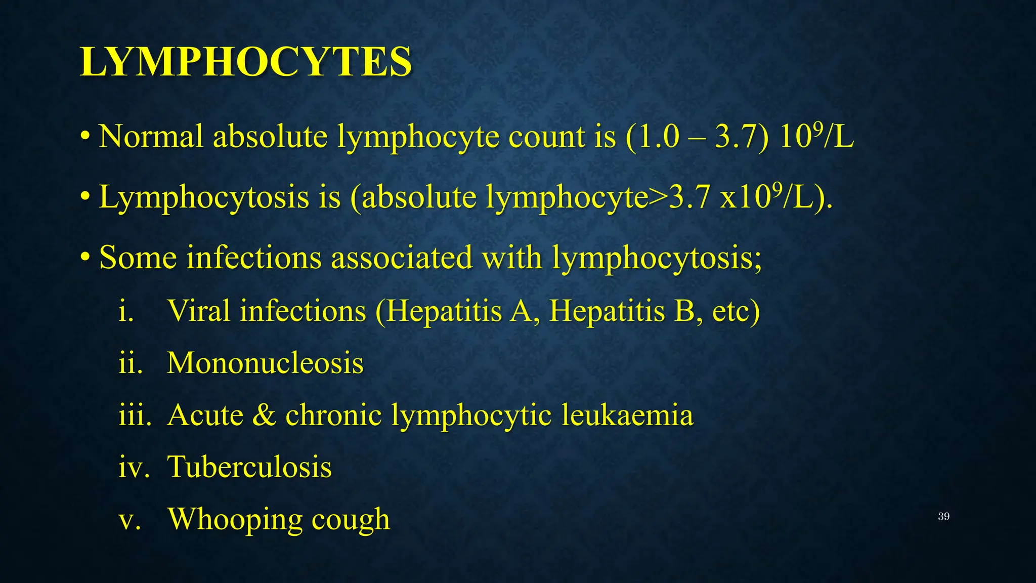
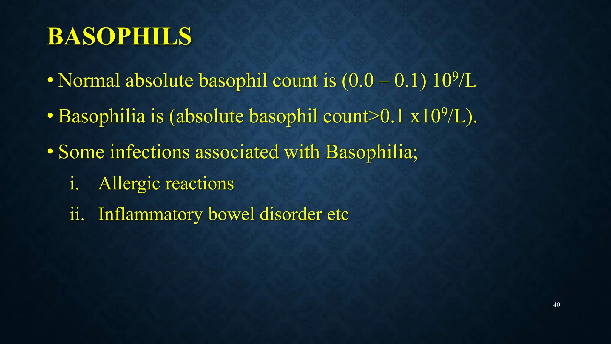
INTERPRETATION OF FULL
BLOOD COUNT
Presented By
 Gamor Kwame Dickson
OUTLINE OF PRESENTATION
• Introduction to full blood count
• Types of full blood count components.
• Importance of full blood count (FBC) results interpretation.
• Methods of obtaining FBC.
• Interpretation of results.
• Conclusion 2
FULL BLOOD COUNT
• One of the most commonly ordered laboratory tests and the most
ordered test in the hematology lab.
• Measure of the cellular components in the blood: white blood cells,
red blood cells, platelets and features of blood.
• It gives useful information about the production of all blood cells.
• It also provides information about the immune system through the
evaluation of the white blood cell (WBC) count.
3
CONT’
• When interpreted carefully and correctly in relation to
clinical history, it provides information that assists;
• Diagnosis (anaemia, cancer, clotting issues, infections &
immune system disorder)
• Drug monitoring
• Management of diseases
4
THE CONSTITUENTS OF THE FULL
BLOOD COUNT
• Red blood cells (RBC) count and its indices.
• White blood cells (WBC) count with differential count of the
various leukocytes.
• The Platelet count.
5
METHOD OBTAINING FULL BLOOD
COUNT
• Manuel Method of testing individual parameters.
• Automated Haematology analyzer.
i. 3-part differential analyzer
ii. 5-part differential analyzer
6
Picture of Beckman-Coulter AcT 3-part differential
haematology analyzer.
Source: https://portal.issn.org/resource/ISSN/2578-501X
Sysmex XN Series 5-Part differential Haematology
Autoanalyzer
Source: https://portal.issn.org/resource/ISSN/2578-501X
7
FULL BLOOD COUNT AND RANGES
PARAMETERS UNIT NORMAL RANGES
White Blood Cells (WBCs) 103/uL
Adult M&F (3.50 – 10.50)
6 – 12yrs (4.5 -14.5)
2 – 6mths (5.0 -15.0)
Birth(8.0 – 24.0)
Red Blood Cells (RBC) 106/uL 2.50 – 5.50
Haemoglobin (Hgb) g/dL
Adult M (13.0 – 18.0)
Adult F (12.0 – 16.0)
1 – 6yrs (11.5 – 15.5)
Birth (14.0 – 24.0)
Hematocrit (Hct) % 36.0 – 54.0
Mean Corpuscular Volume (MCV) fL
Adult M& F (80.0 – 100.0)
1 – 6yrs (70.0 -85.0)
Birth (100 – 125)
Mean corpuscular hemoglobin (MCH) Pg 27.0 – 32.0
Mean Corpuscular Hemoglobin Con (MCHC) g/dL 32.0 – 36.0
Platelets (PLT) 103/uL 150.0 – 400.0
8
FBC AND NORMAL RANGES
Red Cell Distribution Width (RDW) - SD fL 37.0 – 54.0
Red Cell Distribution Width (RDW) - CV % 11.0 – 16.0
Platelet Distribution Width (PDW) fL 11.0 – 16.0
Mean Platelet Volume (MPV) fL 6.0 – 11.0
Platelet Large Cell Ratio (PLCR) % 13.0 – 43.0
PCT % 0.17 – 0.35
NRBC% %
NRBC# 109/L
NEUT% % 40.0 – 75.0
LYMPH% % 21.0 – 40.0
MONO% % 2.0 – 10.0
EO% % 1.0 – 6.0
BASO% % 0.0 – 1.0
Absolute Neutrophil Count (NEUT#) 109/L 1.50 – 7.0
9
FULL BLOOD COUNT AND RANGES
Absolute Lymphocyte Count (LYMPH#) 109/L 1.0 – 3.7
Absolute Monocyte Count (MONO#) 109/L 0.0 – 0.7
Absolute Eosinophil Count (EO#) 109/L 0.0 – 0.4
Absolute Basophil Count (BASO#) 109/L 0.0 – 0.1
Immature Granulocytes percent (IG%) % 0.0 – 72.0
Absolute Immature Granulocytes (IG#) % 0.0 – 7.0
RET% % 0.0 – 99.9
Absolute Reticulocytes count(RET#) 106/uL 0.0 – 0.99
Immature Reticulocyte Fraction(IRF) % 1.6 – 10.6
Low Fluorescence Reticulocytes (LFR) % 86.5 – 98.5
Medium Fluorescence Reticulocytes (MFR) % 1.5 – 11.5
High Fluorescence Reticulocytes (HFR) % 0.0 – 1.7
Reticulocyte Hemoglobin Equivalent(RET-He) Pg 32.1 – 38.8
10
WHITE BLOOD CELL (WBC) COUNT
• White blood cells are cellular elements which play a role in
humoral and cell mediate immunity.
• Normal range for adult male & female– (3.50 – 10.50) 103/uL
• WBC count is most often used to help diagnose disorders
related to having;
• high white blood cell count (leukocytosis)
• low white blood cell count (leucopenia)
11
CONDITIONS ASSOCIATED WITH
LEUKOCYTOSIS & LEUCOPENIA
LEUKOCYTOSIS
• Autoimmune diseases (Lupus)
• Inflammatory diseases
• Bacterial , parasitic or viral infections
• Cancers such as leukemia
• Allergic reactions
• Tissue damage
• Kidney failure
LEUCOPENIA
• Immune suppression -related diseases
(HIV/AIDS)
• Lymphoma
• Diseases of the liver or spleen
• Effect of chemotherapy and
radiotherapy
• Splenomegaly
• Effect of drugs (antibiotics)
12
HEMOGLOBIN (HGB) & RED BLOOD
CELLS
• Haemoglobin (Hb) is a protein within the
cytoplasm of the red blood cells which is
composed of the protoporphyrin ring heme
and globin.
• Haemoglobin plays a role in tissue perfusion.
• It is the most commonly used marker of
anaemia.
Red blood cells on a peripheral
smear
Source: Google
13
CONT’
Reference ranges of Haemoglobin:
• Adult Male (13.0 – 18.0)g/dL
• Adult Female (12.0 – 16.0)g/dL
• Infants1 – 6yrs (11.5 – 15.5)g/dL
• Birth (14.0 – 24.0)g/dL
14
CONT’
• The red cell count reflects the number of circulating red blood cells.
• The red cell count is particularly useful in identifying
erythrocytosis.
• RBC Reference range – ( 2.50 – 5.50 ) 106/uL
• A decrease in the red cell count and/or haemoglobin is an indication
of anaemia, and depending on the following indices;
i. MCV
ii. MCH
iii. MCHC
15
HAEMATOCRIT (HCT) OR PACKED CELL
VOLUME
• It is the percentage (%) of the
concentration of red blood cells in blood.
• Normal Range – (36.0 -54.0)%
Diagram of packed cell column in a
microhematocrit tube.
Source: Basic Clinical Laboratory
Techniques 6th Edition
16
CONDITIONS ASSOCIATED WITH HIGH
& LOW HCT
LOW HCT
• Anaemia
• Associated with long-
term illness, infection or
white blood cell
disorders such as
leukemia or lymphoma.
• Recent or long-term
hemorrhage.
HIGH HCT
• Dehydration
• Polycythemia vera
• Lung or heart disease
• Chronic obstructive
pulmonary disease (COPD)
• Doping or erythropoietin
(EPO)
• Recent blood transfusion.
17
MEAN CELL VOLUME (MCV)
• The mean cell volume (MCV) is the volume of an average RBC in
a blood sample.
• MCV (fL) = Haematocrit (percent) x 10
RBC
• It is a measure of the average size of the red cells.
• Normal range – (80.0 – 100.0) fL
• Classification of different types of anaemia.
NORMAL MCV LOW MCV HIGH MCV
Normocytic anaemia Microcytic anaemia Macrocytic anaemia
18
CONDITIONS ASSOCIATED WITH HIGH
& LOW MCV
LOW MCV
• Iron deficiency anaemia
• Anaemia of chronic disease
• Sideroblastic anaemia
• Lead poisoning
• Spherocytosis
HIGH MCV
• Vitamin B12 deficiency
• Folate deficiency
• Liver disease
• Alcoholism
• Hypothyroidism
• Myelodysplastic syndromes
• Aplastic anaemia 19
MEAN CELL HEMOGLOBIN CONCENTRATION
(MCHC)
• MCHC is the average concentration of haemoglobin in a given
volume of red blood cells.
• For diagnosing anaemia particularly when used in conjunction
with MCV and RDW.
• MCHC = Haemoglobin / Haematocrit x 100 (g/dL)
• MCHC = MCH/MCV
• Normal Range: (32.0 – 36.0 ) g/dL 20
CONDITIONS ASSOCIATED WITH,
NORMAL, LOW & HIGH MCHC
NORMAL MCHC
• Blood loss anaemia
• Anaemia due to
kidney disease
• Mixed anaemia
• Bone marrow
failure
LOW MCHC
• Iron deficiency
• Lead poisoning
• Thalassemia
• Sideroblastic anaemia
• Anaemia of chronic
disease
HIGH MCHC
• Autoimmune
hemolytic anaemia
• Hereditary
spherocytosis
• Cold agglutinin
• Liver disease
• Sickle cell disease
21
RELATIONSHIP BETWEEN MCV AND
MCHC AND ANAEMIA
MCV MCHC
EXAMPLE OF
CONDITON
Low (Microcytic) Low (Hypochromic) Iron deficiency anaemia
Low (Microcytic) Normal (Normochromic)
Thalassemia
Normal (Normocytic) Normal (Normochromic) Anaemia of chronic disease
High (Macrocytic) Normal (Normochromic) Vitamin B12 deficiency
22
MEAN CELL HEMOGLOBIN(MCH)
• It is the average amount of hemoglobin in a person’s red cells.
• MCH = Haemoglobin/RBC x 10
• Normal Range - (27.0 – 32.0) pg
• MCH value is low in the following conditions;
i. Hypochromic anaemia
ii. Iron Deficiency Anaemia.
23
RED CELL DISTRIBUTION WIDTH (RDW)
• The RDW is a measurement of the degree of anisocytosis.
• Normal RDW is about 11.0%–16.0% in normal population
and tend to be increased under some physiological and
pathological conditions.
• RDW(%) = Standard deviation (SD) of MCV x 100
Mean MCV 24
ANAEMIA
TYPE
NORMAL RDW HIGH RDW
Microcytic
Thalassemia Iron deficiency anemia
Some hemolytic anemias
Normocytic
Acute blood loss anaemia
Anaemia of chronic disease Sickle cell anaemia
Spherocytosis Sideroblastic anaemia
Chronic blood loss
Macrocytic
Aplastic anaemia B12/folate deficiency
Preleukaemia Cold agglutinin disease
ANAEMIA BASED ON MCV AND RDW
25
PLATELET COUNT(PLT)
• Measures the average number of platelets in
the blood.
• Normal Range – (150.0 – 400.0)103/uL
• Initiate blood clotting following injuries to
blood vessels.
• Low platelet count – thrombocytopenia.
• High platelet count - thrombocytosis.
Megakaryocyte and platelets
Source: Basic Clinical
Laboratory Techniques 6th
Edition 26
COMMON CAUSES OF THROMBOCYTOSIS
AND THROMBOCYTOPENIA
COMMON CAUSES OF
THROMBOCYTOSIS.
COMMON CAUSES OF
THROMBOCYTOPENIA.
Chronic infection Aplastic anaemia
Acute blood loss
Drug-induced thrombocytopenic
purpura (heparininduced
thrombocytopenia (HIT)
Hyposplenism (post-splenectomy) Hemolytic–uremic syndrome
Malignancy Immune thrombocytopenia (ITP)
Chronic inflammation Idiopathic thrombocytopenic purpura
Polycythemia vera Pregnancy
27
CONDITIONS ASSOCIATED WITH
THROMBOCYTOSIS AND THROMBOCYTOPENIA
THROMBOCYTOSIS. THROMBOCYTOPENIA.
Splenectomy Thrombotic Thrombocytopenic
Purpura (TTP)
Inflammation Disseminated Intravascular
Coagulation (DIC)
Myeloproliferative disease Immune Thrombocytopenia (ITP)
Iron deficiency anemia Splenomegaly
Blood loss 28
PLATELET DISTRIBUTION WIDTH(PDW)
• Platelet distribution width (PDW) is a component of a
normal FBC. It reflects a variation in the size distribution
(platelet anisocytosis).
• Normal range of PDW – (11.0 – 16.0)fL
• PDW tend to be increased in inflammatory reaction and
clinical diseases
• Diabetes, Cancer etc
29
RETICULOCYTE COUNT
• Reticulocytes are immature and anucleated red blood cell
produced in the bone marrow and released into the
peripheral blood
• They contain remnants of RNA and micro-organelles.
• RET% - (0.0 – 99.9) %
• Absolute Reticulocyte Count – (0.0 – 0.99) 106/uL
• Reticulocyte count measures the compensatory ability of
the bone marrow to replenish red cells in circulation. 30
CONDITIONS ASSOCIATED WITH
RETICULOCYTE COUNT
LOW RETICULOCYTE
COUNT
HIGH RETICULOCYTE
COUNT
Iron deficiency anaemia Increased premature destruction
(haemolytic anaemia)
Aplastic anaemia At high altitudes due to hypoxia
Bone marrow failure due to effect of
drug toxicity, infection or cancer
Haemolytic Disease of the Foetus
and Newborn (HDFN)
Kidney disease Chronic Blood Loss
Side effects from chemotherapy and
radiotherapy
31
DIFFERENTIAL LEUCOCYTES COUNT
• The white cells or leukocytes in the peripheral blood
comprise of five types of cell:
• The Neutrophils
• The Eosinophils
• The Basophils
• The Monocytes
• The lymphocytes
32
NEUTROPHILIA
• Neutrophilia is defined as absolute neutrophil count above
7.0 x 10⁹/L.
• Normal absolute neutrophil count is 1.5 - 7.0 x 10⁹/L
• Neutrophilia is commonly seen in patients with bacterial
infection.
• Physiological Neutrophilia can occur as a normal
physiological process.
33
CONT’
CONDITIONS ASSOCIATED
WITH NEUTROPHILIA
CONDITIONS ASSOCIATED
PHYSIOLOGICAL
NEUTROPHILIA
Acute and chronic bacterial infection
(pyogenic bacteria including miliary
TB).
Neonates have a higher neutrophil
count
Chronic Myeloid leukaemia
Women of childbearing age have
higher neutrophil counts than men
Inflammation
Pregnancy causes a marked rise in the
neutrophil Count
Malignancy: carcinoma, lymphoma,
melanoma, sarcoma
Vigorous exercise can double the
neutrophil count
34
NEUTROPENIA
• Neutropenia is defined as absolute neutrophil count less
than 1.5 x 10⁹/L.
• Neutropenic patients are more susceptible to bacterial
infections.
• Neutropenia is commonly associated with;
i. deficiencies of vit B12,
ii. folate,
iii. aplastic anaemia,
iv. tumors,
v. effect of certain drug etc
35
EOSINOPHIL
• Normal absolute eosinophil count is (0.0 - 0.4) 109/L
• Common cause of eosinophilia:
i. Asthma
ii. hay fever
iii. parasitic infection,
iv. allergic condition
v. Hypersensitivity reaction,
vi. vasculitis and eczema. 36
MONOCYTE
• Normal absolute monocyte count is (0.0 – 0.7) 109/L
• Monocytosis is an increase in the number of monocytes
circulating in the blood.
• Monocytosis can be a feature in chronic infection.
• A persistent monocytosis that is unexplained, particularly if
associated with anaemia or thrombocytopenia, may be a
feature of myelodysplastic and myeloproliferative disorders
37
CONT’
Conditions associated with monocytosis
• Infections: TB, bacterial, viral etc
• Blood & Immune causes: chronic neutropenia and
myeloproliferative disorders.
• Autoimmune diseases and vasculitis
• Malignancies: Hodgkin’s disease, chronic myelomococytic
leukemia etc
38
LYMPHOCYTES
• Normal absolute lymphocyte count is (1.0 – 3.7) 109/L
• Lymphocytosis is (absolute lymphocyte>3.7 x10⁹/L).
• Some infections associated with lymphocytosis;
i. Viral infections (Hepatitis A, Hepatitis B, etc)
ii. Mononucleosis
iii. Acute & chronic lymphocytic leukaemia
iv. Tuberculosis
v. Whooping cough 39
BASOPHILS
• Normal absolute basophil count is (0.0 – 0.1) 109/L
• Basophilia is (absolute basophil count>0.1 x10⁹/L).
• Some infections associated with Basophilia;
i. Allergic reactions
ii. Inflammatory bowel disorder etc
40
CONCLUSION
• National service personnel should be able to interpret all
full blood count (FBC) for both normal and abnormal
results.
41
REFERENCES
• Erhabor, O., Ha, M., Muhammad, K., Onwuchekwa, C., &
Nb, E. (2021). Interpretation of Full Blood Count
Parameters in Health and Disease.
https://doi.org/10.23880/hij-16000180
• Hematologist, C. (2020). The Basic Principles &
Interpretation of Automated Full Blood Coun t.
• Learning, C., Reserved, A. R., & Learning, C. (n.d.). B asic
C linical L aboratory T echniques. 42
THANK YOU
43

Full Blood Count (FBC) Interpretation.pptx

  • 1.
    INTERPRETATION OF FULL BLOODCOUNT Presented By  Gamor Kwame Dickson
  • 2.
    OUTLINE OF PRESENTATION •Introduction to full blood count • Types of full blood count components. • Importance of full blood count (FBC) results interpretation. • Methods of obtaining FBC. • Interpretation of results. • Conclusion 2
  • 3.
    FULL BLOOD COUNT •One of the most commonly ordered laboratory tests and the most ordered test in the hematology lab. • Measure of the cellular components in the blood: white blood cells, red blood cells, platelets and features of blood. • It gives useful information about the production of all blood cells. • It also provides information about the immune system through the evaluation of the white blood cell (WBC) count. 3
  • 4.
    CONT’ • When interpretedcarefully and correctly in relation to clinical history, it provides information that assists; • Diagnosis (anaemia, cancer, clotting issues, infections & immune system disorder) • Drug monitoring • Management of diseases 4
  • 5.
    THE CONSTITUENTS OFTHE FULL BLOOD COUNT • Red blood cells (RBC) count and its indices. • White blood cells (WBC) count with differential count of the various leukocytes. • The Platelet count. 5
  • 6.
    METHOD OBTAINING FULLBLOOD COUNT • Manuel Method of testing individual parameters. • Automated Haematology analyzer. i. 3-part differential analyzer ii. 5-part differential analyzer 6
  • 7.
    Picture of Beckman-CoulterAcT 3-part differential haematology analyzer. Source: https://portal.issn.org/resource/ISSN/2578-501X Sysmex XN Series 5-Part differential Haematology Autoanalyzer Source: https://portal.issn.org/resource/ISSN/2578-501X 7
  • 8.
    FULL BLOOD COUNTAND RANGES PARAMETERS UNIT NORMAL RANGES White Blood Cells (WBCs) 103/uL Adult M&F (3.50 – 10.50) 6 – 12yrs (4.5 -14.5) 2 – 6mths (5.0 -15.0) Birth(8.0 – 24.0) Red Blood Cells (RBC) 106/uL 2.50 – 5.50 Haemoglobin (Hgb) g/dL Adult M (13.0 – 18.0) Adult F (12.0 – 16.0) 1 – 6yrs (11.5 – 15.5) Birth (14.0 – 24.0) Hematocrit (Hct) % 36.0 – 54.0 Mean Corpuscular Volume (MCV) fL Adult M& F (80.0 – 100.0) 1 – 6yrs (70.0 -85.0) Birth (100 – 125) Mean corpuscular hemoglobin (MCH) Pg 27.0 – 32.0 Mean Corpuscular Hemoglobin Con (MCHC) g/dL 32.0 – 36.0 Platelets (PLT) 103/uL 150.0 – 400.0 8
  • 9.
    FBC AND NORMALRANGES Red Cell Distribution Width (RDW) - SD fL 37.0 – 54.0 Red Cell Distribution Width (RDW) - CV % 11.0 – 16.0 Platelet Distribution Width (PDW) fL 11.0 – 16.0 Mean Platelet Volume (MPV) fL 6.0 – 11.0 Platelet Large Cell Ratio (PLCR) % 13.0 – 43.0 PCT % 0.17 – 0.35 NRBC% % NRBC# 109/L NEUT% % 40.0 – 75.0 LYMPH% % 21.0 – 40.0 MONO% % 2.0 – 10.0 EO% % 1.0 – 6.0 BASO% % 0.0 – 1.0 Absolute Neutrophil Count (NEUT#) 109/L 1.50 – 7.0 9
  • 10.
    FULL BLOOD COUNTAND RANGES Absolute Lymphocyte Count (LYMPH#) 109/L 1.0 – 3.7 Absolute Monocyte Count (MONO#) 109/L 0.0 – 0.7 Absolute Eosinophil Count (EO#) 109/L 0.0 – 0.4 Absolute Basophil Count (BASO#) 109/L 0.0 – 0.1 Immature Granulocytes percent (IG%) % 0.0 – 72.0 Absolute Immature Granulocytes (IG#) % 0.0 – 7.0 RET% % 0.0 – 99.9 Absolute Reticulocytes count(RET#) 106/uL 0.0 – 0.99 Immature Reticulocyte Fraction(IRF) % 1.6 – 10.6 Low Fluorescence Reticulocytes (LFR) % 86.5 – 98.5 Medium Fluorescence Reticulocytes (MFR) % 1.5 – 11.5 High Fluorescence Reticulocytes (HFR) % 0.0 – 1.7 Reticulocyte Hemoglobin Equivalent(RET-He) Pg 32.1 – 38.8 10
  • 11.
    WHITE BLOOD CELL(WBC) COUNT • White blood cells are cellular elements which play a role in humoral and cell mediate immunity. • Normal range for adult male & female– (3.50 – 10.50) 103/uL • WBC count is most often used to help diagnose disorders related to having; • high white blood cell count (leukocytosis) • low white blood cell count (leucopenia) 11
  • 12.
    CONDITIONS ASSOCIATED WITH LEUKOCYTOSIS& LEUCOPENIA LEUKOCYTOSIS • Autoimmune diseases (Lupus) • Inflammatory diseases • Bacterial , parasitic or viral infections • Cancers such as leukemia • Allergic reactions • Tissue damage • Kidney failure LEUCOPENIA • Immune suppression -related diseases (HIV/AIDS) • Lymphoma • Diseases of the liver or spleen • Effect of chemotherapy and radiotherapy • Splenomegaly • Effect of drugs (antibiotics) 12
  • 13.
    HEMOGLOBIN (HGB) &RED BLOOD CELLS • Haemoglobin (Hb) is a protein within the cytoplasm of the red blood cells which is composed of the protoporphyrin ring heme and globin. • Haemoglobin plays a role in tissue perfusion. • It is the most commonly used marker of anaemia. Red blood cells on a peripheral smear Source: Google 13
  • 14.
    CONT’ Reference ranges ofHaemoglobin: • Adult Male (13.0 – 18.0)g/dL • Adult Female (12.0 – 16.0)g/dL • Infants1 – 6yrs (11.5 – 15.5)g/dL • Birth (14.0 – 24.0)g/dL 14
  • 15.
    CONT’ • The redcell count reflects the number of circulating red blood cells. • The red cell count is particularly useful in identifying erythrocytosis. • RBC Reference range – ( 2.50 – 5.50 ) 106/uL • A decrease in the red cell count and/or haemoglobin is an indication of anaemia, and depending on the following indices; i. MCV ii. MCH iii. MCHC 15
  • 16.
    HAEMATOCRIT (HCT) ORPACKED CELL VOLUME • It is the percentage (%) of the concentration of red blood cells in blood. • Normal Range – (36.0 -54.0)% Diagram of packed cell column in a microhematocrit tube. Source: Basic Clinical Laboratory Techniques 6th Edition 16
  • 17.
    CONDITIONS ASSOCIATED WITHHIGH & LOW HCT LOW HCT • Anaemia • Associated with long- term illness, infection or white blood cell disorders such as leukemia or lymphoma. • Recent or long-term hemorrhage. HIGH HCT • Dehydration • Polycythemia vera • Lung or heart disease • Chronic obstructive pulmonary disease (COPD) • Doping or erythropoietin (EPO) • Recent blood transfusion. 17
  • 18.
    MEAN CELL VOLUME(MCV) • The mean cell volume (MCV) is the volume of an average RBC in a blood sample. • MCV (fL) = Haematocrit (percent) x 10 RBC • It is a measure of the average size of the red cells. • Normal range – (80.0 – 100.0) fL • Classification of different types of anaemia. NORMAL MCV LOW MCV HIGH MCV Normocytic anaemia Microcytic anaemia Macrocytic anaemia 18
  • 19.
    CONDITIONS ASSOCIATED WITHHIGH & LOW MCV LOW MCV • Iron deficiency anaemia • Anaemia of chronic disease • Sideroblastic anaemia • Lead poisoning • Spherocytosis HIGH MCV • Vitamin B12 deficiency • Folate deficiency • Liver disease • Alcoholism • Hypothyroidism • Myelodysplastic syndromes • Aplastic anaemia 19
  • 20.
    MEAN CELL HEMOGLOBINCONCENTRATION (MCHC) • MCHC is the average concentration of haemoglobin in a given volume of red blood cells. • For diagnosing anaemia particularly when used in conjunction with MCV and RDW. • MCHC = Haemoglobin / Haematocrit x 100 (g/dL) • MCHC = MCH/MCV • Normal Range: (32.0 – 36.0 ) g/dL 20
  • 21.
    CONDITIONS ASSOCIATED WITH, NORMAL,LOW & HIGH MCHC NORMAL MCHC • Blood loss anaemia • Anaemia due to kidney disease • Mixed anaemia • Bone marrow failure LOW MCHC • Iron deficiency • Lead poisoning • Thalassemia • Sideroblastic anaemia • Anaemia of chronic disease HIGH MCHC • Autoimmune hemolytic anaemia • Hereditary spherocytosis • Cold agglutinin • Liver disease • Sickle cell disease 21
  • 22.
    RELATIONSHIP BETWEEN MCVAND MCHC AND ANAEMIA MCV MCHC EXAMPLE OF CONDITON Low (Microcytic) Low (Hypochromic) Iron deficiency anaemia Low (Microcytic) Normal (Normochromic) Thalassemia Normal (Normocytic) Normal (Normochromic) Anaemia of chronic disease High (Macrocytic) Normal (Normochromic) Vitamin B12 deficiency 22
  • 23.
    MEAN CELL HEMOGLOBIN(MCH) •It is the average amount of hemoglobin in a person’s red cells. • MCH = Haemoglobin/RBC x 10 • Normal Range - (27.0 – 32.0) pg • MCH value is low in the following conditions; i. Hypochromic anaemia ii. Iron Deficiency Anaemia. 23
  • 24.
    RED CELL DISTRIBUTIONWIDTH (RDW) • The RDW is a measurement of the degree of anisocytosis. • Normal RDW is about 11.0%–16.0% in normal population and tend to be increased under some physiological and pathological conditions. • RDW(%) = Standard deviation (SD) of MCV x 100 Mean MCV 24
  • 25.
    ANAEMIA TYPE NORMAL RDW HIGHRDW Microcytic Thalassemia Iron deficiency anemia Some hemolytic anemias Normocytic Acute blood loss anaemia Anaemia of chronic disease Sickle cell anaemia Spherocytosis Sideroblastic anaemia Chronic blood loss Macrocytic Aplastic anaemia B12/folate deficiency Preleukaemia Cold agglutinin disease ANAEMIA BASED ON MCV AND RDW 25
  • 26.
    PLATELET COUNT(PLT) • Measuresthe average number of platelets in the blood. • Normal Range – (150.0 – 400.0)103/uL • Initiate blood clotting following injuries to blood vessels. • Low platelet count – thrombocytopenia. • High platelet count - thrombocytosis. Megakaryocyte and platelets Source: Basic Clinical Laboratory Techniques 6th Edition 26
  • 27.
    COMMON CAUSES OFTHROMBOCYTOSIS AND THROMBOCYTOPENIA COMMON CAUSES OF THROMBOCYTOSIS. COMMON CAUSES OF THROMBOCYTOPENIA. Chronic infection Aplastic anaemia Acute blood loss Drug-induced thrombocytopenic purpura (heparininduced thrombocytopenia (HIT) Hyposplenism (post-splenectomy) Hemolytic–uremic syndrome Malignancy Immune thrombocytopenia (ITP) Chronic inflammation Idiopathic thrombocytopenic purpura Polycythemia vera Pregnancy 27
  • 28.
    CONDITIONS ASSOCIATED WITH THROMBOCYTOSISAND THROMBOCYTOPENIA THROMBOCYTOSIS. THROMBOCYTOPENIA. Splenectomy Thrombotic Thrombocytopenic Purpura (TTP) Inflammation Disseminated Intravascular Coagulation (DIC) Myeloproliferative disease Immune Thrombocytopenia (ITP) Iron deficiency anemia Splenomegaly Blood loss 28
  • 29.
    PLATELET DISTRIBUTION WIDTH(PDW) •Platelet distribution width (PDW) is a component of a normal FBC. It reflects a variation in the size distribution (platelet anisocytosis). • Normal range of PDW – (11.0 – 16.0)fL • PDW tend to be increased in inflammatory reaction and clinical diseases • Diabetes, Cancer etc 29
  • 30.
    RETICULOCYTE COUNT • Reticulocytesare immature and anucleated red blood cell produced in the bone marrow and released into the peripheral blood • They contain remnants of RNA and micro-organelles. • RET% - (0.0 – 99.9) % • Absolute Reticulocyte Count – (0.0 – 0.99) 106/uL • Reticulocyte count measures the compensatory ability of the bone marrow to replenish red cells in circulation. 30
  • 31.
    CONDITIONS ASSOCIATED WITH RETICULOCYTECOUNT LOW RETICULOCYTE COUNT HIGH RETICULOCYTE COUNT Iron deficiency anaemia Increased premature destruction (haemolytic anaemia) Aplastic anaemia At high altitudes due to hypoxia Bone marrow failure due to effect of drug toxicity, infection or cancer Haemolytic Disease of the Foetus and Newborn (HDFN) Kidney disease Chronic Blood Loss Side effects from chemotherapy and radiotherapy 31
  • 32.
    DIFFERENTIAL LEUCOCYTES COUNT •The white cells or leukocytes in the peripheral blood comprise of five types of cell: • The Neutrophils • The Eosinophils • The Basophils • The Monocytes • The lymphocytes 32
  • 33.
    NEUTROPHILIA • Neutrophilia isdefined as absolute neutrophil count above 7.0 x 10⁹/L. • Normal absolute neutrophil count is 1.5 - 7.0 x 10⁹/L • Neutrophilia is commonly seen in patients with bacterial infection. • Physiological Neutrophilia can occur as a normal physiological process. 33
  • 34.
    CONT’ CONDITIONS ASSOCIATED WITH NEUTROPHILIA CONDITIONSASSOCIATED PHYSIOLOGICAL NEUTROPHILIA Acute and chronic bacterial infection (pyogenic bacteria including miliary TB). Neonates have a higher neutrophil count Chronic Myeloid leukaemia Women of childbearing age have higher neutrophil counts than men Inflammation Pregnancy causes a marked rise in the neutrophil Count Malignancy: carcinoma, lymphoma, melanoma, sarcoma Vigorous exercise can double the neutrophil count 34
  • 35.
    NEUTROPENIA • Neutropenia isdefined as absolute neutrophil count less than 1.5 x 10⁹/L. • Neutropenic patients are more susceptible to bacterial infections. • Neutropenia is commonly associated with; i. deficiencies of vit B12, ii. folate, iii. aplastic anaemia, iv. tumors, v. effect of certain drug etc 35
  • 36.
    EOSINOPHIL • Normal absoluteeosinophil count is (0.0 - 0.4) 109/L • Common cause of eosinophilia: i. Asthma ii. hay fever iii. parasitic infection, iv. allergic condition v. Hypersensitivity reaction, vi. vasculitis and eczema. 36
  • 37.
    MONOCYTE • Normal absolutemonocyte count is (0.0 – 0.7) 109/L • Monocytosis is an increase in the number of monocytes circulating in the blood. • Monocytosis can be a feature in chronic infection. • A persistent monocytosis that is unexplained, particularly if associated with anaemia or thrombocytopenia, may be a feature of myelodysplastic and myeloproliferative disorders 37
  • 38.
    CONT’ Conditions associated withmonocytosis • Infections: TB, bacterial, viral etc • Blood & Immune causes: chronic neutropenia and myeloproliferative disorders. • Autoimmune diseases and vasculitis • Malignancies: Hodgkin’s disease, chronic myelomococytic leukemia etc 38
  • 39.
    LYMPHOCYTES • Normal absolutelymphocyte count is (1.0 – 3.7) 109/L • Lymphocytosis is (absolute lymphocyte>3.7 x10⁹/L). • Some infections associated with lymphocytosis; i. Viral infections (Hepatitis A, Hepatitis B, etc) ii. Mononucleosis iii. Acute & chronic lymphocytic leukaemia iv. Tuberculosis v. Whooping cough 39
  • 40.
    BASOPHILS • Normal absolutebasophil count is (0.0 – 0.1) 109/L • Basophilia is (absolute basophil count>0.1 x10⁹/L). • Some infections associated with Basophilia; i. Allergic reactions ii. Inflammatory bowel disorder etc 40
  • 41.
    CONCLUSION • National servicepersonnel should be able to interpret all full blood count (FBC) for both normal and abnormal results. 41
  • 42.
    REFERENCES • Erhabor, O.,Ha, M., Muhammad, K., Onwuchekwa, C., & Nb, E. (2021). Interpretation of Full Blood Count Parameters in Health and Disease. https://doi.org/10.23880/hij-16000180 • Hematologist, C. (2020). The Basic Principles & Interpretation of Automated Full Blood Coun t. • Learning, C., Reserved, A. R., & Learning, C. (n.d.). B asic C linical L aboratory T echniques. 42
  • 43.