Downloaded 65 times


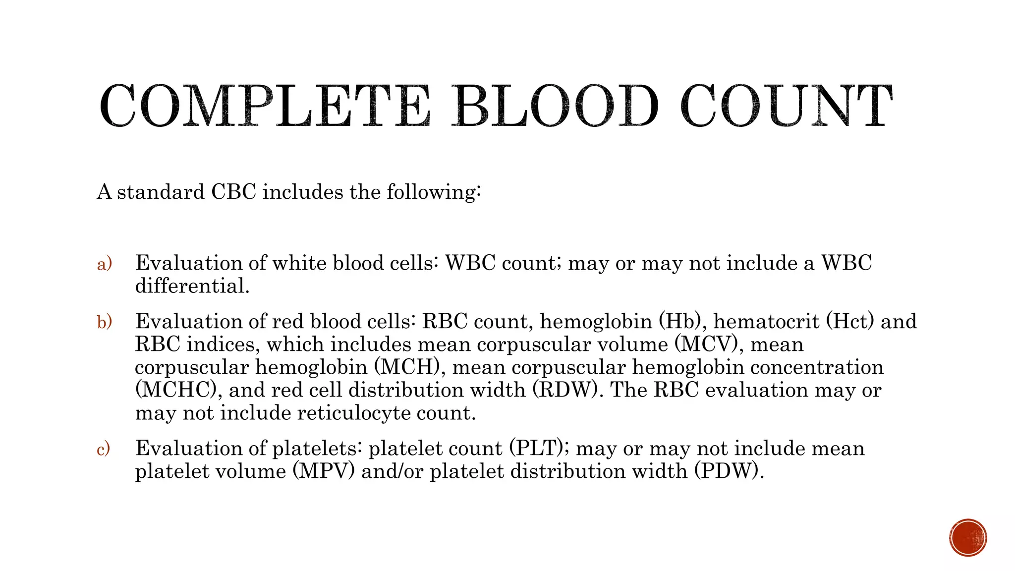


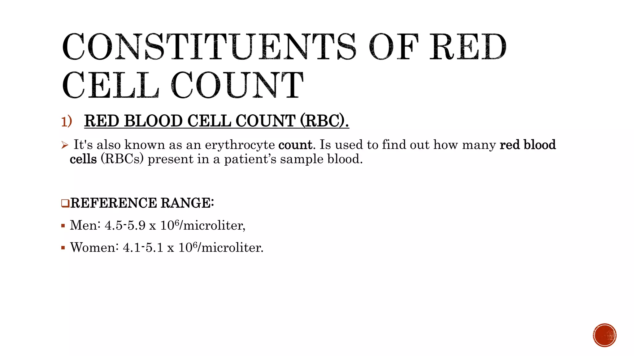


A complete blood count (CBC) is a blood test used to evaluate overall health and detect disorders like anemia and infection. It measures red blood cells, white blood cells, hemoglobin, hematocrit, and platelets, providing insights into various health conditions. The CBC results can indicate abnormalities in blood cell populations, which may suggest the presence of underlying medical issues.