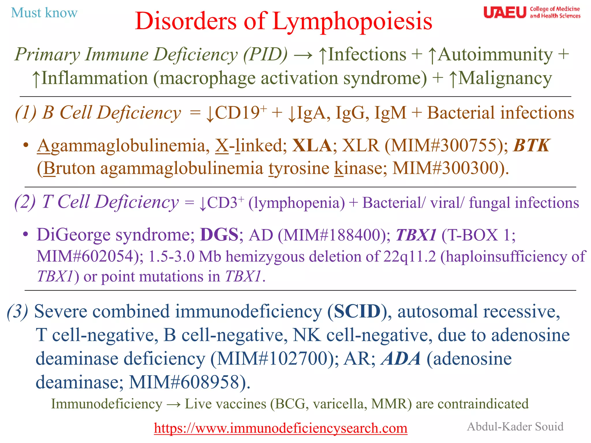
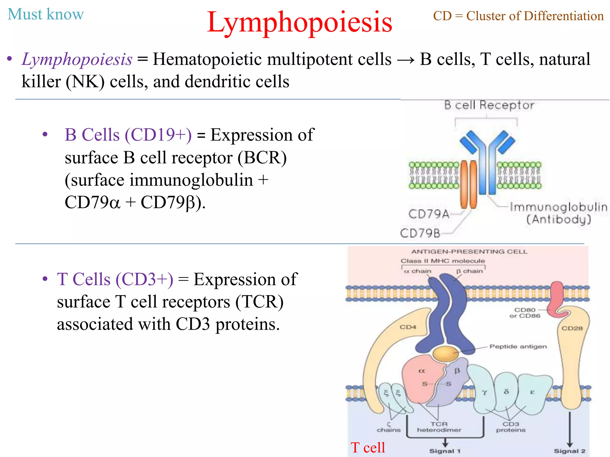
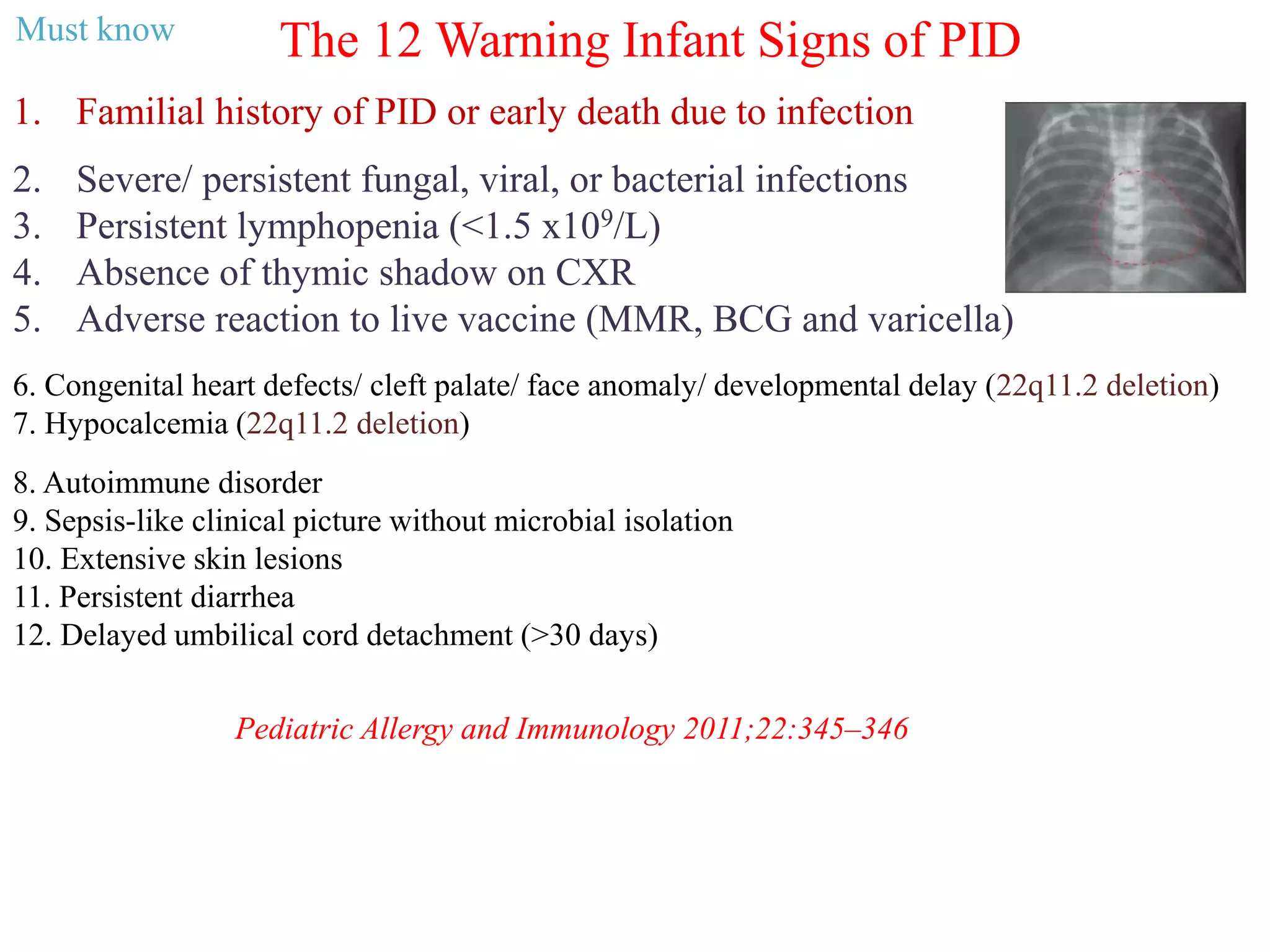
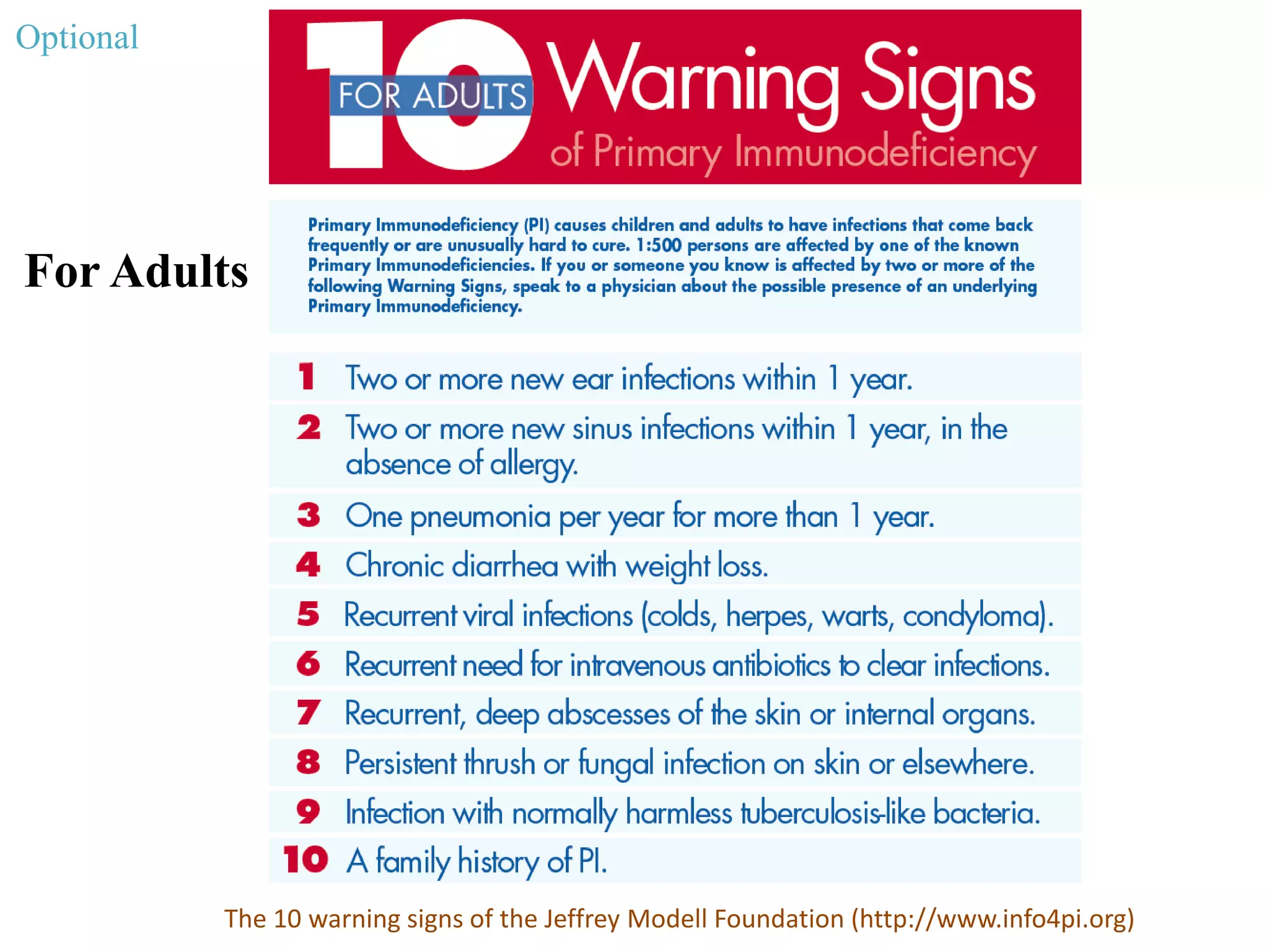
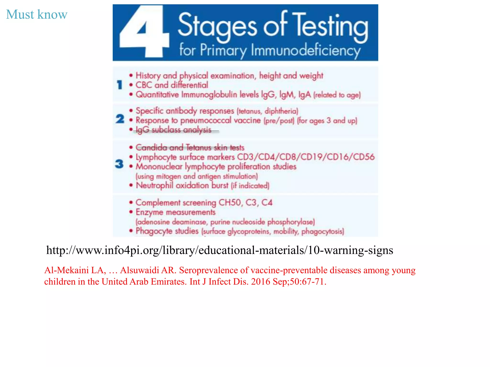
Abdul-Kader Souid
Primary Immune Deficiency (PID) → ↑Infections + ↑Autoimmunity +
↑Inflammation (macrophage activation syndrome) + ↑Malignancy
Disorders of Lymphopoiesis
(1) B Cell Deficiency = ↓CD19+ + ↓IgA, IgG, IgM + Bacterial infections
• Agammaglobulinemia, X-linked; XLA; XLR (MIM#300755); BTK
(Bruton agammaglobulinemia tyrosine kinase; MIM#300300).
(2) T Cell Deficiency = ↓CD3+ (lymphopenia) + Bacterial/ viral/ fungal infections
• DiGeorge syndrome; DGS; AD (MIM#188400); TBX1 (T-BOX 1;
MIM#602054); 1.5-3.0 Mb hemizygous deletion of 22q11.2 (haploinsufficiency of
TBX1) or point mutations in TBX1.
Immunodeficiency → Live vaccines (BCG, varicella, MMR) are contraindicated
(3) Severe combined immunodeficiency (SCID), autosomal recessive,
T cell-negative, B cell-negative, NK cell-negative, due to adenosine
deaminase deficiency (MIM#102700); AR; ADA (adenosine
deaminase; MIM#608958).
Must know
https://www.immunodeficiencysearch.com
Lymphopoiesis
• Lymphopoiesis = Hematopoietic multipotent cells → B cells, T cells, natural
killer (NK) cells, and dendritic cells
• B Cells (CD19+) = Expression of
surface B cell receptor (BCR)
(surface immunoglobulin +
CD79a + CD79b).
• T Cells (CD3+) = Expression of
surface T cell receptors (TCR)
associated with CD3 proteins.
CD = Cluster of DifferentiationMust know
T cell
Primary Immune Deficiency → ↑Infections + ↑Autoimmunity +
↑Inflammation (MAS) + ↑Malignancies
• Recurrent respiratory infections (otitis, sinusitis,
bronchiectasis, pneumonia)
• Invasive infections (bones, joints, liver)
• Purulent lymphadenitis
• Skin/ deep tissue abscesses
• Infections by poorly virulent / opportunistic
pathogens (Pneumocystis jiroveci, cytomegalovirus,
mycobacteria, Cryptosporidium, Giardia lamblia)
• Persistent/ recurrent candidiasis (T cell deficiency)
• Failure to thrive
Primary Immune Deficiency (PID)
Dilated bronchi (bronchiectasis)
in a 13-year-old boy with
agammaglobulinemia
Pneumocystis jiroveci
pneumonia
Must know
https://www.immunodeficiencysearch.com
The 12 Warning Infant Signs of PID
1. Familial history of PID or early death due to infection
2. Severe/ persistent fungal, viral, or bacterial infections
3. Persistent lymphopenia (<1.5 x10⁹/L)
4. Absence of thymic shadow on CXR
5. Adverse reaction to live vaccine (MMR, BCG and varicella)
Pediatric Allergy and Immunology 2011;22:345–346
6. Congenital heart defects/ cleft palate/ face anomaly/ developmental delay (22q11.2 deletion)
7. Hypocalcemia (22q11.2 deletion)
8. Autoimmune disorder
9. Sepsis-like clinical picture without microbial isolation
10. Extensive skin lesions
11. Persistent diarrhea
12. Delayed umbilical cord detachment (>30 days)
Must know
Panel A: Patient skin lesions showing erythematous nodules. Panels A-C. H&E stain of a skin excisional
lesion showing infiltration by histiocytes in the dermis. Panels D-E :H&E stain of bone marrow
biopsy showing infiltration by histiocytes. Panel F .Bone marrow specimen revealing infiltration by
histiocytes ingesting acid-fast bacilli.
A B C
D E F
A female infant received BCG at birth. She presents at four months of age with fever and
erythematous skin nodules. Family history is significant for familial histiocytic
lymphohistocytosis (HLH) involving her brother. Histology of skin and marrow revealed
infiltration by histiocytes ingesting acid-fast bacilli.
How can we avoid administering BCG to neonates with immunodeficiency?
Al-Hammadi S, Alsuwaidi AR. Disseminated Bacillus Calmette-Guérin (BCG) infections in infants with
immunodeficiency. BMC Res Notes. 2017 May 5;10(1):177.
Optional
For Adults
The 10 warning signs of the Jeffrey Modell Foundation (http://www.info4pi.org)
Optional
For Children
The 10 warning signs of the Jeffrey Modell Foundation (http://www.info4pi.org)
Optional
http://www.info4pi.org/library/educational-materials/10-warning-signs
Must know
Al-Mekaini LA, … Alsuwaidi AR. Seroprevalence of vaccine-preventable diseases among young
children in the United Arab Emirates. Int J Infect Dis. 2016 Sep;50:67-71.
A 5-month-old male infant with fever
His lymphocyte count is 1.1 x10⁹/L (= 1,100/mL). Lymphocyte surface
markers (immunophenotyping) shows:
CD3+ T cells = 800/µL (10th percentile = 2,500/µL)
• CD4+ T cells = 50/µL (10th percentile = 1,600/µL)
• CD8+ T cells = 750/µL (10th percentile = 560/µL)
CD19+ B cells = 60/µL (10th percentile = 300/µL)
NK cells = 102/µL (normal)
T cells constitute ~70% of circulating lymphocytes. Thus, T cell immune
deficiency is associated with lymphopenia.
Newborn screening for T cell immunodeficiency
is based on absence of small circles (TREC, T
cell receptor excision circles) of DNA created in
T cells during V(D)J recombination.
RSS, recombination signal sequences
TREC
Must know
BTK encodes a cytoplasmic protein
involved in B cell maturation. BTK binding
to phosphatidylinositol (3,4,5)-
trisphosphate (PIP3) → ↑activity of downstream
proteins involved in B cell signaling.
Ibrutinib is a selective inhibitor of Bruton tyrosine kinase (Btk).
It is approved for treatment of B-cell malignancies.
Ibrutinib
B Cell Deficiency = ↓CD19+ + ↓IgA, IgG, IgM + Bacterial infections
• Agammaglobulinemia, X-linked; XLA; XLR (OMIM#300755);
BTK (Bruton agammaglobulinemia tyrosine kinase; OMIM#300300).
Cytoplasm
Extracellular
Must know
T Cell Deficiency = ↓CD3+ (lymphopenia) + Bacterial/ viral/ fungal infections
• DiGeorge syndrome; DGS; AD (OMIM#188400); TBX1 (T-BOX 1;
OMIM#602054); 1.5-3.0 Mb hemizygous deletion of 22q11.2 (haploinsufficiency
of TBX1) or point mutations in TBX1.
DGS presents with a variety of phenotypes, that include Cardiac
defect (e.g., tetralogy of Fallot), Abnormal face, T cell (thymic)
hypoplasia, Cleft palate, and Hypocalcemia (neonatal
hypoparathyroidism) as a result of 22q11 deletion (CATCH22).
The T-box genes are transcription factors involved in the regulation
of developmental processes.
Must know
In ADA deficiency, T and B cells accumulate the apoptosis-inducing toxins
adenosine, deoxyadenosine, and deoxyadenosine triphosphate (dATP).
ribonucleotide reductase
Severe combined immunodeficiency (SCID), autosomal recessive, T cell-
negative, B cell-negative, NK cell-negative, due to adenosine deaminase deficiency
(MIM#102700); AR; ADA (adenosine deaminase; MIM#608958).
The diagnosis is made by ↓enzyme activity in erythrocytes or ADA gene sequencing.
Must know
Must Know Pearls
• Primary immune deficiency (PID) increases susceptibility to infection,
autoimmunity (AHA, ITP, T1D, thyroiditis), inflammation
(macrophage activation syndrome), and malignancy.
• Live vaccines (BCG, varicella, MMR) are contraindicated in PID.
• Work-up includes CBC, chest radiograph, immunoglobulin levels,
specific antibody response (tetanus, diphtheria), and lymphocyte
surface marker.
• T cell immunodeficiency is associated with lymphopenia.
• TREC is a screening marker for T cell immunodeficiency.
9/26/2018 13
Required Reading
1. Wood P, Stanworth S, Burton J, et al. Recognition, clinical diagnosis and
management of patients with primary antibody deficiencies: a systematic
review. Clin Exp Immunol 2007;149:410.
2. Ballow M. Approach to the patient with recurrent infections. Clin Rev Allergy
Immunol 2008;34:129.
3. Carneiro-Sampaio M, Jacob CM, Leone CR. A proposal of warning signs for
primary immunodeficiencies in the first year of life. Pediatr Allergy Immunol
2011;22:345.
9/26/2018 14

Disorders of lymphobioesis.ppt

  • 1.
    Abdul-Kader Souid Primary ImmuneDeficiency (PID) → ↑Infections + ↑Autoimmunity + ↑Inflammation (macrophage activation syndrome) + ↑Malignancy Disorders of Lymphopoiesis (1) B Cell Deficiency = ↓CD19+ + ↓IgA, IgG, IgM + Bacterial infections • Agammaglobulinemia, X-linked; XLA; XLR (MIM#300755); BTK (Bruton agammaglobulinemia tyrosine kinase; MIM#300300). (2) T Cell Deficiency = ↓CD3+ (lymphopenia) + Bacterial/ viral/ fungal infections • DiGeorge syndrome; DGS; AD (MIM#188400); TBX1 (T-BOX 1; MIM#602054); 1.5-3.0 Mb hemizygous deletion of 22q11.2 (haploinsufficiency of TBX1) or point mutations in TBX1. Immunodeficiency → Live vaccines (BCG, varicella, MMR) are contraindicated (3) Severe combined immunodeficiency (SCID), autosomal recessive, T cell-negative, B cell-negative, NK cell-negative, due to adenosine deaminase deficiency (MIM#102700); AR; ADA (adenosine deaminase; MIM#608958). Must know https://www.immunodeficiencysearch.com
  • 2.
    Lymphopoiesis • Lymphopoiesis =Hematopoietic multipotent cells → B cells, T cells, natural killer (NK) cells, and dendritic cells • B Cells (CD19+) = Expression of surface B cell receptor (BCR) (surface immunoglobulin + CD79a + CD79b). • T Cells (CD3+) = Expression of surface T cell receptors (TCR) associated with CD3 proteins. CD = Cluster of DifferentiationMust know T cell
  • 3.
    Primary Immune Deficiency→ ↑Infections + ↑Autoimmunity + ↑Inflammation (MAS) + ↑Malignancies • Recurrent respiratory infections (otitis, sinusitis, bronchiectasis, pneumonia) • Invasive infections (bones, joints, liver) • Purulent lymphadenitis • Skin/ deep tissue abscesses • Infections by poorly virulent / opportunistic pathogens (Pneumocystis jiroveci, cytomegalovirus, mycobacteria, Cryptosporidium, Giardia lamblia) • Persistent/ recurrent candidiasis (T cell deficiency) • Failure to thrive Primary Immune Deficiency (PID) Dilated bronchi (bronchiectasis) in a 13-year-old boy with agammaglobulinemia Pneumocystis jiroveci pneumonia Must know https://www.immunodeficiencysearch.com
  • 4.
    The 12 WarningInfant Signs of PID 1. Familial history of PID or early death due to infection 2. Severe/ persistent fungal, viral, or bacterial infections 3. Persistent lymphopenia (<1.5 x10⁹/L) 4. Absence of thymic shadow on CXR 5. Adverse reaction to live vaccine (MMR, BCG and varicella) Pediatric Allergy and Immunology 2011;22:345–346 6. Congenital heart defects/ cleft palate/ face anomaly/ developmental delay (22q11.2 deletion) 7. Hypocalcemia (22q11.2 deletion) 8. Autoimmune disorder 9. Sepsis-like clinical picture without microbial isolation 10. Extensive skin lesions 11. Persistent diarrhea 12. Delayed umbilical cord detachment (>30 days) Must know
  • 5.
    Panel A: Patientskin lesions showing erythematous nodules. Panels A-C. H&E stain of a skin excisional lesion showing infiltration by histiocytes in the dermis. Panels D-E :H&E stain of bone marrow biopsy showing infiltration by histiocytes. Panel F .Bone marrow specimen revealing infiltration by histiocytes ingesting acid-fast bacilli. A B C D E F A female infant received BCG at birth. She presents at four months of age with fever and erythematous skin nodules. Family history is significant for familial histiocytic lymphohistocytosis (HLH) involving her brother. Histology of skin and marrow revealed infiltration by histiocytes ingesting acid-fast bacilli. How can we avoid administering BCG to neonates with immunodeficiency? Al-Hammadi S, Alsuwaidi AR. Disseminated Bacillus Calmette-Guérin (BCG) infections in infants with immunodeficiency. BMC Res Notes. 2017 May 5;10(1):177. Optional
  • 6.
    For Adults The 10warning signs of the Jeffrey Modell Foundation (http://www.info4pi.org) Optional
  • 7.
    For Children The 10warning signs of the Jeffrey Modell Foundation (http://www.info4pi.org) Optional
  • 8.
    http://www.info4pi.org/library/educational-materials/10-warning-signs Must know Al-Mekaini LA,… Alsuwaidi AR. Seroprevalence of vaccine-preventable diseases among young children in the United Arab Emirates. Int J Infect Dis. 2016 Sep;50:67-71.
  • 9.
    A 5-month-old maleinfant with fever His lymphocyte count is 1.1 x10⁹/L (= 1,100/mL). Lymphocyte surface markers (immunophenotyping) shows: CD3+ T cells = 800/µL (10th percentile = 2,500/µL) • CD4+ T cells = 50/µL (10th percentile = 1,600/µL) • CD8+ T cells = 750/µL (10th percentile = 560/µL) CD19+ B cells = 60/µL (10th percentile = 300/µL) NK cells = 102/µL (normal) T cells constitute ~70% of circulating lymphocytes. Thus, T cell immune deficiency is associated with lymphopenia. Newborn screening for T cell immunodeficiency is based on absence of small circles (TREC, T cell receptor excision circles) of DNA created in T cells during V(D)J recombination. RSS, recombination signal sequences TREC Must know
  • 10.
    BTK encodes acytoplasmic protein involved in B cell maturation. BTK binding to phosphatidylinositol (3,4,5)- trisphosphate (PIP3) → ↑activity of downstream proteins involved in B cell signaling. Ibrutinib is a selective inhibitor of Bruton tyrosine kinase (Btk). It is approved for treatment of B-cell malignancies. Ibrutinib B Cell Deficiency = ↓CD19+ + ↓IgA, IgG, IgM + Bacterial infections • Agammaglobulinemia, X-linked; XLA; XLR (OMIM#300755); BTK (Bruton agammaglobulinemia tyrosine kinase; OMIM#300300). Cytoplasm Extracellular Must know
  • 11.
    T Cell Deficiency= ↓CD3+ (lymphopenia) + Bacterial/ viral/ fungal infections • DiGeorge syndrome; DGS; AD (OMIM#188400); TBX1 (T-BOX 1; OMIM#602054); 1.5-3.0 Mb hemizygous deletion of 22q11.2 (haploinsufficiency of TBX1) or point mutations in TBX1. DGS presents with a variety of phenotypes, that include Cardiac defect (e.g., tetralogy of Fallot), Abnormal face, T cell (thymic) hypoplasia, Cleft palate, and Hypocalcemia (neonatal hypoparathyroidism) as a result of 22q11 deletion (CATCH22). The T-box genes are transcription factors involved in the regulation of developmental processes. Must know
  • 12.
    In ADA deficiency,T and B cells accumulate the apoptosis-inducing toxins adenosine, deoxyadenosine, and deoxyadenosine triphosphate (dATP). ribonucleotide reductase Severe combined immunodeficiency (SCID), autosomal recessive, T cell- negative, B cell-negative, NK cell-negative, due to adenosine deaminase deficiency (MIM#102700); AR; ADA (adenosine deaminase; MIM#608958). The diagnosis is made by ↓enzyme activity in erythrocytes or ADA gene sequencing. Must know
  • 13.
    Must Know Pearls •Primary immune deficiency (PID) increases susceptibility to infection, autoimmunity (AHA, ITP, T1D, thyroiditis), inflammation (macrophage activation syndrome), and malignancy. • Live vaccines (BCG, varicella, MMR) are contraindicated in PID. • Work-up includes CBC, chest radiograph, immunoglobulin levels, specific antibody response (tetanus, diphtheria), and lymphocyte surface marker. • T cell immunodeficiency is associated with lymphopenia. • TREC is a screening marker for T cell immunodeficiency. 9/26/2018 13
  • 14.
    Required Reading 1. WoodP, Stanworth S, Burton J, et al. Recognition, clinical diagnosis and management of patients with primary antibody deficiencies: a systematic review. Clin Exp Immunol 2007;149:410. 2. Ballow M. Approach to the patient with recurrent infections. Clin Rev Allergy Immunol 2008;34:129. 3. Carneiro-Sampaio M, Jacob CM, Leone CR. A proposal of warning signs for primary immunodeficiencies in the first year of life. Pediatr Allergy Immunol 2011;22:345. 9/26/2018 14

Editor's Notes

  • #10 T cell Receptor Excision Circles (TRECs). TRECs are episomal DNA circles produced in thymocytes by excisional rearrangements of T cell receptor (TCR) genes; they are stable, not duplicated during mitosis, diluted out with each cell division, and therefore higher in thymocytes, recent thymic emigrants (RTEs) and naïve T cells. Quantitative polymerase chain reaction (PCR) of coding-joint (cj) δRec ψJα TREC, produced at TCRα/δ locus within chromosome 14 (14q11) by > 70% of developing human α:β T cells, counts in the peripheral blood naïve α:β T lymphocytes recently dismetted by thymus: in newborn, values < 25 TRECs/μL indicate SCID. Italian Journal of Pediatrics 2010 36:76   doi:10.1186/1824-7288-36-76.