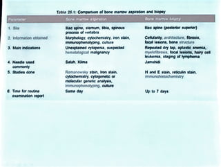
Examination of Bone Marrow
Bone marrow is the site of hematopolesls in postnatal life.
Bone marrow is one of the largest organs in the body, and
although distributed widely in the skeleton, functlons as a single
unit
It is located within the cavities of the bones and mainly consists
of hematopoletlc cells, vascular sinusolds, fibroblasts, fat cells,
and macrophages.
There are no lymphatlc channels in the bone marrow.
There are two types of marrow: red and yellow.
Red marrow refers to the active hematopolotic tissue while fatty
tissue comprises the yellow (inactive) marrow
Red coloration is due to presence of large amounts of developing
red cells.
Megakaryocytes, the largest of the hematopoietic cells.
There are two techniques for sampling of bone marrow:
aspiration and biopsy.
INDICATIONS FOR BONE MARROW ASPIRATION-
Unexplained cytopenia: If the cause of cytopenia (anemia,
leukopenia, or thrombocytopenia) is not apparent from
blood investigations and clinical details, bone marrow
examination is indicated.
Suspected acute leukemia
Suspected myelodysplastic syndrome.
Suspected myeloproliferative disorders
Investigation of pyrexia of unknown origin
Suspected storagedisorder like Gaucher's diseaseor
Neimann-Pick disease.
Suspected infections like kala azar, miliary tuberculosis, or
histoplasmosis.
SITES FOR BONE MARROW ASPIRATION OR BIOPSY
Jliac spines or crest:
Sternum
Spinous processes oflumbar vertebra
Tibia:
Poaterior superiorlactpine n
Vertebra Tiia
Fig. 25.1: Sites of bono marow aspiration
(shaded areas)
METHOD
An informed consent should be obtained before the
procedure.
Bone marrow aspiration or biopsy should be performed
by the physician.
autoclaved bone marrow aspiration needle, sterile
disposable syringes with needles, local anesthetic
solution, clean and dry glass slides, spreader slide,
gloves, drapes, gauze, and a skin antiseptic solution.
Salah and Klima needles are commonly used.
Jamshidi needle, which is longer, can be used for both
aspiration and biopsy from iliac crest
Salah and Klima needle
JAMSHEDI NEEDLE
For aspiration from posterior superior iliac spine,
patient should lie on one side with back towards the
physician, knees and hips flexed, and the knees drawn
towards the chest.
Skin and periosteum areinfiltratedwith a local
anesthetic.
After waiting for 5 minutes for anesthesia to take effect,
bone marrow aspiration needle is inserted along with
the fitted stylet.
(Stylet prevents blockage of lumen of needle by tissues
through which needle passes).
When the bone is reached, the needle is rotated
clockwise and anticlockwise and slowly advanced into
the bone, maintaining steady and firm pressure
A5 or 10 ml syringe is attached to the needle anda
small 3mount of mamow is aspirated (till the first drop
of blood appears i.e. 0.25-0.50 ml) by quickly pulling
the plunger of the syringe.
The syringe should be handed over to the assistant for
preparation of smears on glass slides.
The smears should be made promptly, before clotting
occurs, by putting one drop of the aspirated material
near one end of a glass slide and spreadingit similar to
a blood film.
tf immunophenotyping or cytogenetic -3spirated in a
tube containing heparin anticoagulant
A5 or 10 ml syringe is attached to the needle and a
small amount of marrow is aspirated (till the first drop
of blood appears i.e. 0.25-0.50 ml) by quickly pulling
the plunger of the syringe.
The syringe should be handed over to the assistant for
preparation of smears on glassslides
The smears should be made promptly, before clotting
occurs, by putting one drop of the aspirated material
near one end of a glass slide and spreading it similar to
a blood film.
If immunophenotyping orcytogenetic-aspirated in a
tube containing heparin anticoagulant
PROCESSING OF MARROW SPECIMEN
A drop of aspirated marrow sample is put on each of
the several glass slides, excess blood is removed by
sucking with a Pasteur pipette, and smears are
prepared with a 'spreader'.
The smears are allowed to dry in the air and are
labeled.
They are stained with one of the Romanowsky stains.
Routinely, one marrow smear (containing one or more
marrow particles) should also be stained with Perl's
stain for assessment of bone marrow storage iron.
Table 25.1: Comparison of bone mamrowaspiration andbiopay
Parameter Bone marrow aspirabon Bone marow biopsy
ac spino (posterior superior)
iac spine, stemum, tbia, spinous
process of vertebra
1. Site
Morphology. cytochemistry, iron stain,
immunophenotyping. cuturo
Unexplained cylopenia, suspectoed
hematological matignancy
Celludanty. architecture, fbrosis,
focal lesions, bono structuro
2. Information obtained
3.Mainindications Repeated dry tap. aplastic anemia,
myelofibrosis, focal lesions, hairy cel
leukemia, staging of lymphoma
4. Needle used Salah, Kma Jamshidi
commonly
5. Studies done Romanowsky stain, Iron stain,
cylochemistry, cytogeneic or
molecular genotic analysis,
immunophenotyping. cutture
Same dayY
H and E stain, reticulin stain,
immunohistochemistry
6. Time for routine Up to 7 days
examinaton report
Tabio 25.1: Compartson of bone mamow aspirationandbiopy
Parameter Bone marow aspiration 8one marrow biopsy
iac splno (posteror superior)
liao spine, stemum, bdia, spinous
process of vertebra
Morpholagy. cytochemistry, iron stain,
immunophenotyping. culture
Unexplained cylopenis, suspected
hematological malignancy
1. Site
Cellulanty, architecture, fibrosis,
focal lesions, bone structure
2. Information obtained
3.Main indlcations Repeated dry tap. aplastic anemia,
myelafibrosis, focal lesions, hairy cell
leukemla,staging of bymphoma
4. Needie vsed
commonly
Satah, Klima Jamshidi
H and E stain, reticulin stain,
immunahistochemistry
5. Studies done Romanowsky stain, Iron stain,
cylochemistry. oytogenetic or
molecular genetic analysis,
Immunophenotyping. culuro
Same day
6. Time for routine Up to 7 days
examinstion report
Iron Staining of Bone Marrow Aspiration Smears
Presence of iron in marrow aspiration smears can be
demonstrated by Perls' Prussian blue reaction.
ionic iron reacts with acid ferrrocyanide solution to form
blue-colored ferric ferrocyanide.
FIXATIVE OF CHOICE:
FORMOLMETHANOL(ratio1:9)
Methanol alone is also adequate.
STAINING SOLUTION:
Equal quantities of
2% HCL and
4% potassium ferrocyanide.
Procedure
OFx the air dried bone marrow specimen in methanol for
15 min.
aDythe slides in room temperature.
Mk equai parts of HCL and potassium ferrocyanide
prepared immediatelybetore use.
Oimmerse slides in this working solution for 15 min. in roon
temperature.
ONow wash the slides well with distilled water for 5 min.
OCounter stain with aqueous solution of safranin (0.1% nu
fast red) for 10-15 sec.
OWash with distiled water. Let the sides dry.
RESULTS
FERRIC 1AONBLUE
NUCLEI
CYTOPLASM
RED
PINK
Procedure:
OFix the air dried bone marrow specimen in methanol for
15 min.
ODry the slides in room temperature.
OMix equal parts of HCLand potassium ferrocyanide
prepared immediately before use.
Olmmerse slides in this working solution for 15 min. in roon
temperature.
ONow wash the slides well with distilled water for 5 min.
OCounter stain with aqueous solution of safranin (0.1%nu
fast red) for 10-15 sec.
OWash with distilled water. Let theslidesdry.
RESULTS:
FERRIC IRON BLUE
NUCLEI
CYTOPLASM
RED
PINK
Types of Cytochemical stains
ENZYMATIC NON-ENZYMATIC
Myeloperoxidase A
4. Sudan black B
2. Esterase 5. Periodic acid Schiff
a) specific 6. Toluidine blue
b) non specific
3. Phosphatase
7. Perl' stain
a) leucocyte alkaline phosphatase
b)acid phosphatase
MYELOPEROXIDASE (MPO)
tisan enzyme presentin primaryandsecondarygranulesofneutrophils
and theirprecursors
Also present in eosinophl granules and in azurophillicgranulesof
monocytes
Purpose
Differentiate betweenmyelogencus and monacyticleulaemia from acute
lymphoblastic leukaemia(ALL}.
Olagnose congenitaldefidencyofneutrophil MPO.
Principle.
Myeloperaxidase splits H202 in the presence of chromogenic
electron donor (e-g. DA8) and forms an insolubie reaction product.
The reaction product is stable, insolubie and non diffusible.
Reagents
Fixative:- Buffered Formal acetone (BFE)
Acetone-40ml
Buffer 30ml
Formalin- 25 ml
Substrate:-3,3- DAB(Diaminobenzidine)
Buffer:- Sorensen's phosphate buffer, PH-7.3
Hydrogen peroxide(H202, 30% w/v)
Counterstain:- Aqueous haematoxylin.
Procedure-
1) Fix air dried smear in cold buffered acetone for 30 sec.
2) Rinse in running tap water and dry.
3) Incubate for 10 min. in working substrate solution.
Working substrate solution:-30mg of DAB in 60ml buffer,
and add 120ul H202 just before use.
4) Wash, counter stain with haematoxylin for 3-5 min.
5) Rinse in running tap water and air dry.
Results:
The reaction product is browm and granular.
All nuclei are blue.
SUDANBLACK B (SBB)
Sudan blak B is a lipophilic dye that stains intra cellular
phospholipids and ctherlipids.
Purpose-
Same significant as MPO Le. to differentiate between ALL and AML
Done in old smears in witch MPO can not be performed.
Principle:
The S88 is a lipophilic dye binds irreversibly to an unidentified
undefined granule in granulocytes and eosinophils.
Reagents
Fixative:-40% formaldehyde vapours
Stain:-0.3%SB8in absolute alcohol
Phenolic buffer- 16gm crystalline phenol in 30mlofabsolute
ethanol and final volume up to 100ml with buffer.
Working stain solution- add 40ml phenolic buffer to 60ml SBS
solution.
Counter stain:-Leishmanstain
Procedure
1) Fix air dried smear in in formalin vapours for 5-10 min.
2) Then air wash for 15 min.
3) Now stain with working solution of SBB stain solution for 1 hour.
4) Now give 3 washings of ethanol for 30 sec each.
Sudan Black B
5) Wash with water and air dry.
6) Now counter stain with Leishman stain. Positive sudan blackB(596)
stain in a patient with AML,
Results:- Not the biack suining
cyloplasmicgranudesin the
myeloblasts
The reaction product is black and granular.
Ali nuclei are blue.
Interpretation
The results aresimilarto MPO staining
both in normal and ieukemic cells.
ses positive prominent Auer rods in bone
marrow smear
The differences are:
The eosinophil granules are SBB negative
In rare cases (1-2%} ofALL shows non
granular smudgy positivity not seen in
MPO staining e.g. in 8urkett's lymphoma.

Examination of Bone marrow.pdf

  • 1.
    Examination of BoneMarrow Bone marrow is the site of hematopolesls in postnatal life. Bone marrow is one of the largest organs in the body, and although distributed widely in the skeleton, functlons as a single unit It is located within the cavities of the bones and mainly consists of hematopoletlc cells, vascular sinusolds, fibroblasts, fat cells, and macrophages. There are no lymphatlc channels in the bone marrow. There are two types of marrow: red and yellow. Red marrow refers to the active hematopolotic tissue while fatty tissue comprises the yellow (inactive) marrow Red coloration is due to presence of large amounts of developing red cells.
  • 2.
    Megakaryocytes, the largestof the hematopoietic cells. There are two techniques for sampling of bone marrow: aspiration and biopsy. INDICATIONS FOR BONE MARROW ASPIRATION- Unexplained cytopenia: If the cause of cytopenia (anemia, leukopenia, or thrombocytopenia) is not apparent from blood investigations and clinical details, bone marrow examination is indicated. Suspected acute leukemia Suspected myelodysplastic syndrome. Suspected myeloproliferative disorders Investigation of pyrexia of unknown origin Suspected storagedisorder like Gaucher's diseaseor Neimann-Pick disease. Suspected infections like kala azar, miliary tuberculosis, or histoplasmosis.
  • 3.
    SITES FOR BONEMARROW ASPIRATION OR BIOPSY Jliac spines or crest: Sternum Spinous processes oflumbar vertebra Tibia: Poaterior superiorlactpine n Vertebra Tiia Fig. 25.1: Sites of bono marow aspiration (shaded areas)
  • 4.
    METHOD An informed consentshould be obtained before the procedure. Bone marrow aspiration or biopsy should be performed by the physician. autoclaved bone marrow aspiration needle, sterile disposable syringes with needles, local anesthetic solution, clean and dry glass slides, spreader slide, gloves, drapes, gauze, and a skin antiseptic solution. Salah and Klima needles are commonly used. Jamshidi needle, which is longer, can be used for both aspiration and biopsy from iliac crest
  • 5.
    Salah and Klimaneedle JAMSHEDI NEEDLE
  • 6.
    For aspiration fromposterior superior iliac spine, patient should lie on one side with back towards the physician, knees and hips flexed, and the knees drawn towards the chest. Skin and periosteum areinfiltratedwith a local anesthetic. After waiting for 5 minutes for anesthesia to take effect, bone marrow aspiration needle is inserted along with the fitted stylet. (Stylet prevents blockage of lumen of needle by tissues through which needle passes). When the bone is reached, the needle is rotated clockwise and anticlockwise and slowly advanced into the bone, maintaining steady and firm pressure
  • 7.
    A5 or 10ml syringe is attached to the needle anda small 3mount of mamow is aspirated (till the first drop of blood appears i.e. 0.25-0.50 ml) by quickly pulling the plunger of the syringe. The syringe should be handed over to the assistant for preparation of smears on glass slides. The smears should be made promptly, before clotting occurs, by putting one drop of the aspirated material near one end of a glass slide and spreadingit similar to a blood film. tf immunophenotyping or cytogenetic -3spirated in a tube containing heparin anticoagulant
  • 8.
    A5 or 10ml syringe is attached to the needle and a small amount of marrow is aspirated (till the first drop of blood appears i.e. 0.25-0.50 ml) by quickly pulling the plunger of the syringe. The syringe should be handed over to the assistant for preparation of smears on glassslides The smears should be made promptly, before clotting occurs, by putting one drop of the aspirated material near one end of a glass slide and spreading it similar to a blood film. If immunophenotyping orcytogenetic-aspirated in a tube containing heparin anticoagulant
  • 9.
    PROCESSING OF MARROWSPECIMEN A drop of aspirated marrow sample is put on each of the several glass slides, excess blood is removed by sucking with a Pasteur pipette, and smears are prepared with a 'spreader'. The smears are allowed to dry in the air and are labeled. They are stained with one of the Romanowsky stains. Routinely, one marrow smear (containing one or more marrow particles) should also be stained with Perl's stain for assessment of bone marrow storage iron.
  • 10.
    Table 25.1: Comparisonof bone mamrowaspiration andbiopay Parameter Bone marrow aspirabon Bone marow biopsy ac spino (posterior superior) iac spine, stemum, tbia, spinous process of vertebra 1. Site Morphology. cytochemistry, iron stain, immunophenotyping. cuturo Unexplained cylopenia, suspectoed hematological matignancy Celludanty. architecture, fbrosis, focal lesions, bono structuro 2. Information obtained 3.Mainindications Repeated dry tap. aplastic anemia, myelofibrosis, focal lesions, hairy cel leukemia, staging of lymphoma 4. Needle used Salah, Kma Jamshidi commonly 5. Studies done Romanowsky stain, Iron stain, cylochemistry, cytogeneic or molecular genotic analysis, immunophenotyping. cutture Same dayY H and E stain, reticulin stain, immunohistochemistry 6. Time for routine Up to 7 days examinaton report
  • 11.
    Tabio 25.1: Compartsonof bone mamow aspirationandbiopy Parameter Bone marow aspiration 8one marrow biopsy iac splno (posteror superior) liao spine, stemum, bdia, spinous process of vertebra Morpholagy. cytochemistry, iron stain, immunophenotyping. culture Unexplained cylopenis, suspected hematological malignancy 1. Site Cellulanty, architecture, fibrosis, focal lesions, bone structure 2. Information obtained 3.Main indlcations Repeated dry tap. aplastic anemia, myelafibrosis, focal lesions, hairy cell leukemla,staging of bymphoma 4. Needie vsed commonly Satah, Klima Jamshidi H and E stain, reticulin stain, immunahistochemistry 5. Studies done Romanowsky stain, Iron stain, cylochemistry. oytogenetic or molecular genetic analysis, Immunophenotyping. culuro Same day 6. Time for routine Up to 7 days examinstion report
  • 12.
    Iron Staining ofBone Marrow Aspiration Smears Presence of iron in marrow aspiration smears can be demonstrated by Perls' Prussian blue reaction. ionic iron reacts with acid ferrrocyanide solution to form blue-colored ferric ferrocyanide. FIXATIVE OF CHOICE: FORMOLMETHANOL(ratio1:9) Methanol alone is also adequate. STAINING SOLUTION: Equal quantities of 2% HCL and 4% potassium ferrocyanide.
  • 13.
    Procedure OFx the airdried bone marrow specimen in methanol for 15 min. aDythe slides in room temperature. Mk equai parts of HCL and potassium ferrocyanide prepared immediatelybetore use. Oimmerse slides in this working solution for 15 min. in roon temperature. ONow wash the slides well with distilled water for 5 min. OCounter stain with aqueous solution of safranin (0.1% nu fast red) for 10-15 sec. OWash with distiled water. Let the sides dry. RESULTS FERRIC 1AONBLUE NUCLEI CYTOPLASM RED PINK
  • 14.
    Procedure: OFix the airdried bone marrow specimen in methanol for 15 min. ODry the slides in room temperature. OMix equal parts of HCLand potassium ferrocyanide prepared immediately before use. Olmmerse slides in this working solution for 15 min. in roon temperature. ONow wash the slides well with distilled water for 5 min. OCounter stain with aqueous solution of safranin (0.1%nu fast red) for 10-15 sec. OWash with distilled water. Let theslidesdry. RESULTS: FERRIC IRON BLUE NUCLEI CYTOPLASM RED PINK
  • 16.
    Types of Cytochemicalstains ENZYMATIC NON-ENZYMATIC Myeloperoxidase A 4. Sudan black B 2. Esterase 5. Periodic acid Schiff a) specific 6. Toluidine blue b) non specific 3. Phosphatase 7. Perl' stain a) leucocyte alkaline phosphatase b)acid phosphatase
  • 17.
    MYELOPEROXIDASE (MPO) tisan enzymepresentin primaryandsecondarygranulesofneutrophils and theirprecursors Also present in eosinophl granules and in azurophillicgranulesof monocytes Purpose Differentiate betweenmyelogencus and monacyticleulaemia from acute lymphoblastic leukaemia(ALL}. Olagnose congenitaldefidencyofneutrophil MPO. Principle. Myeloperaxidase splits H202 in the presence of chromogenic electron donor (e-g. DA8) and forms an insolubie reaction product. The reaction product is stable, insolubie and non diffusible.
  • 18.
    Reagents Fixative:- Buffered Formalacetone (BFE) Acetone-40ml Buffer 30ml Formalin- 25 ml Substrate:-3,3- DAB(Diaminobenzidine) Buffer:- Sorensen's phosphate buffer, PH-7.3 Hydrogen peroxide(H202, 30% w/v) Counterstain:- Aqueous haematoxylin. Procedure- 1) Fix air dried smear in cold buffered acetone for 30 sec. 2) Rinse in running tap water and dry. 3) Incubate for 10 min. in working substrate solution. Working substrate solution:-30mg of DAB in 60ml buffer, and add 120ul H202 just before use. 4) Wash, counter stain with haematoxylin for 3-5 min. 5) Rinse in running tap water and air dry. Results: The reaction product is browm and granular. All nuclei are blue.
  • 19.
    SUDANBLACK B (SBB) Sudanblak B is a lipophilic dye that stains intra cellular phospholipids and ctherlipids. Purpose- Same significant as MPO Le. to differentiate between ALL and AML Done in old smears in witch MPO can not be performed. Principle: The S88 is a lipophilic dye binds irreversibly to an unidentified undefined granule in granulocytes and eosinophils. Reagents Fixative:-40% formaldehyde vapours Stain:-0.3%SB8in absolute alcohol Phenolic buffer- 16gm crystalline phenol in 30mlofabsolute ethanol and final volume up to 100ml with buffer. Working stain solution- add 40ml phenolic buffer to 60ml SBS solution. Counter stain:-Leishmanstain
  • 20.
    Procedure 1) Fix airdried smear in in formalin vapours for 5-10 min. 2) Then air wash for 15 min. 3) Now stain with working solution of SBB stain solution for 1 hour. 4) Now give 3 washings of ethanol for 30 sec each. Sudan Black B 5) Wash with water and air dry. 6) Now counter stain with Leishman stain. Positive sudan blackB(596) stain in a patient with AML, Results:- Not the biack suining cyloplasmicgranudesin the myeloblasts The reaction product is black and granular. Ali nuclei are blue. Interpretation The results aresimilarto MPO staining both in normal and ieukemic cells. ses positive prominent Auer rods in bone marrow smear The differences are: The eosinophil granules are SBB negative In rare cases (1-2%} ofALL shows non granular smudgy positivity not seen in MPO staining e.g. in 8urkett's lymphoma.