This document discusses various types of apheresis procedures including plateletpheresis, erythrocytopheresis, and leukapheresis. It provides definitions and principles of plateletpheresis including donor selection criteria, product requirements, advantages of single donor platelets, and complications. Therapeutic indications for plateletpheresis and leukapheresis are mentioned. Guidelines for donor selection, product requirements, and procedures for erythrocytopheresis are summarized as well.
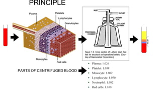
The CDC crossmatch is the traditional method for detecting donor-specific HLA antibodies before transplantation. It detects complement-fixing antibodies but has low sensitivity and specificity. Modifications like adding anti-human globulin increase its sensitivity but it still has limitations. More sensitive techniques like flow cytometry crossmatch, solid phase immunoassays like Luminex single antigen beads, and virtual crossmatches based on donor HLA profiles are now commonly used before transplantation to better assess antibody risk. Performing multiple complementary tests provides the most accurate assessment of donor-specific antibody levels and risk.
Bleeding disorders are caused by abnormalities in hemostasis and coagulation, characterized by skin or mucous membrane bleeding from small blood vessels. The bleeding is usually spontaneous or from minor trauma. Hemostasis involves platelet plug formation and the coagulation cascade, which produces fibrin. Testing such as aPTT, PT, fibrinogen levels, and platelet counts are used to assess coagulation function and identify the cause of bleeding disorders. Treatment depends on the underlying cause, such as replacing deficient clotting factors.
Flow cytometry can be used for several applications in transfusion medicine, including quantifying residual white blood cells and detecting antibody-bound platelets. It is useful for phenotyping red blood cells, such as detecting and quantifying RBC-bound IgG, IgM, and complement in a direct antiglobulin test. Flow cytometry can also measure CD34-positive stem cells for bone marrow transplantation and monitor engraftment. It allows for ex vivo T-cell depletion prior to transplantation. Overall, flow cytometry provides several applications for quality control of blood components and detecting various cell populations.
This patient has a partial D phenotype, not a true weak D phenotype, based on the following:
- Her RBCs reacted weakly with some anti-D reagents but strongly with others, indicating a qualitative antigenic variation (partial D) rather than purely quantitative variation (weak D).
- She produced alloanti-D, which is uncommon for true weak D phenotypes since all D epitopes are present, even at low levels. Production of alloanti-D suggests one or more epitopes are missing (partial D).
- DNA analysis predicted amino acid changes in the external portion of RhD, not just the internal/transmembrane regions as is typical for true weak D. External changes are more likely to result in a
This document provides an overview of blood component therapy. It discusses the composition of blood and history of blood transfusion. It describes the preparation of various blood components like red blood cells, platelets, plasma, and cryoprecipitate. It outlines the indications and guidelines for transfusion of these components. It also reviews trials on restrictive versus liberal transfusion strategies and discusses adverse effects and management of transfusion reactions.
most controversial topic in the field of transfusion medicine, most of the transfusions worldwide are associated with the deleterious effects of immunomodulation, simplified for PG students with latest article support
Blood can be separated into components to meet most transfusion needs while minimizing risks. Effective separation requires centrifugation based on differences in specific gravity of components. Common components prepared include packed red blood cells, platelet concentrates, fresh frozen plasma, and granulocyte concentrates. Preparation of blood components allows for optimal use of donated blood by providing only the required constituent to the patient in need.
Preanalytical variables in coagulation testingShabab Ali
This document discusses pre-analytical variables that can affect coagulation testing. It emphasizes the importance of proper sample collection, including using the correct anticoagulant (sodium citrate), collection technique (slow draw with mixing), and storage requirements. Errors in collection such as short draws, improper mixing or storage, excessive mixing, or hemolysis can falsely affect test results. Proper centrifugation is also needed to obtain platelet-poor plasma for certain tests.
This document discusses apheresis, which involves separating blood into its components. There are different methods of apheresis including intermittent flow centrifugation, continuous flow centrifugation, and membrane filtration. Apheresis can be used to collect various blood components from donors like platelets, plasma, red cells, and leukocytes. It has therapeutic applications like therapeutic plateletpheresis to treat high platelet counts and therapeutic plasmapheresis to remove pathological substances from the plasma like immune complexes. The document outlines the principles, methods, components that can be collected, and indications of apheresis.
One unit of whole blood can be separated into various blood components through centrifugation and other processing steps. This allows each component to be stored optimally and transfused only as needed by patients. Red blood cells can be stored for 21 days in CPD or 42 days when added to ADSOL. Platelets are stored at room temperature while plasma products like FFP and cryoprecipitate must be frozen and thawed as needed. Whole blood is rarely used today due to non-functional platelets and labile coagulation factors after collection.
This document discusses the processes of hemostasis, thrombosis, and fibrinolysis. It defines key terms like blood clot, platelet, fibrin, coagulation cascade, and anticoagulants. The document describes the steps of primary hemostasis which involves platelet adhesion and activation at the site of injury. It also outlines the secondary hemostasis process known as the coagulation cascade that generates thrombin and ultimately forms a fibrin clot to stop bleeding. The roles of fibrinolysis and anticoagulant pathways in regulating clot formation are also summarized.
This document discusses blood collection procedures and anticoagulants. It explains that blood can be collected from veins, capillaries, or arteries for various tests like hematological, biochemical, serological, and molecular examinations. The venipuncture procedure and necessary supplies are outlined. Common anticoagulants like EDTA are described which prevent clotting by chelating calcium. Effects of storage temperature and time on blood cell counts and morphology are also summarized.
Platelets are non-nucleated cell fragments produced by megakaryocytes that play a key role in primary hemostasis and clot retraction. They can be collected through pooled random donor units or apheresis of a single donor. Platelets are stored at 22°C on an agitator for 3-5 days and indications for transfusion include platelet counts <10x109/L for stable patients or <50x109/L for surgical patients. Fresh frozen plasma contains all coagulation factors and is used to treat coagulopathies or reverse anticoagulation in cases of bleeding or prior to procedures.
Stem cell enumeration involves quantifying stem cells, typically through CD34+ cell counting. Key methods include mononuclear cell counts, colony forming unit assays, and CD34+ enumeration via flow cytometry. For flow cytometry, the ISHAGE protocol is commonly used and involves sequential gating of lymphocytes, CD34+ cells, and viable cells. Accurate stem cell quantification is important for determining transplant timing and adequacy.
Clinical Applications Of Therapeutic ApheresisRHMBONCO
This document discusses therapeutic apheresis, which involves separating blood components using centrifugation. It describes how plasma exchange is used to selectively remove plasma constituents like immunoglobulins, proteins, and metabolic waste from the blood to treat various conditions. Conditions treated include autoimmune diseases, renal diseases, hematologic diseases, and neurological disorders. The document outlines the ASFA guidelines for therapeutic apheresis indications and categories, procedures like plasma exchange and photopheresis, and considerations for evaluating new patients and managing risks.
leucodepletion is the removal of 99% leucocytes from the whole blood, pcv or platelets before transfusing into the donor.
this process many infections, transfusion reactions..
Antibody mediated rejection in kidney transplantationimrana tanvir
Antibody mediated rejection (AMR) in kidney transplants can occur in several forms, including hyperacute, acute, and chronic. AMR is defined by the presence of donor-specific antibodies, C4d staining on biopsy, and histological features of antibody injury. C4d staining detects complement activation and is a marker for AMR, though it has low sensitivity. Treatment for AMR includes antibody removal by plasmapheresis, immunosuppression with antilymphocyte therapies, and terminal complement inhibitors. Plasmapheresis is effective but requires multiple sessions and carries risks like infections. No single treatment consistently reverses AMR due to the complex nature of the condition.
- The document discusses histograms generated by cell counters that graphically represent cell population data. It focuses on histograms for red blood cells (RBCs), white blood cells (WBCs), and platelets.
- Key parameters for each type of histogram are defined, such as how cells are counted and measured, normal ranges, and flags that indicate potential issues. Common causes for shifts or abnormalities in the histograms are also outlined.
- The information provides guidance on interpreting cell counter histograms and histograms to help evaluate a patient's blood cell counts and detect possible blood disorders or interferences.
Acute Promyelocytic Leukemia (APL) is a subtype of AML characterized by the t(15;17) translocation resulting in the PML-RARA fusion gene. APL has a high cure rate with all-trans retinoic acid (ATRA) and chemotherapy due to its differentiation of promyelocytes. Complications include disseminated intravascular coagulation, ATRA syndrome, and pseudotumor cerebri. Modern treatment protocols using risk stratification and ATRA with chemotherapy have increased survival to over 80% for APL.
Pretransfusion testing involves several important steps to ensure blood compatibility and prevent transfusion reactions:
1) Blood typing to determine the patient's ABO and Rh blood group is performed along with antibody screening to detect any unexpected antibodies.
2) Crossmatching tests the patient's serum against donor red blood cells to identify any antibodies that could cause a transfusion reaction.
3) Computerized crossmatching can detect ABO incompatibility but requires strict data entry and confirmation of patient and donor blood types to ensure accuracy.
This document discusses blood transfusion and its components. It covers whole blood, packed red blood cells, platelets, fresh frozen plasma, cryoprecipitates, and plasma products. It describes their indications, storage, and administration. The document also discusses transfusion reactions like acute hemolytic reactions, allergic reactions, febrile reactions, anaphylaxis, TRALI, and late hemolytic reactions. It provides treatment protocols for various complications of transfusion like circulatory overload, hypothermia, and infections that can be transmitted through blood products.
This document provides information on various blood products and massive blood transfusion. It defines massive blood transfusion as replacing one entire blood volume within 24 hours or transfusing over 10 units of packed red blood cells in 24 hours. It describes components of blood like whole blood, packed red blood cells, plasma, platelets, and plasma derivatives. It discusses indications, storage, and risks of these products. It also outlines complications of massive transfusion like coagulopathy, hypothermia, acidosis, and circulatory overload and targets for resuscitation like maintaining hemoglobin, coagulation factors, platelets, pH, and temperature.
Megakaryopoiesis and thrombopoiesis involve the production of megakaryocytes and platelets from hematopoietic stem cells in the bone marrow. Megakaryoblasts are early stage megakaryocytes that are 8-24 μm in size with minimal nuclear lobulation and scant cytoplasm. They express surface adhesion molecules like integrin αIIbβ3 and glycoprotein Ib-IX complex. Megakaryoblasts undergo endomitosis, replicating their DNA without cell division to become polyploid. Cytokines such as thrombopoietin are critical for megakaryoblast survival and proliferation.
This document outlines the plasmapheresis protocol at the New Mansoura General Hospital Nephrology Department in Egypt. It defines plasmapheresis as removing, treating, and returning blood plasma from circulation. Examples of diseases treated with plasmapheresis include idiopathic pulmonary fibrosis, Guillain-Barre syndrome, and thrombotic thrombocytopenic purpura. The procedure, complications, replacement solutions, and post-procedure care are described.
Blood components preparation and therapeutic uses finalglobalsoin
This document discusses the preparation of blood components and their therapeutic uses. It begins by explaining how whole blood can be separated into various components to provide targeted replacement therapies. The main blood components and derivatives discussed include packed red blood cells, platelets, fresh frozen plasma, cryoprecipitate, and plasma derivatives obtained through fractionation. The document then goes into details about the history of blood transfusions and developments in blood component preparation methods, types of components, preparation processes, storage and usage guidelines. It provides information on specific blood products like platelet-rich plasma and platelet concentrates.
This document discusses blood components and their uses. It begins by explaining that effective blood transfusion now relies on separating whole blood into components. These components can meet most patient transfusion needs while minimizing risks. The document then discusses the various cellular and plasma components that can be derived from whole blood, including red blood cells, platelets, fresh frozen plasma, cryoprecipitate, and more specialized components. It provides details on the preparation methods, storage, and clinical indications for each component type.
This document discusses plasmapheresis, which is a therapeutic apheresis procedure that removes plasma from the blood. There are two main techniques used: membrane apheresis, which is fast but limited in substance removal, and centrifugal devices, which are more expensive but efficient. Complications can include hypotension, bleeding, and allergic reactions. Plasmapheresis is used to treat autoimmune disorders by removing autoantibodies, and other conditions involving abnormal circulating factors. Care must be taken with anticoagulation and replacement fluids during the procedure.
BLOOD COMPONENTS TRANSFUSION AND ITS COMPLICATIONS.pptxKhushbooGarg61
1) Blood can be separated into components through whole blood collection or apheresis. The main components are packed red blood cells (PRBC), platelet concentrates, fresh frozen plasma (FFP), and cryoprecipitate.
2) Platelet concentrates can be prepared from single donor apheresis or from random donors. Random donor platelets contain lower platelet counts but expose recipients to fewer donors.
3) Transfusion of PRBCs is indicated when patients show signs of anemia such as low hemoglobin levels or oxygen delivery issues. The decision to transfuse depends on multiple clinical factors rather than hemoglobin level alone.
This document discusses various blood components used in transfusion therapy, including their properties, collection, storage, and clinical indications. It covers red blood cells, platelets, fresh frozen plasma, and cryoprecipitate. Key points include how each component is prepared from whole blood, typical volumes and contents, storage guidelines, dosage recommendations, and common clinical scenarios where their use is appropriate or contraindicated.
This document provides guidelines for blood transfusion, including:
1. It discusses the selection and preparation of blood products such as whole blood, platelet concentrates, fresh frozen plasma, and packed red blood cells. Proper donor requirements, collection procedures, and storage conditions are outlined.
2. Indications, dosing, and expected responses to transfusions of various blood components are covered. Red blood cell and platelet transfusion thresholds and dosing are provided.
3. Safety procedures for blood typing, cross-matching, and transfusion monitoring are described. Special considerations for patients with conditions like autoimmune hemolytic anemia that could cause transfusion reactions are highlighted.
4. Two case illustrations demonstrate the
1) Blood conservation strategies are important in cardiac surgery to reduce bleeding and transfusions which can increase mortality and morbidity.
2) Preoperative interventions include managing antiplatelet drugs and anticoagulants, correcting anemia, and using drugs to increase red cell mass.
3) Intraoperative techniques involve autologous blood donation, maintaining normothermia, pharmacological agents like tranexamic acid, and restrictive transfusion triggers.
4) Close monitoring of bleeding and vital signs is also important intraoperatively to guide transfusions which should be a last resort.
This document discusses the management of coagulopathy and bleeding risk in a patient with chronic liver disease undergoing surgery. It notes that the patient has mildly abnormal coagulation tests including an INR of 2.2, but that such tests do not reliably predict bleeding risk. It emphasizes that prophylactic transfusions are not recommended and that bleeding is best managed by addressing its underlying cause rather than by correcting coagulation parameters. Active bleeding should be treated with vasoconstrictors, endoscopic therapies, and restrictive transfusions as needed while avoiding volume overload.
Blood and blood components therapy involves separating whole blood into individual components for targeted transfusion based on a patient's needs. The main components are packed red blood cells (PRBC), fresh frozen plasma (FFP), platelets, and cryoprecipitate. PRBC is used to treat anemia, FFP contains clotting factors for replacing multiple deficient factors, platelets treat thrombocytopenia, and cryoprecipitate contains fibrinogen for bleeding disorders. Component therapy has advantages over whole blood transfusion by targeting specific deficiencies and allowing one donation to treat multiple patients.
It contains indications of blood and blood products and perioperative blood therapy that we usually follow in Aiims Patna ..its is most recent one made in April 2020
This document provides an overview of blood transfusion including indications, measurement of blood loss, types of blood transfusion, donation and collection, administration of blood, and complications. It discusses indications for different blood components like whole blood, packed red blood cells, platelet rich plasma, fresh frozen plasma, cryoprecipitate, and various factor concentrates. Methods of measuring blood loss and amount of transfusion are described. The process of blood donation, collection, storage and administration is outlined. Immediate complications like febrile reactions, allergic reactions, hemolytic reactions, bacterial contamination, circulatory overload, and air embolism are summarized. Delayed complications of thrombophlebitis, delayed hemolytic reactions, and post-trans
Intra dialytic hypotension ,,, prof Alaa SabryFarragBahbah
This document describes a case of intradialytic hypotension in a 65-year-old man on hemodialysis. During one of his dialysis treatments, he developed hypotension with symptoms of feeling poorly and diaphoresis. His dry weight was increased in response, but he experienced another episode of hypotension several days later. The document then discusses intradialytic hypotension in general, including definitions, mechanisms, complications, and approaches to assessing volume status in hemodialysis patients.
Blood transfusion involves carefully matching donor and recipient blood types to prevent dangerous antigen-antibody reactions. Key aspects include typing for the ABO and RH blood group systems, compatibility testing between donor and recipient blood, and cross-matching to confirm safety prior to transfusion. Proper collection, testing, and storage of blood is also important to ensure its viability and safety for transfusion. Guidelines provide triggers for transfusing specific blood components like red blood cells, platelets, and plasma based on clinical factors and lab values. Massive bleeding requires rapid volume replacement and transfusion to maintain adequate blood volume and composition.
Blood transfusion guidelines provide recommendations for appropriate clinical use of blood and components to reduce risks. The risks of transfusion can be lowered through effective donor selection, screening, testing, component separation, storage, and clinical use. Transfusion is recommended when the hemoglobin is less than 7g/dL or the platelet count is less than 10,000/uL, depending on the clinical situation. Alternatives to allogenic transfusion include autologous donation prior to surgery, acute normovolemic hemodilution, erythropoietin, and blood salvage to reduce transfusion needs.
1) Hemodialysis and CRRT are renal replacement therapies used to treat acute kidney injury by removing waste and fluid from the blood. CRRT provides more gradual and continuous treatment compared to intermittent hemodialysis.
2) Indications for RRT include fluid overload, hyperkalemia, acidosis, rising urea/creatinine, uraemia symptoms, and sepsis. The choice of RRT depends on what needs to be removed, the patient's stability, resources, and clinical factors.
3) CRRT modalities include CVVH, CVVHD, and CVVHDF. Anticoagulation is usually needed but can be avoided if the patient is already coagulo
Management of upper gi bleeding email copynadiagulnaz
- Acute upper gastrointestinal bleeding is a common medical emergency that can have high mortality.
- Risk assessment using tools like the Blatchford and Rockall scores helps determine urgency of endoscopy and predict outcomes.
- Endoscopy within 24 hours of admission is recommended to treat bleeding lesions through methods like injection, thermal coagulation, or band ligation of varices.
- Post-endoscopic management involves PPIs, transfusions targeting a hemoglobin of 70-90 g/L, H. pylori treatment if present, and continued medications like terlipressin or beta blockers to prevent rebleeding.
Blood transfusion involves introducing donor blood into a recipient's bloodstream. It is used to increase oxygen-carrying capacity, reverse tissue hypoxia, restore circulating volume, and provide clotting factors. Blood products include whole blood, packed red blood cells, platelets, fresh frozen plasma, and cryoprecipitate. Transfusions aim to treat anemia and coagulation disorders while minimizing complications like reactions, infections, or electrolyte abnormalities through careful screening, storage, and monitoring during the procedure.
Dr. Osama El-Shahat is a consultant nephrologist and head of the nephrology department at New Mansoura General Hospital. He is also an educational ambassador for the International Society of Nephrology. The document discusses the stage-based management of acute kidney injury (AKI) including monitoring for stage 1 risk, conservative therapy for stage 2 injury, and considering renal replacement therapy for stage 3 failure. It provides recommendations at each stage to monitor patients, discontinue nephrotoxic agents, and consider ICU admission or invasive diagnostic workup as needed.
Renal Replacement Therapy: modes and evidenceMohd Saif Khan
This document discusses various modes of renal replacement therapy (RRT) for acute kidney injury (AKI) patients, including their principles, advantages, disadvantages, and evidence regarding optimal dosing. It summarizes that while early RRT initiation and higher RRT doses were associated with better outcomes in some studies, large randomized controlled trials found no significant differences in mortality between early versus late initiation or higher versus lower RRT doses. The optimal RRT modality and timing remains unclear based on current evidence.
Dialysis without anticoagulation (Heparin Free Dialysis)Mahmoud Eid
This document discusses techniques for performing dialysis without anticoagulation. It describes indications for heparin-free dialysis such as recent surgery or bleeding risks. Techniques mentioned include regional citrate anticoagulation, saline flushes, heparin-coated membranes, and citrasate dialysate. Signs of clotting and scoring systems are provided. Tips for priming, high blood flows, and alternatives to heparin locking are also outlined. The key recommendations are to prime properly, have no rushing, follow a written protocol, and focus on patient safety above all else.
This document provides guidelines for blood and blood component transfusion. It discusses the various blood components, quality control parameters, rationale for transfusion in different clinical settings, transfusion guidelines including blood group shifting, use of leukoreduced and irradiated blood products, and the impact of oral antiplatelet therapy. It aims to update best practices for blood transfusion safety.
1. Ascites is the accumulation of fluid in the peritoneal cavity, most commonly caused by cirrhosis.
2. Initial tests of ascitic fluid include appearance, serum-to-ascites albumin gradient, cell count, and total protein to determine if the fluid is infected and if portal hypertension is present.
3. Malignant ascites indicates late-stage cancer and a poor prognosis, accounting for 10% of ascites cases. It results from an imbalance of fluid production and absorption in the peritoneal cavity caused by factors like lymphatic obstruction or increased vascular permeability from the tumor.
Muktapishti is a traditional Ayurvedic preparation made from Shoditha Mukta (Purified Pearl), is believed to help regulate thyroid function and reduce symptoms of hyperthyroidism due to its cooling and balancing properties. Clinical evidence on its efficacy remains limited, necessitating further research to validate its therapeutic benefits.
share - Lions, tigers, AI and health misinformation, oh my!.pptxTina Purnat
• Pitfalls and pivots needed to use AI effectively in public health
• Evidence-based strategies to address health misinformation effectively
• Building trust with communities online and offline
• Equipping health professionals to address questions, concerns and health misinformation
• Assessing risk and mitigating harm from adverse health narratives in communities, health workforce and health system
These lecture slides, by Dr Sidra Arshad, offer a quick overview of the physiological basis of a normal electrocardiogram.
Learning objectives:
1. Define an electrocardiogram (ECG) and electrocardiography
2. Describe how dipoles generated by the heart produce the waveforms of the ECG
3. Describe the components of a normal electrocardiogram of a typical bipolar lead (limb II)
4. Differentiate between intervals and segments
5. Enlist some common indications for obtaining an ECG
6. Describe the flow of current around the heart during the cardiac cycle
7. Discuss the placement and polarity of the leads of electrocardiograph
8. Describe the normal electrocardiograms recorded from the limb leads and explain the physiological basis of the different records that are obtained
9. Define mean electrical vector (axis) of the heart and give the normal range
10. Define the mean QRS vector
11. Describe the axes of leads (hexagonal reference system)
12. Comprehend the vectorial analysis of the normal ECG
13. Determine the mean electrical axis of the ventricular QRS and appreciate the mean axis deviation
14. Explain the concepts of current of injury, J point, and their significance
Study Resources:
1. Chapter 11, Guyton and Hall Textbook of Medical Physiology, 14th edition
2. Chapter 9, Human Physiology - From Cells to Systems, Lauralee Sherwood, 9th edition
3. Chapter 29, Ganong’s Review of Medical Physiology, 26th edition
4. Electrocardiogram, StatPearls - https://www.ncbi.nlm.nih.gov/books/NBK549803/
5. ECG in Medical Practice by ABM Abdullah, 4th edition
6. Chapter 3, Cardiology Explained, https://www.ncbi.nlm.nih.gov/books/NBK2214/
7. ECG Basics, http://www.nataliescasebook.com/tag/e-c-g-basics
One health condition that is becoming more common day by day is diabetes.
According to research conducted by the National Family Health Survey of India, diabetic cases show a projection which might increase to 10.4% by 2030.
Histololgy of Female Reproductive System.pptxAyeshaZaid1
Dive into an in-depth exploration of the histological structure of female reproductive system with this comprehensive lecture. Presented by Dr. Ayesha Irfan, Assistant Professor of Anatomy, this presentation covers the Gross anatomy and functional histology of the female reproductive organs. Ideal for students, educators, and anyone interested in medical science, this lecture provides clear explanations, detailed diagrams, and valuable insights into female reproductive system. Enhance your knowledge and understanding of this essential aspect of human biology.
- Video recording of this lecture in English language: https://youtu.be/Pt1nA32sdHQ
- Video recording of this lecture in Arabic language: https://youtu.be/uFdc9F0rlP0
- Link to download the book free: https://nephrotube.blogspot.com/p/nephrotube-nephrology-books.html
- Link to NephroTube website: www.NephroTube.com
- Link to NephroTube social media accounts: https://nephrotube.blogspot.com/p/join-nephrotube-on-social-media.html
Integrating Ayurveda into Parkinson’s Management: A Holistic ApproachAyurveda ForAll
Explore the benefits of combining Ayurveda with conventional Parkinson's treatments. Learn how a holistic approach can manage symptoms, enhance well-being, and balance body energies. Discover the steps to safely integrate Ayurvedic practices into your Parkinson’s care plan, including expert guidance on diet, herbal remedies, and lifestyle modifications.
Osteoporosis - Definition , Evaluation and Management .pdfJim Jacob Roy
Osteoporosis is an increasing cause of morbidity among the elderly.
In this document , a brief outline of osteoporosis is given , including the risk factors of osteoporosis fractures , the indications for testing bone mineral density and the management of osteoporosis
4. DEFINITION
• Plateletpheresis referred to a procedure in which a portion of
the donor's platelet and some plasma is removed with the
return of the donor's red blood cells (RBCs), white blood cells
(WBCs), and remaining plasma. The product is also known as
single donor platelet (SDP).
6. GENERAL CRITERIA
• Donor screening similar to whole blood collection.( Weight - above 60kg)
• Written informed consent
• Repeat donor
• Venous access- vein examination
• TTI screening - history & testing
• Investigation - hemoglobin, calcium, blood grouping & typing
• Medical examination.
DGHS GUIDELINES
7. SELECTION CRITERIA FOR PLATELETPHERESIS
1. Donor weight should be more than 60 Kg.
2. The interval between procedures should be at least 48 hours. A donor should not undergo the procedure more
than 2 times a week or 24 times a year. After the whole blood donation plateletpheresis, the donor should be
accepted only after 28 days of interval.
3. Platelets may be collected from donors who do not meet the requirement if the component is of particular value
to the patient - HLA matched donors.
4. Donors who have taken aspirin-containing medication within 3 days / 72 hours are deferred.
5. A complete haematological profile, including platelet count, should be done before all plateletpheresis
procedures, and platelet count and haemoglobin must be more than 150,000/μl and > 12.5 g/dl before starting
the procedure.
6. If extra plasma is collected and if the procedure is performed more than once in every 4 weeks, the procedure
should not be done if the total serum protein is less than 6.0 g/dl or if there has been an unexplained weight
loss.
7. Double unit apheresis can be taken in donors whose platelet count is more than 250,000/ul and weight more
than 60kg and those who are not first-time apheresis donors.
AABB GUIDELINES
9. THERAPEUTIC INDICATION
• For therapeutic procedure, the American Society for Apheresis (ASFA) delineates
plateletpheresis based on the acuity of the clinical presentation
◦ Symptomatic thrombocytosis: ASFA category II
◦ Prophylactic or secondary thrombocystosis: ASFA category III
DONOR
THROMBOCYTOPENIA DUE TO LEUKEMIA , APLASTIC ANEMIA , BONE MARROW TRANSPLANT
PLATELET DYSFUNCTION
NEWBORN&POST PARTUM BLEED
TRAUMA - SURGICAL CASES
AS A PART OF MASSIVE TRANSFUSION PROTOCOL
12. CHARACTERISTICS OF TUBING USED IN APHERESIS PROCEDURE
Heparin should be avoided as an anticoagulant because of its tendency to cause platelet clumping within the
apheresis circuit.
14. WHAT HAPPENS POST DONATION ?
•The typical platelet donor experiences a 20% to 29% drop in platelet count (Szymanski et al.,
1973; Heyns et al., 1985), with that among females typically being greater (Rogers et al.,
1995; Dettke et al., 1998).
•Glowitz & Slichter, 1980-In donors undergoing alternate-day collections, platelet count and
apheresis yields have been shown to return to baseline levels by day 10 of collection
PLATELET MOBILISATION FROM SPLEEN OCCURS
IMMEDIETLY MAINTAINING THE COUNT
MALE RETURN TO BASELINE PLT COUNT 4 DAYS
FEMALE INCREASE IN THROMBOPOIETIN
GREATER RECOVERY TIME THAN MALE/calcium level is low normally.
15. ROLE OF ACD IN APHERESIS PROCEDURE
SYMPTOMS OF HYPOCALCEMIA
CIRCUMORAL PARASTHESIA
NAUSEA
VOMITING
DIARRHOEA
TETANY
QT PROLONGATION IN ECG
16. COMPLICATIONS
• Citrate toxicity- mild -perioral tingling and paresthesias, chills, nausea, twitching, and
tremors. severe-carpopedal spasm, seizures, tetany, and cardiac arrhythmias.
• Vascular complications- sepsis,hematoma,phlebitis
• Vasovagal reaction- counselling & hydration.
• Hypovolemia
• Allergic reaction .
• Air embolism
• Depletion of clotting factors
• Transfusion transmitted infections
17. PLATELET PRODUCT REQUIREMENT
•Standards for Blood Banks and Transfusion Services require that the mini- mum
number of platelets in an apheresis product should be 3.0 × 10¹¹ in 90% of units tested
(Standards Committee of the AABB, 2018).
•The U.S. Food and Drug Administration (FDA) requires a minimum of 3.0 × 10¹¹ in 75%
of units tested (Code of Federal Regulations [CFR], 2014).
•The result of these requirements, however, is that an apheresis platelet product
contains at least as many platelets as a pool of platelets from six whole- blood
donations.
18. ADVANTAGES OF SDP
• Routinely, the number of platelets in an
apheresis product is equivalent to 6–8
random platelet concentrates.
• SDP has unique advantages
leukoreduced product, can be human
leukocyte antigen matched, and
platelet-antigen-compatible phenotypes.
• collect large numbers of platelets from
-fewer donor exposures for the patient.
19. ADVANTAGES OF SDP
The potential advantages of SDP that were considered included the following:
• Reduction of infectious complications- SEPTIC PLT TRANSFUSION REACTION DUE
TO BACTERIAL CONTAMINATION IN STORED PLT.
• Reduction of transfusion reactions;
• Ease of leukodepletion- CMV TRANSMISSION
• Reduction in transfusion frequency;
• Prevention of alloimmunization;
• Enhancement of platelet quality;
• Elimination of the need to pool WBDP in transfusion service. (Whole blood derived
platelet)
• SHELF LIFE -5 DAYS
20. RATIONALE
•The goal of therapeutic plateletpheresis in patients with acute thrombotic or
hemorrhagic disorders is reduction of the platelet count to a normal or near-normal
level (typically 400,000/μL); such a procedure can be expected to reduce the platelet
count by 30% to 60% .
• VOLUME : The current ASFA guidelines5
recommend processing anywhere from 1.5 to
2 total blood volumes (TBV), with the anticoagulant-to-blood ratio ranging from 1:8 to
1:12.
21. THERAPEUTIC
INDICATIONS
Thrombocytosis,
It is defined as a platelet count
exceeding 450,000 to
500,000/μL, may be caused by a
reactive process (second- ary
thrombocytosis) or by an
underlying clonal marrow
disorder (primary )
22. • Therapeutic plateletpheresis of patients can rapidly reduce an elevated platelet count
to help mitigate either thrombotic or hemorrhagic complications. Such short-term
reduction can provide symptomatic relief and a therapeutic bridge to the delayed
effects of cytoreductive therapy.
24. •STANDARD CRITERIA for granulocyte transfusion IS NOT ESTABLISHED:
•It is typically agreed on as the minimum necessary to proceed (Schiffer, 1990).
•These include:
1. A proven (by clinical testing) bacterial or fungal infection
2. No response to a trial of appropriate antibiotics/antifungals
3. Absolute neutropenia (<500 cells/μL)
4. A reasonable expectation of patient marrow recovery
Since their first infusion in 1964, GRANULOCYTE TRANSFUSION - adjunct to
antibiotic therapy in neutropenic patients with bacterial and/or fungal infections that are
otherwise refractory to treatment.
25. DONOR LEUKOCYTAPHERESIS
•leukocytapheresis donors must fulfill all of the
requirements applicable to whole-blood donation.
•150,000/μL platelet count requirement .
•ABO type of both recipient and donor should match.
•cytomegalovirus (CMV) status of the donor and
recipient .
• In alloimmunized patients, it is critical that HLA-
• crossmatch-compatible donors be selected.
26. GRANULOCYTE DONOR STIMULATION
1.administer steroids to donors 10 to 12 hours before collection -ORAL PREDNISOLONE
OR DEXAMETHASONE
? WHY - The effects of steroids appear to result from mobilization of granulocytes from
marrow stores as well as delayed granulocyte apoptosis .
2.granulocyte colony stimulating factor (G-CSF) -
Administration of 5 μg/kg of G-CSF every other day resulted in granulocyte
collections four to five times those seen in unstimulated donors and greater than those
seen with prednisone stimulation (Jendiroba et al., 1998).
The optimum timing for granulocyte collection -12 hours after administration of G-CSF
27. GRANULOCYTE PRODUCT
REQUIREMENT
The current adult collection dose requires that 75% of granulocyte components
collected have a minimum of 1.0 × 10¹⁰ granulocytes-(Standards Committee of the AABB,
2018).
granulocytes are stored at room temperature (20°C–24°C) without agitation and have an
expiration date of 24 hours (Standards Committee of the AABB, 2018)
EARLY TRANSFUSION SUGGESTED - APOPTOSIS OCCURS RAPIDLY IN
STORAGE PERIOD .
28. GRANULOCYTE COLLECTION PROCEDURE
• Red blood cells and granulocytes have similar densities and sedimentation rates,
resulting in poor separation during centrifugation .
• hydroxyethyl starch (HES )-The mechanism of action of HES is the induction of
rouleaux formation among red blood cells.
29. GRANULOCYTE DONOR
CONCERN
• granulocytes, platelets and significant numbers of red blood cells are present in the
leukocytapheresis product.
• The donor’s hematocrit typically drops by 7% following a granulocyte collection
(Hester et al., 1995).- loss of red blood cells& dilutional effects of volume expansion
caused by the HES.
• Platelet count typically drops by 22% after each procedure
30. SIDE EFFECTS OF PROCEDURE
• PROLONGED PLATELET RECOVERY.
• G-CSF SIDE EFFECTS-Nausea, vomiting ,headache,splenic rupture, anaphylaxis,
acute iritis, marginal keratitis, gouty arthritis, autoimmune thyroiditis, rheumatoid
arthritis exacerbaTion, thrombosis, erythema multiforme, acute lung injury, and
capillary leak syndrome .
• CORTICOSTERIODS side effects
31. HYPERLEUKOCYTOSIS
WBC count exceeding 50,000 to 100,000/μL,
may occur in acute or chronic leukemias of either myeloid or lymphocytic lineage.
WBC ARE MORE RIGID THAN WBC
MICROVASCULATURE OCCLUSIVE COMPLICATION
HYPERLEUKOSTASIS
ISCHEMIA
TUMOUR LYSIS SYNDROME
CHEMOTHERAPY RELEASE INTRACELLULAR TUMOUR CONTENTS
DIC
MULTI-ORGAN DAMAGE
THERAPEUTIC ASPECT :::
32. The use of an erythrocyte sedimenting agent such as hydroxyethyl starch (HES) to
enhance separation of immature and mature myeloid cells from red cells .
34. CLINICAL APPLICATION OF Therapeutic Leukapheresis
The role of leukapheresis is to reduce the leukocyte concentration in the peripheral blood.
1. AML patients with HL-then leukapheresis offers the potential to decrease morbidity and
mortality by removing large quantities of these activated cells- CUTRTAILS THE RELEASE
OF INFLAMMATORY CYTOKINES & PROLIFERATION
2.Leukapheresis may also have an adjunctive role in chronic disorders characterized by
leukocytosis such as CML, CMML, and CLL - chemotherapy is contraindicated.
3.use of leukapheresis to avoid the teratogenic effects of chemotherapy in early pregnancy
when complicated by myeloproliferative disorders .
35. SELECTIVE LEUKOCYTE APHERESIS
This procedures the removal of more specific pathologic subsets
of leukocytes that may be relevant to the pathogenesis of clinical
disease, with the goal of modulating or suppressing the disease
process using specialized columns or filters to remove leukocytes
from whole blood
DEFINITION
36. TYPES
1. 2-stage filter composed of nonwoven polyester
fabric that removes leukocytes through filtration and
adhesion.
2.The efficiency of removal is related to the small
diameter (0.8 to 2.8 microns) of the inner filter fibers.
3.Remove about 99% of granulocytes and
monocytes and 40% to 60% of lymphocytes from
processed blood.35,36
4.USED IN Active ulcerative colitis and rheumatoid
arthritis
1.filter component consists of a column (335-mL
capacity) filled with 220 g of cellulose acetate
beads (2 mm in diameter) and immersed in 130
mL of isotonic saline.37
2.selectively adsorbs approximately 65% of
granulocytes and 55% of monocytes without
significantly adsorbing lymphocytes.38
3.treatment of Crohn disease, ulcerative colitis
(UC), rheumatoid arthritis, systemic lupus
erythematosus, and ocular Behçet disease, and in
Japan for ulcerative colitis.
leukocyte adsorptive apheresis system granulocyte/monocyte adsorptive apheresis system
37. CONCLUSION
• Clinical signs and symptoms arising from HL or thrombocytosis are variable and
unpredictable.
• Without specific laboratory assays to identify patients at risk for complications,
guidelines for therapeutic apheresis procedures to lower peripheral blood
concentrations of platelets and leukocytes may have to continue to rely on diagnosis,
clinical findings, and peripheral blood counts alone.
38. • CLAUDIA S COHN,MEGHAN DALANEY, AABB TEXTBOOK OF APHERESIS PRINCIPLES AND
PRACTICE - 20EDITION,PAGE NO-705-720
• MACKROO.R.N, PRINCIPLES &PRACTICE OF TRANSFUSION MEDICINE , SECOND EDITION,
CHAPTER 7
• TOBY L SIMON,JEFFERY MACCULOUGH, EDWARD ,ROSSI OF PRINCIPLES OF TRANSFUSION
MEDICINE-5TH EDITION
• NBEMS WEBINAR
• MAGRET DIGUARDO , ELEH BOBR,HENRYS CLINICAL DIAGNOSIS & MANAGMENT BY LABORATORY
METHODS , 24TH EDITION, CHAPTER-38,ELSEIVER
REFERENCES
Thank you
40. RED CELL APHERESIS
This term particularly applies to the removal of red cells using
automated blood-processing instruments that are capable of
selectively removing erythrocytes while returning the plasma,
buffy coat cells, and additional isotonic saline to the patient
DEFINITION
41. DONOR SELECTION
• The FDA and the AABB do not define a minimum hemoglobin/hematocrit for double
red cell donation.
• The AABB limits the total volume of red blood cells removed, such that the donor’s
hematocrit and hemoglobin are not <30% and <10 g/ dL, respectively, after volume
replacement (
42. Erythrocytopheresis Product Requirements
• The Standards for Blood Banks and Transfusion Services - mean hemoglobin >60 g
or a packed red cell volume of 180 mL.
• 95% of the units sampled must have >50 g of hemoglobin or 150 mL packed red cell
volume (Standards Commit- tee of the AABB, 2018).
• If the red blood cell units are leukocyte reduced, then the mean hemoglobin must be
>51 g or 153 mL packed red cell volume. Here, at least 95% of tested units must have
>42.5 g of hemoglobin or 128 mL packed red blood cell volume (Standards
Committee of the AABB, 2018).
43. SICKLE CELL DISEASE
ASFA- CATEGORY I
-ability to rapidly reduce the burden of parasitemia
-reduce the level of toxic mediators such as cytokines.
- IMPROVED SURVIVAL OF RED CELLS
- malaria with parasitemia >10%.
-babesiosis with >5% parasitemia
MALARIA
ASFA CATEGORY-II
BABESIA
ASFA CATEGORY-II
INDICATIONS
POLYCYTHEMIA VERA
ASFA- CATEGORY I
-rapidly decrease hematocrit for extended intervals relative to simple phlebotomy
-acute thrombotic or microvascular complications
HEREDITARY
HEMOCHROMATOSIS
ASFA CATEGORY-I
-more rapid decline and normalization of ferritin;
--erythrocytapheresis procedure every two weeks and weekly whole blood phlebotomy
are equiv alent