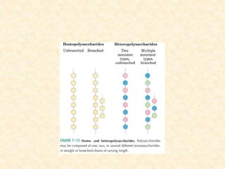
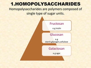
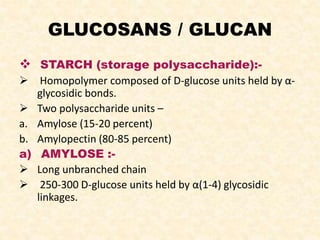
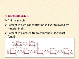
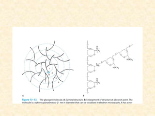
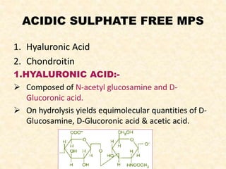
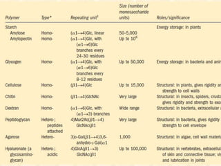
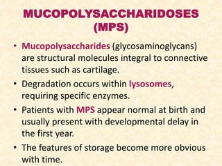
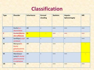
POLYSACCHARIDES
POLYSACCHARIDES
• Simply glycans.
• Repeat units of monosaccharides or their
derivatives,held together by glycosidic bonds.
• Linear as well as branched polymers.
• Two types-
1. Homopolysaccharides
2. Heteropolysaccharides
1.HOMOPOLYSACCHARIDES
Homopolysaccharides are polymers composed of
single type of sugar units.
Fructosan
e.g inulin
Glucosan
e.g
starch,glycogen,cellulose
Galactosan
e.g agar
GLUCOSANS / GLUCAN
 STARCH (storage polysaccharide):-
 Homopolymer composed of D-glucose units held by α-
glycosidic bonds.
 Two polysaccharide units –
a. Amylose (15-20 percent)
b. Amylopectin (80-85 percent)
a) AMYLOSE :-
 Long unbranched chain
 250-300 D-glucose units held by α(1-4) glycosidic
linkages.
b) AMYLOPECTIN:-
 Branched chain with α(1-6) glycosidic bonds at the
branching points and α(1-4) linkages everywhere
else.
 Contains few thousand glucose units looks like a
branched tree (20-30) glucose units per branch.
STRUCTURE OF STARCH
(AMYLOSE & AMYLOPECTIN)
Amylose Amylopectin
1) Soluble in water
2)Gives Blue colour with dilute
iodine solution
3)Structure –unbranched
4)250 to 300 D-glucose units
linked by alpha 1→4 linkages
5)Mol. wt approx.60000
6)Occurs at the extent of 15 to
20%
1)Insoluble in water
2)Gives reddish colour with iodine
solution
3)Structure – highly branched
4)Units joined together
by Alpha 1→4glycosidic bond and at
branch point with alpha 1→6
glycosidic linkages
5)Mol. wt. approx 500000
6)0ccurs at 80 to 85%
GLYCOGEN:-
 Animal starch.
 Present in high concentration in liver followed by
muscle, brain.
 Present in plants with no chlorophyll (eg.yeast ,
fungi)
 Structure of glycogen is similar to that of
amylopectin with more number of branches.
 Glucose is the repeating unit in glycogen joined
together by α(1-4) glycosidic bonds,and α(1-6)
glycosidic bonds at branching points.
 Present in cells as granules with high molecular
weight.
 Complete hydrolysis yields glucose.
 In the liver, glycogen synthesis and degradation are
regulated to maintain blood-glucose levels . Glycogen
serves as a buffer to maintain blood glucose level.
 In contrast, in muscle, these processes are regulated
to meet the energy needs of the muscle itself.
 - The concentration of glycogen is higher in the liver
than in muscle , but more glycogen is stored in
skeletal muscle overall because of its much greater
mass.
CELLULOSE:-
 Polymer of glucose.
 On heating with high concentrations of acids yields
disaccharide cellobiose and D-glucose.
 Cellobiose is made up of two molecules of D-glucose
linked together by β -Glucosidic linkages between C1
and C4 of adjacent glucose units.
 Most abundant of all carbohydrates.
 Very stable insoluble compound.
 Herbivorous animals utilise cellulose with the help
of bacteria.
 Human beings lack any enzyme that hydrolyzes the β
(1→ 4) bonds, and so cannot digest cellulose. It is an
important source of "bulk" in the diet, and the major
component of dietary fiber stimulating peristalsis
and elimination of indigestible food residues.
CHITIN:-
 It is linear homopolysaccharide composed of N-
acetyl glucosamine residues in b-linkage.
 Only difference from cellulose is the replacement of
the hydroxyl group at C-2 with an acetylated amino
group.
 Principal component of the hard exoskeleton of
arthopods.
 Second most abundant in nature.
DEXTRINS:-
 Produced by the partial hydrolysis of starch along
with maltose and glucose.
 Dextrins are often referred to as either
amylodextrins, erythrodextrins or achrodextrins.
 Used as mucilages (glues).
 Used in infant formulas .
 Indigestible dextrin are developed as soluble fiber
supplements for food products.
 Also Used as thickening agents in food processing
DEXTRANS:-
 Polymer of D-glucose.
 Synthesised by the action of Leuconostoc
mesenteroides.
 Exocellular enzyme produced by the organisms bring
about polymerisation of glucose moiety of sucrose
molecule DEXTRANS.
 They differ from dextrins in structure.
 Contains α (1,4), α(1,6) and α (1,3) linkages.
 Molecular weight : 40,000; 70,000; 75,000
 Used as plasma expanders .
Disadvantage:-
 It interferes with grouping and cross matching.
FRUCTOSANS
INULIN:-
 Polymer of D-fructose.
 Low molecular weight -5000
 Occurs in tubers of the Dehlia, in the roots of
Jerusalem artichoke, dandelion and in the bulbs of
onion and garlic.
 b-(1,2) linked fructofuranoses.
 Linear ,no branching.
 Hydrolysis yields fructose.
 Used for the evaluation of glomerular filtration rate.
 It has no dietary importance in human beings as
inulinase is absent.
GALACTOSANS
AGAR:-
 Agar is a galactose polymer.
 Obtained from the cell walls of some species of red
algae (Sphaerococcus Euchema ) and species of
Gelidium.
 Dissolved in hot water and cooled, agar becomes
gelatinous.
 Used in microbiology .
-.
-
 As laxative.
 A vegetarian gelatin substitute.
 A thickener for soups, jellies, ice cream and Japanese
desserts.
 As a clarifying agent in brewing, and for sizing
fabrics.
HETEROPOLYSACCHARIDES/
HETEROGYLCANS
 Mucopolysaccharides/Glycosaminoglycans(GAGS)
 Amino sugar +ss uronic acid units.
 Some contain amino sugar and monosaccharide units
without the presence of uronic acid.
 Amino group is generally acetylated.
 Carbohydrate content more than 4% -Mucoproteins.
 Carbohydrate content less than 4% -Glycoproteins.
CLASSIFICATION
MPS
Acidic
Sulphate
free
Sulphate
contain
Neutr
al
--
ACIDIC SULPHATE FREE MPS
1. Hyaluronic Acid
2. Chondroitin
1.HYALURONIC ACID:-
 Composed of N-acetyl glucosamine and D-
Glucoronic acid.
 On hydrolysis yields equimolecular quantities of D-
Glucosamine, D-Glucoronic acid & acetic acid.
 Occurrence:- Synovial fluid, ECM of loose connective
tissue. Serves as a lubricant and shock absorber.
 HYALURONIDASE –An enzyme catalyses the
depolymerisation of hyaluronic acid and by reducing
its viscosity facilitates diffusion of materials into
tissue spaces.
 Clinically the enzyme is used to increase the
efficiency of absorption of solutions administered by
clysis.
2.CHONDROITIN
-Another Sulphate free acid mucoplosaccharide.
-It is found in cornea and also in cranial
cartilages.
- N-acetyl galactosamine +D-Glucoronic acid.
ACIDIC SULPHATE CONTAIN
1.Chondroitin sulphate
2.Keratan sulphate
3.Heparin
4.Heparitin sulphate
1.CHONDROITIN SULPHATE
It is a principle MPS in ground substance of
mammalian tissues and cartilages.
 It is a sulphate containing MPs.
 Four chondroitin sulphate are isolated which are
chondritin Sulphate A,B,C,D.
- 1.Chondritin Sulphate A - Consists of
repeating units of N-acetyl-D-Galactosamine and
D-Glucuronic acid. N-Acetylgalactosamine is
esterified with sulphate in position 4 of
galactosamine .
- It is present in cartilages ,bone and cornea.
2.Chondroitin Sulphate B:-
 It is present in skin ,cardiac valve and tendon.
 It has L-iduronic acid in place of glucuronic
acid which is found in other chondritin sulphate.
 L-iduronic acid is an epimer of D-
Glucuronic acid.
 It consist of repeating units of L-iduronic
acid and N-acetyl galactosamine at c4
sulphate moity present.
 It has weak anticoagulent property.
 Sometimes it is found in skin hence called as
3.Chondroitin sulphate C
• It is found in cartilage and tendon.
• Structure of chondritin sulphate c is similar to
chondritin sulphate A. Except that sulphate group is
present at position 6 of galactosamine molecule
instead of position 4.
4.Chondroitin Sulphate D
It is isolated from cartilage of shark
It resembles in structure to chondroitin
sulphate C,except that it has second SO4
attached at carbon 2 or 3 of uronic acid.
2.KERATAN SULPHATE
It is a sulphate containg acid MPS.
Found in costalcartilage,cornea,aorta,nucleus
pulposus.
KERATAN SULPHATE - It consist of repeating
diassacharide units of N-aceyl glucosamine
and galactose.
No uronic acids in molecule.
• There are 2 types as follows -
• Keratan Sulphate type 1- found in
cornea.In this type linkage are between N-
acetylglucosamine and aspargine.
• Keratan Sulphate type 2- found in
skeletal muscle. In this type linkage to protein
is through –OH group on serine and threonine
residue of protein.
3.HEPARIN
 Has Molecular weight 17000 to 20000.
 It is anticoagulant present in liver produced by
mast cells present in liver .
It is found in lungs ,thymus,spleen,walls of large
arteries ,skin ,blood.
 It is a polymer of repeating diasaccharide unit of
D-Glucosamine and either of the two uronic acid
–D-Glucuronic acid and L-Iduronic acid.
 In fully formed heparin molecule 90% or more of
uronic acid residues are L-Iduronic acid.
Heparin
4.HEPARITIN SULPHATE
It is isolated from amyloid liver and spleen of pts
with Hurler’s syndrome.
It has negligible anticoagulant activity.
Unlike heparin it’s predominant uronic acid is D-
Glucuronic acid.
Some of the amino group carry acetyl groups and
% of –SO4 group are smaller.
Recent study shows that it is present on cell
surface as proteoglycans and is extracellular.
It has low molecular weight.
NEUTRAL MPS
 Found in Pneumococci capsule.
Act as blood group substances. Four
monosaccharides , Galactose, Fucose,
Galactosamine(Acetylated) and Acetylated
Glucosamine are present in all types of blood
group substances.
 Also found in egg protein- ovalbumin.
PROTEOGYLCANS
Proteoglycans are conjugate proteins.
Proteins called ‘core’proteins are covalently
linked to glycosaminoglycans.
Amount of carbohydrates in proteoglycans are
95% more as compared to glycoproteins.
 LINKAGES – Three types
1) O-glycosidic linkage-formed between N-acetyl
glucosamine and serine or threonine. seen in
keratan so4 .
2)N-glycosylamine linkage –formed between N-acetyl
glucosamine and amide N of aspargine of core
protein.
• seen in keratan so4and N-linked glycoprotein
3) O-Glycosidic linkage – formed between xylose and
serine of protein.This bond is unique to
proteoglycans.
FUNCTIONS
 It acts as a costituent of extracellular
matrix or ground substances .
 Proteoglycans acts as barrier in tissue..
 Proteoglycans play a vital role in release of
hormones.
 It plays a essentiel role in cell migratio of
embryonic tissues.
 It is useful in glomerular filtration rate
 It acts as anticoagulent.
 It acts as coenzyme.
 It acts as receptor for cells.
 It plys a role in compressibility of cartilage.
 It plays a role in sclera of eyes
It act as lubricant in joints.
It plays a role in corneal transparency.
MUCOPOLYSACCHARIDOSES
(MPS)
• Mucopolysaccharides (glycosaminoglycans)
are structural molecules integral to connective
tissues such as cartilage.
• Degradation occurs within lysosomes,
requiring specific enzymes.
• Patients with MPS appear normal at birth and
usually present with developmental delay in
the first year.
• The features of storage become more obvious
with time.
Classification
Type Disorder Inheritance Corneal
clouding
Skeleton Hepato-
Splenomegaly
MR
I Hurler(a-L-
Iduronidase
AR + +++ +++ +++
II Hunter(Iduron
ate sulfatase)
XL - +++ +++ +++
III Sanfillipo(sulph
amidase)
AR + + + +++
IV Morquio(N-
Acetyl
galactosamine
6-sulphatase
AR + +++ + -
VI Maroteaux-
Lamy(N-Acetyl
galactosamine
4-sulphatase
AR + +++ +++ -
VII Sly AR + +++ +++ +++
THANK YOU

POLYSACCHARIDES

  • 1.
  • 2.
    POLYSACCHARIDES • Simply glycans. •Repeat units of monosaccharides or their derivatives,held together by glycosidic bonds. • Linear as well as branched polymers. • Two types- 1. Homopolysaccharides 2. Heteropolysaccharides
  • 4.
    1.HOMOPOLYSACCHARIDES Homopolysaccharides are polymerscomposed of single type of sugar units. Fructosan e.g inulin Glucosan e.g starch,glycogen,cellulose Galactosan e.g agar
  • 5.
    GLUCOSANS / GLUCAN STARCH (storage polysaccharide):-  Homopolymer composed of D-glucose units held by α- glycosidic bonds.  Two polysaccharide units – a. Amylose (15-20 percent) b. Amylopectin (80-85 percent) a) AMYLOSE :-  Long unbranched chain  250-300 D-glucose units held by α(1-4) glycosidic linkages.
  • 6.
    b) AMYLOPECTIN:-  Branchedchain with α(1-6) glycosidic bonds at the branching points and α(1-4) linkages everywhere else.  Contains few thousand glucose units looks like a branched tree (20-30) glucose units per branch.
  • 7.
  • 8.
    Amylose Amylopectin 1) Solublein water 2)Gives Blue colour with dilute iodine solution 3)Structure –unbranched 4)250 to 300 D-glucose units linked by alpha 1→4 linkages 5)Mol. wt approx.60000 6)Occurs at the extent of 15 to 20% 1)Insoluble in water 2)Gives reddish colour with iodine solution 3)Structure – highly branched 4)Units joined together by Alpha 1→4glycosidic bond and at branch point with alpha 1→6 glycosidic linkages 5)Mol. wt. approx 500000 6)0ccurs at 80 to 85%
  • 10.
    GLYCOGEN:-  Animal starch. Present in high concentration in liver followed by muscle, brain.  Present in plants with no chlorophyll (eg.yeast , fungi)
  • 12.
     Structure ofglycogen is similar to that of amylopectin with more number of branches.  Glucose is the repeating unit in glycogen joined together by α(1-4) glycosidic bonds,and α(1-6) glycosidic bonds at branching points.  Present in cells as granules with high molecular weight.  Complete hydrolysis yields glucose.
  • 13.
     In theliver, glycogen synthesis and degradation are regulated to maintain blood-glucose levels . Glycogen serves as a buffer to maintain blood glucose level.  In contrast, in muscle, these processes are regulated to meet the energy needs of the muscle itself.  - The concentration of glycogen is higher in the liver than in muscle , but more glycogen is stored in skeletal muscle overall because of its much greater mass.
  • 14.
    CELLULOSE:-  Polymer ofglucose.  On heating with high concentrations of acids yields disaccharide cellobiose and D-glucose.  Cellobiose is made up of two molecules of D-glucose linked together by β -Glucosidic linkages between C1 and C4 of adjacent glucose units.  Most abundant of all carbohydrates.  Very stable insoluble compound.
  • 15.
     Herbivorous animalsutilise cellulose with the help of bacteria.  Human beings lack any enzyme that hydrolyzes the β (1→ 4) bonds, and so cannot digest cellulose. It is an important source of "bulk" in the diet, and the major component of dietary fiber stimulating peristalsis and elimination of indigestible food residues.
  • 16.
    CHITIN:-  It islinear homopolysaccharide composed of N- acetyl glucosamine residues in b-linkage.  Only difference from cellulose is the replacement of the hydroxyl group at C-2 with an acetylated amino group.  Principal component of the hard exoskeleton of arthopods.  Second most abundant in nature.
  • 18.
    DEXTRINS:-  Produced bythe partial hydrolysis of starch along with maltose and glucose.  Dextrins are often referred to as either amylodextrins, erythrodextrins or achrodextrins.  Used as mucilages (glues).  Used in infant formulas .
  • 19.
     Indigestible dextrinare developed as soluble fiber supplements for food products.  Also Used as thickening agents in food processing
  • 20.
    DEXTRANS:-  Polymer ofD-glucose.  Synthesised by the action of Leuconostoc mesenteroides.  Exocellular enzyme produced by the organisms bring about polymerisation of glucose moiety of sucrose molecule DEXTRANS.  They differ from dextrins in structure.
  • 21.
     Contains α(1,4), α(1,6) and α (1,3) linkages.  Molecular weight : 40,000; 70,000; 75,000  Used as plasma expanders .
  • 22.
    Disadvantage:-  It interfereswith grouping and cross matching.
  • 23.
    FRUCTOSANS INULIN:-  Polymer ofD-fructose.  Low molecular weight -5000  Occurs in tubers of the Dehlia, in the roots of Jerusalem artichoke, dandelion and in the bulbs of onion and garlic.
  • 24.
     b-(1,2) linkedfructofuranoses.  Linear ,no branching.  Hydrolysis yields fructose.  Used for the evaluation of glomerular filtration rate.  It has no dietary importance in human beings as inulinase is absent.
  • 25.
    GALACTOSANS AGAR:-  Agar isa galactose polymer.  Obtained from the cell walls of some species of red algae (Sphaerococcus Euchema ) and species of Gelidium.  Dissolved in hot water and cooled, agar becomes gelatinous.  Used in microbiology . -. -
  • 26.
     As laxative. A vegetarian gelatin substitute.  A thickener for soups, jellies, ice cream and Japanese desserts.  As a clarifying agent in brewing, and for sizing fabrics.
  • 27.
    HETEROPOLYSACCHARIDES/ HETEROGYLCANS  Mucopolysaccharides/Glycosaminoglycans(GAGS)  Aminosugar +ss uronic acid units.  Some contain amino sugar and monosaccharide units without the presence of uronic acid.  Amino group is generally acetylated.  Carbohydrate content more than 4% -Mucoproteins.  Carbohydrate content less than 4% -Glycoproteins.
  • 28.
  • 29.
    ACIDIC SULPHATE FREEMPS 1. Hyaluronic Acid 2. Chondroitin 1.HYALURONIC ACID:-  Composed of N-acetyl glucosamine and D- Glucoronic acid.  On hydrolysis yields equimolecular quantities of D- Glucosamine, D-Glucoronic acid & acetic acid.
  • 30.
     Occurrence:- Synovialfluid, ECM of loose connective tissue. Serves as a lubricant and shock absorber.  HYALURONIDASE –An enzyme catalyses the depolymerisation of hyaluronic acid and by reducing its viscosity facilitates diffusion of materials into tissue spaces.  Clinically the enzyme is used to increase the efficiency of absorption of solutions administered by clysis.
  • 31.
    2.CHONDROITIN -Another Sulphate freeacid mucoplosaccharide. -It is found in cornea and also in cranial cartilages. - N-acetyl galactosamine +D-Glucoronic acid.
  • 32.
    ACIDIC SULPHATE CONTAIN 1.Chondroitinsulphate 2.Keratan sulphate 3.Heparin 4.Heparitin sulphate
  • 33.
    1.CHONDROITIN SULPHATE It isa principle MPS in ground substance of mammalian tissues and cartilages.  It is a sulphate containing MPs.  Four chondroitin sulphate are isolated which are chondritin Sulphate A,B,C,D. - 1.Chondritin Sulphate A - Consists of repeating units of N-acetyl-D-Galactosamine and D-Glucuronic acid. N-Acetylgalactosamine is esterified with sulphate in position 4 of galactosamine . - It is present in cartilages ,bone and cornea.
  • 34.
    2.Chondroitin Sulphate B:- It is present in skin ,cardiac valve and tendon.  It has L-iduronic acid in place of glucuronic acid which is found in other chondritin sulphate.  L-iduronic acid is an epimer of D- Glucuronic acid.  It consist of repeating units of L-iduronic acid and N-acetyl galactosamine at c4 sulphate moity present.  It has weak anticoagulent property.  Sometimes it is found in skin hence called as
  • 35.
    3.Chondroitin sulphate C •It is found in cartilage and tendon. • Structure of chondritin sulphate c is similar to chondritin sulphate A. Except that sulphate group is present at position 6 of galactosamine molecule instead of position 4.
  • 36.
    4.Chondroitin Sulphate D Itis isolated from cartilage of shark It resembles in structure to chondroitin sulphate C,except that it has second SO4 attached at carbon 2 or 3 of uronic acid.
  • 37.
    2.KERATAN SULPHATE It isa sulphate containg acid MPS. Found in costalcartilage,cornea,aorta,nucleus pulposus. KERATAN SULPHATE - It consist of repeating diassacharide units of N-aceyl glucosamine and galactose. No uronic acids in molecule.
  • 39.
    • There are2 types as follows - • Keratan Sulphate type 1- found in cornea.In this type linkage are between N- acetylglucosamine and aspargine. • Keratan Sulphate type 2- found in skeletal muscle. In this type linkage to protein is through –OH group on serine and threonine residue of protein.
  • 40.
    3.HEPARIN  Has Molecularweight 17000 to 20000.  It is anticoagulant present in liver produced by mast cells present in liver . It is found in lungs ,thymus,spleen,walls of large arteries ,skin ,blood.  It is a polymer of repeating diasaccharide unit of D-Glucosamine and either of the two uronic acid –D-Glucuronic acid and L-Iduronic acid.  In fully formed heparin molecule 90% or more of uronic acid residues are L-Iduronic acid.
  • 41.
  • 42.
    4.HEPARITIN SULPHATE It isisolated from amyloid liver and spleen of pts with Hurler’s syndrome. It has negligible anticoagulant activity. Unlike heparin it’s predominant uronic acid is D- Glucuronic acid. Some of the amino group carry acetyl groups and % of –SO4 group are smaller. Recent study shows that it is present on cell surface as proteoglycans and is extracellular. It has low molecular weight.
  • 43.
    NEUTRAL MPS  Foundin Pneumococci capsule. Act as blood group substances. Four monosaccharides , Galactose, Fucose, Galactosamine(Acetylated) and Acetylated Glucosamine are present in all types of blood group substances.  Also found in egg protein- ovalbumin.
  • 44.
    PROTEOGYLCANS Proteoglycans are conjugateproteins. Proteins called ‘core’proteins are covalently linked to glycosaminoglycans. Amount of carbohydrates in proteoglycans are 95% more as compared to glycoproteins.  LINKAGES – Three types 1) O-glycosidic linkage-formed between N-acetyl glucosamine and serine or threonine. seen in keratan so4 .
  • 45.
    2)N-glycosylamine linkage –formedbetween N-acetyl glucosamine and amide N of aspargine of core protein. • seen in keratan so4and N-linked glycoprotein 3) O-Glycosidic linkage – formed between xylose and serine of protein.This bond is unique to proteoglycans. FUNCTIONS  It acts as a costituent of extracellular matrix or ground substances .  Proteoglycans acts as barrier in tissue..
  • 46.
     Proteoglycans playa vital role in release of hormones.  It plays a essentiel role in cell migratio of embryonic tissues.  It is useful in glomerular filtration rate  It acts as anticoagulent.  It acts as coenzyme.  It acts as receptor for cells.  It plys a role in compressibility of cartilage.  It plays a role in sclera of eyes
  • 47.
    It act aslubricant in joints. It plays a role in corneal transparency.
  • 49.
    MUCOPOLYSACCHARIDOSES (MPS) • Mucopolysaccharides (glycosaminoglycans) arestructural molecules integral to connective tissues such as cartilage. • Degradation occurs within lysosomes, requiring specific enzymes. • Patients with MPS appear normal at birth and usually present with developmental delay in the first year. • The features of storage become more obvious with time.
  • 50.
    Classification Type Disorder InheritanceCorneal clouding Skeleton Hepato- Splenomegaly MR I Hurler(a-L- Iduronidase AR + +++ +++ +++ II Hunter(Iduron ate sulfatase) XL - +++ +++ +++ III Sanfillipo(sulph amidase) AR + + + +++ IV Morquio(N- Acetyl galactosamine 6-sulphatase AR + +++ + - VI Maroteaux- Lamy(N-Acetyl galactosamine 4-sulphatase AR + +++ +++ - VII Sly AR + +++ +++ +++
  • 51.