SlideShare a Scribd company logo
RESPIRATORY SYSTEM
The respiratory system provides the route
by which the supply of oxygen present in
the atmospheric air gains entry to the body
and it provides the route of excretion of
carbon dioxide
• External respiration- Exchange of gases
between the blood and the lungs
• Internal respiration- Exchange of gases
between the blood and the cells
Organs of Respiratory System
• Nose
• Pharynx Upper Respiratory tract
• Larynx
• Trachea
• Two bronchi
• Bronchioles Lower respiratory tract
• Two lungs
• Coverings of Lungs-Pleura
• Muscles of Respiration- Intercostal muscles &
Diaphragm
Functional Zones of Respiratory System
NOSE
• Made up of external nose & an inner
chamber-Nasal Cavity
Anterior nare
Posterior nare
Opening of
Auditory tube
Nasal Cavity:
• Consists of a large irregular cavity divided into
two equal passages by a septum
• The posterior bony part of the septum -
perpendicular plate of the ethmoid bone and the
vomer. Anteriorly - hyaline cartilage.
• roof -cribriform plate of the ethmoid bone, the
sphenoid bone, frontal bone and nasal bones.
• floor- hard palate in front and the soft palate behind. The
hard palate is composed of the maxilla and palatine
bones and the soft palate consists of involuntary muscle.
• medial wall - septum.
• lateral walls - maxilla, the ethmoid bone and the inferior
conchae.
• posterior wall - posterior wall of the pharynx.
Openings into the nasal cavity
• The anterior nares, or nostrils, are the openings from the
exterior into the nasal cavity. Hairs are present in this
area.
• The posterior nares are the openings from the nasal
cavity into the pharynx.
• The paranasal sinuses are cavities in the bones of the
face and the cranium which contain air. There are tiny
openings between the paranasal sinuses and the nasal
cavity. They are lined with mucous membrane,
continuous with that of the nasal cavity. The main
sinuses are:
• maxillary sinuses in the lateral walls
• frontal and sphenoidal sinuses in the roof
• ethmoidal sinuses in the upper part of the lateral walls
Respiratory function of the nose
• The function of the nose is to begin the
process by which the air is warmed,
moistened and 'filtered'.
• The projecting conchae increase the
surface area and cause turbulence,
spreading inspired air over the whole
nasal surface. The large surface area
maximises warming, humidification and
filtering.
Warming: This is due to the immense vascularity of
the mucosa. This explains the large blood loss when
a nosebleed (epistaxis) occurs.
Filtering and cleaning of air: This occurs as hairs at
the anterior nares trap larger particles. Smaller
particles such as dust and microbes settle and
adhere to the mucus. Mucus protects the underlying
epithelium from irritation and prevents drying.
Synchronous beating of the cilia wafts the mucus
towards the throat where it is swallowed or
expectorated.
Humidification: This occurs as air travels over the
moist mucosa and becomes saturated with water
vapour. Irritation of the nasal mucosa results in sneezing, a
reflex action that forcibly expels an irritant.
PHARYNX
PHARYNX-ANATOMY
• Pharynx is a tube 12 to 14 cm long that extends
from the base of the skull to the level of the 6th
cervical vertebra.
• Anatomical regions of pharynx:
Name Region
Nasopharynx Behind nasal cavity-internal
nare to soft palate
Oropharynx Behind roof of tongue-soft
palate to epiglottis & hyoid
Laryngopharynx Behind larynx-epiglottis to
cricoid cartilage of larynx
The pharynx is composed of three layers of tissue:
1. Mucous membrane lining: The mucosa varies slightly
in the different parts. In the nasopharynx it is continuous
with the lining of the nose and consists of ciliated columnar
epithelium; in the oropharynx and laryngopharynx it is
formed by tougher stratified squamous epithelium which is
continuous with the lining of the mouth and oesophagus.
2. Fibrous tissue: This forms the intermediate layer. It is
thicker in the nasopharynx, where there is little muscle, and
becomes thinner towards the lower end, where the muscle
layer is thicker.
3. Muscle tissue: This consists of several involuntary
constrictor muscles that play an important part in the
mechanism of swallowing (deglutition) which, in the
pharynx, is not under voluntary control. The upper end of
the oesophagus is closed by the lower constrictor muscle,
except during swallowing.
Functions:
Passageway for air and food: The pharynx is an organ involved
in both the respiratory and the digestive systems: air passes through the
nasal and oral parts, and food through the oral and laryngeal parts.
Warming and humidifying: By the same methods as in the nose,
the air is further warmed and moistened as it passes through the
pharynx.
Taste: There are olfactory nerve endings of the sense of taste in the
epithelium of the oral and pharyngeal parts.
Hearing: The auditory tube, extending from the nasal part to each middle ear,
allows air to enter the middle ear.
Satisfactory hearing depends on the presence of air at atmospheric pressure on
each side of the tympanic membrane (ear drum)
Protection: The lymphatic tissue of the pharyngeal and laryngeal tonsils
produces antibodies in response to antigens, e.g. microbes. The tonsils are
larger in children and tend to atrophy in adults.
Speech: The pharynx functions in speech; by acting as a resonating chamber
for the sound ascending from the larynx, it helps (together with the sinuses) to
give the voice its individual characteristics.
LARYNX (voice box)
Anatomy:
• The larynx or 'voice box' extends from the root of the
tongue and the hyoid bone to the trachea.
• It lies in front of the laryngopharynx at the level of the
3rd, 4th, 5th and 6th
cervical vertebrae.
• In males, it grows larger in size during puberty.
• The larynx is composed of several irregularly shaped
cartilages attached to each other by ligaments and
membranes. The main cartilages are:
• Thyroid, Cricoid, 2 Arytenoid,
• 2 Corniculate, 2Cuneiform and Epiglottis
• Vocal Folds: medially into lumen of larynx 2 sets of vocal
folds, extend from back of thyroid cartilage to front of
arytenoid cartilage
Functions:
Production of Speech: This occurs during expiration
when the sounds produced by the vocal cords are
manipulated by the tongue, cheeks and lips.
Protection of the lower respiratory tract: During
swallowing (deglutition) the larynx moves upwards,
occluding the opening into it from the pharynx and the
hinged epiglottis closes over the larynx. This ensures
that food passes into the esophagus and not into the
lower respiratory passages.
Passageway for air: between the pharynx and
trachea.
Humidifying, filtering and warming: These continue
as inspired air travels through the larynx.
TRACHEA / WINDPIPE
• The trachea or windpipe is a continuation
of the larynx & extends downwards to
about the level of the 5th
thoracic vertebra
where it bifurcates at the carina into the
right and left bronchi, one bronchus going
to each lung.
• It is approximately 10 to 12 cm long and
lies in front of the esophagus.
• It is composed of from 16 to 20 posteriorly
incomplete (C-shaped) rings of hyaline
cartilages lying one above the other.
•Connective tissue and involuntary muscle join the
cartilages and form the posterior wall where they are
incomplete.
•Trachea composed of three layers of tissue which
'clothe' the cartilages.
The outer layer: consists of fibrous and elastic
tissue and encloses the cartilages.
The middle layer: consists of cartilages and bands
of smooth muscle that wind round the trachea in a
helical arrangement. There is some areolar tissue,
containing blood and lymph vessels and autonomic
nerves.
The inner lining: consists of ciliated columnar
epithelium, containing mucus-secreting goblet cells.
Functions:
Support and patency: The arrangement of cartilage and elastic tissue
prevents kinking and obstruction of the airway as the head and neck move.
The absence of cartilage posteriorly allows the trachea to dilate and constrict
in response to nerve stimulation, and for indentation as the esophagus
distends during swallowing. The cartilages prevent collapse of the tube when
the internal pressure is less than intrathoracic pressure, i.e. at the end of
forced expiration.
Mucociliary escalator: cilia wafts mucus with adherent articles upwards
towards the larynx where it is swallowed or expectorated
Cough reflex: Nerve endings in the larynx, trachea and bronchi are
sensitive to irritation that generates nerve impulses which are conducted by
the vagus nerves to the respiratory centre in the brain stem. The reflex
motor response is deep inspiration followed by closure of the glottis. The
abdominal and respiratory muscles then contract and suddenly the air is
released under pressure expelling mucus and/or foreign material from the
mouth.
Warming, humidifying and filtering of air: These continue as in the
nose, although air is normally saturated and at body temperature when it
reaches the trachea.
BRONCHI & BRONCHIOLES
• At the level of 5th
thoracic vertebra trachea
bifurcates into 2 primary(1o
) bronchus:
• Right & Left Primary bronchus
• The right bronchus: is wider, shorter and more
vertical than the left bronchus. It is
approximately 2.5 cm long. After entering the
right lung at the hilum it divides into three
branches one to each lobe [lobar bronchus(2o
)]
• The left bronchus: This is about 5 cm long and
is narrower than the right. After entering the lung
at the hilum it divides into two branches, one to
each lobe. [lobar bronchus (2o
)]
• Within a lobe, tertiary (3o
) bronchi branch from
secondary bronchi. Each tertiary bronchus
conducts air to & from a bronchopulmonary
segment.
• There are 10 bronchopulmonary segments in
right lung & 8 in left lung.
• The bronchi are composed of the same tissues
as the trachea. They are lined with ciliated
columnar epithelium.
• The bronchi progressively subdivide into
bronchioles, terminal bronchioles, respiratory
bronchioles, alveolar ducts and finally, alveoli.
(1o
)
(2o
)
(3o
)
Functions of air passages not
involved in gaseous exchange
• Control of air entry. The diameter of the
respiratory passages may be altered by
contraction or relaxation of the involuntary
muscles in their walls, thus regulating the
volume of air entering the lungs. These changes
are controlled by the autonomic nerve supply:
parasympathetic stimulation causes constriction
and sympathetic stimulation causes dilatation
• Following functions continue as in the upper
airways:
warming and humidifying; support and patency
removal of particulate matter; cough reflex.
Respiratory bronchioles and
alveoli
• Each bronchopulmonary segment is partitioned by
connective tissue into lobules. Each lobule measure
about 3.5 mm in diameter.
• Lobule consisting of: respiratory bronchioles, alveolar
ducts and alveoli
• Walls gradually become thinner until muscle and
connective tissue fade out leaving a single layer of
simple squamous epithelial cells in the alveolar ducts
and alveoli.
• These respiratory passages are supported by a loose
network of elastic connective tissue in which
macrophages, fibroblasts, nerves and blood and lymph
vessels are embedded.
Functions of respiratory bronchioles
and alveoli
• External respiration:
• Defense against microbes: protective cells
present within the lung tissue- lymphocytes and
plasma cells, which produce antibodies in the
presence of antigens, and macrophages and
polymorphonuclear lymphocytes, which are
phagocytic..
• Warming and humidifying. These continue as
in the upper airways.
LUNGS
• Two lungs, one lying on each side of the
midline in the thoracic cavity
• cone-shaped and are described as having
an apex, a base, costal surface and
medial surface
• Structures enter and leave at the hilum
include the primary bronchus, the
pulmonary artery supplying the lung and
the two pulmonary veins draining it, the
bronchial artery and veins, and the
lymphatic and nerve supply
Pulmonary veins
Right bronchus
Pulmonary artery
Apex
Superior lobe
Middle lobe
Inferior lobe
Base
Pulmonary veins
Left bronchus
Pulmonary artery
Apex
Superior lobe
Inferior lobe
Base
Organisation of Lungs:
• The right lung -3 lobes & left lung -2 lobes
• In right lung, superior lobe and middle lobe
are separated by depression –horizontal
fissure. Inferiorly, an oblique fissure
separates middle & inferior lobe.
• In left lung oblique fissure separates
superior & inferior lobe.
• The lungs are composed of the bronchi
and smaller air passages, alveoli,
connective tissue, blood vessels, lymph
vessels and nerves.
Pleura & Pleural cavity
• The pleura: sac of serous membrane (one for
each lung) which contains a small amount of
serous fluid. The lung is invaginated into this sac
so that it forms two layers: The visceral pleura:
adherent to the lung, covering each lobe and
passing into the fissures which separate them.
• The parietal pleura: adherent to the inside of the chest
wall and the thoracic surface of the diaphragm. It is
continuous with the visceral pleura round the edges of
the hilum.
• The pleural cavity: This is only a potential space. In
health, the two layers of pleura are separated by only a
thin film of serous fluid which allows them to glide over
each other, preventing friction between them during
breathing. The serous fluid is secreted by the epithelial
cells of the membrane.
RESPIRATION
• Inflation and deflation of the lungs occurring with each
breath ensures that regular exchange of gases takes
place between the alveoli and the external air.
• Muscles of respiration:
Intercostal muscles -11 pairs of external & internal
Intercostal muscles supplied by Intercostal nerves
Diaphragm-Dome-shaped muscle supplied by Phrenic
nerve
• The Intercostal muscles and the diaphragm
contract simultaneously ensuring the
enlargement of the thoracic cavity in all
directions, that is from back to front, side to side
and top to bottom
Cycle of Respiration:
• It occurs 12 -15 per minute & consists of 3 phases:
inspiration, expiration & pause
• Inspiration:
• Simultaneous contraction of the intercostal muscles and
the diaphragm capacity of the thoracic cavity is
increased parietal pleura moves with the walls of the
thorax and the diaphragm pressure in the pleural
cavity to a level considerably lower than atmospheric
pressure.
• The visceral pleura follows the parietal pleura pulling the
lung with it. This stretches the lungs and the pressure
within the alveoli and in the air passages falls, drawing
air into the lungs in an attempt to equalize the
atmospheric and alveolar air pressures.
•Expiration:
• Relaxation of the intercostal muscles and the
diaphragm in downward and inward
movement of the rib cage and elastic recoil of
the lungs.
• As this occurs, pressure inside the lungs
exceeds that in the atmosphere and therefore air
is expelled from the respiratory tract.
• The lungs still contain some air and are
prevented from complete collapse by the intact
pleura.
• This process is passive as it does not require
the expenditure of energy.
• After expiration, there is a pause before the next
cycle begins.
Physiological variables affecting
respiration:
• Elasticity: Ability of the lung to return to its normal
shape after each breath.
• Loss of elasticity of the connective tissue in the lungs
necessitates forced expiration and increased effort on
inspiration.
• Compliance: a measure of the distensibility of the
lungs, i.e. the effort required to inflate the alveoli. When
compliance is low the effort needed to inflate the lungs is
greater than normal, e.g. in some diseases where
elasticity is reduced or when insufficient surfactant is
present. Compliance and Elasticity are opposing forces.
• Airflow resistance: When this is increased, e.g. in
bronchoconstriction, more respiratory effort is required to
inflate the lungs.
Lung Volumes & Capacities
Lung Volumes:
• Tidal volume (TV): amount of air which passes
into and out of the lungs during each cycle of
quiet breathing ( 500 ml).
• Inspiratory reserve volume (IRV): This is the
extra volume of air that can be inhaled into the
lungs during maximal inspiration (3100 ml).
• Expiratory reserve volume (ERV): This is the
largest volume of air which can be expelled from
the lungs during maximal expiration (1200 ml).
• Residual volume (RV): This is the volume of air
remaining in the lungs after forced expiration.
Lung Capacities:
• Inspiratory capacity (IC): This is the amount of
air that can be inspired with maximum effort. It
consists of the tidal volume plus the inspiratory
reserve volume.(3600 ml)
• Functional residual capacity (FRC): This is the
amount of air remaining in the air passages and
alveoli at the end of quiet expiration.(2400 ml)
• Vital capacity (VC): This is the maximum
volume of air which can be moved into and out
of the lungs (4800 ml)
VC = Tidal volume + IRV + ERV
• Total Lung Capacity ???
External Respiration
• This is exchange of gases by diffusion between the
alveoli and the blood. Each alveolar wall is one cell thick
and is surrounded by a network of tiny capillaries. The
total area for gas exchange in the lungs is 70 to 80 sq.m.
• Venous blood arriving at the lungs has traveled from all
the active tissues of the body, and contains high levels of
CO2 and low levels of O2.
• Carbon dioxide diffuses from venous blood down its
concentration gradient into the alveoli until equilibrium
with alveolar air is reached. By the same process,
oxygen diffuses from the alveoli into the blood. The slow
flow of blood through the capillaries increases the time
available for diffusion to occur. When blood leaves the
alveolar capillaries, the oxygen and carbon dioxide
concentrations are in equilibrium with those of alveolar
air.
External respiration: exchange of gases
between alveolar air and capillary blood.
Internal Respiration
• This is exchange of gases by diffusion between
blood in the capillaries and the body cells.
• Blood arriving at the tissues has a higher PO2
and a lower PCO2 than the tissues. This creates
concentration gradients between the blood and
the tissues, and gaseous exchange therefore
occurs by diffusion.
• O2 diffuses from the bloodstream through the
capillary wall into the tissues. CO2 diffuses from
the cells into the extracellular fluid then into the
bloodstream towards the venous end of the
capillary.
Internal respiration: exchange of gases
between capillary blood and tissue cells
Disorders of Respiratory
System
• Sinusitis
This is usually caused by spread of microbes
from the nose and pharynx to the mucous
membrane lining the paranasal sinuses in
maxillary, sphenoidal, ethmoidal and frontal
bones. The primary viral infection is usually
followed by bacterial infection, e.g.
Streptococcus pyogenes, Streptococcus
pneumoniae, Staphylococcus aureus. The
congested mucosa may block the openings
between the nose and the sinuses, preventing
drainage of mucopurulent discharge.
Bronchitis
• This is usually a secondary bacterial infection of
the bronchi. It is usually preceded by a common
cold or influenza
Asthma
• Asthma is an inflammatory disease of the
airways in which the mucous membrane and
muscle layers of the bronchi become thickened
and the mucous glands enlarge, reducing airflow
in the lower respiratory tract.

More Related Content

What's hot

CARDIAC CYCLE
CARDIAC CYCLECARDIAC CYCLE
CARDIAC CYCLE
Dr Nilesh Kate
 
Pleura
PleuraPleura
Pleura
Rohit Paswan
 
11th blood supply of a long bone.pptx
11th blood supply of a long bone.pptx11th blood supply of a long bone.pptx
11th blood supply of a long bone.pptx
ChangezKhan33
 
Kidney structure (anatomy)
Kidney structure (anatomy)Kidney structure (anatomy)
Kidney structure (anatomy)D.A.B.M
 
The pleurae
The pleuraeThe pleurae
The pleurae
Idris Siddiqui
 
Blood and Nerve supply of the heart
Blood and Nerve supply of the heartBlood and Nerve supply of the heart
Blood and Nerve supply of the heart
Mohammad shoeb ansari
 
Anatomy of peritoneum
Anatomy of peritoneumAnatomy of peritoneum
Anatomy of peritoneumAhmed Salah
 
The heart chambers
The heart chambersThe heart chambers
The heart chambers
Idris Siddiqui
 
1. pericardium
1. pericardium1. pericardium
1. pericardium
Dr. Mohammad Mahmoud
 
The heart walls
The heart wallsThe heart walls
The heart walls
Idris Siddiqui
 
Respiratory System - Physiology
Respiratory System - PhysiologyRespiratory System - Physiology
Respiratory System - Physiology
Dr. Binu Babu Nursing Lectures Incredibly Easy
 
Lungs.pptx
Lungs.pptxLungs.pptx
Lungs.pptx
Mathew Joseph
 
Trachea
TracheaTrachea
Non respiratory functions of lung ( The Guyton and Hall physiology)
Non respiratory functions of lung ( The Guyton and Hall physiology)Non respiratory functions of lung ( The Guyton and Hall physiology)
Non respiratory functions of lung ( The Guyton and Hall physiology)
Maryam Fida
 
Bronchial tree
Bronchial treeBronchial tree
Bronchial tree
Medical Students
 
Functional Anatomy of Respiratory system
Functional Anatomy of Respiratory systemFunctional Anatomy of Respiratory system
Functional Anatomy of Respiratory system
Sai Sailesh Kumar Goothy
 
Pulmonary circulation
Pulmonary circulationPulmonary circulation
Pulmonary circulation
Dr Nilesh Kate
 
Conducting system of the heart
Conducting system of the heartConducting system of the heart
Conducting system of the heart
Dr. Manish Kharel
 
REGULATION OF RESPIRATION
REGULATION OF RESPIRATIONREGULATION OF RESPIRATION
REGULATION OF RESPIRATION
Dr Nilesh Kate
 
Regulation of respiration
Regulation of respirationRegulation of respiration
Regulation of respirationLawrence James
 

What's hot (20)

CARDIAC CYCLE
CARDIAC CYCLECARDIAC CYCLE
CARDIAC CYCLE
 
Pleura
PleuraPleura
Pleura
 
11th blood supply of a long bone.pptx
11th blood supply of a long bone.pptx11th blood supply of a long bone.pptx
11th blood supply of a long bone.pptx
 
Kidney structure (anatomy)
Kidney structure (anatomy)Kidney structure (anatomy)
Kidney structure (anatomy)
 
The pleurae
The pleuraeThe pleurae
The pleurae
 
Blood and Nerve supply of the heart
Blood and Nerve supply of the heartBlood and Nerve supply of the heart
Blood and Nerve supply of the heart
 
Anatomy of peritoneum
Anatomy of peritoneumAnatomy of peritoneum
Anatomy of peritoneum
 
The heart chambers
The heart chambersThe heart chambers
The heart chambers
 
1. pericardium
1. pericardium1. pericardium
1. pericardium
 
The heart walls
The heart wallsThe heart walls
The heart walls
 
Respiratory System - Physiology
Respiratory System - PhysiologyRespiratory System - Physiology
Respiratory System - Physiology
 
Lungs.pptx
Lungs.pptxLungs.pptx
Lungs.pptx
 
Trachea
TracheaTrachea
Trachea
 
Non respiratory functions of lung ( The Guyton and Hall physiology)
Non respiratory functions of lung ( The Guyton and Hall physiology)Non respiratory functions of lung ( The Guyton and Hall physiology)
Non respiratory functions of lung ( The Guyton and Hall physiology)
 
Bronchial tree
Bronchial treeBronchial tree
Bronchial tree
 
Functional Anatomy of Respiratory system
Functional Anatomy of Respiratory systemFunctional Anatomy of Respiratory system
Functional Anatomy of Respiratory system
 
Pulmonary circulation
Pulmonary circulationPulmonary circulation
Pulmonary circulation
 
Conducting system of the heart
Conducting system of the heartConducting system of the heart
Conducting system of the heart
 
REGULATION OF RESPIRATION
REGULATION OF RESPIRATIONREGULATION OF RESPIRATION
REGULATION OF RESPIRATION
 
Regulation of respiration
Regulation of respirationRegulation of respiration
Regulation of respiration
 

Similar to 08 respiratory system

RESPIRATORY SYSTEM.pptxv gnm first year students
RESPIRATORY SYSTEM.pptxv gnm first year studentsRESPIRATORY SYSTEM.pptxv gnm first year students
RESPIRATORY SYSTEM.pptxv gnm first year students
roy456393
 
Johny's A&P structure and function of respiratory system
Johny's A&P structure and function of respiratory systemJohny's A&P structure and function of respiratory system
Johny's A&P structure and function of respiratory system
Johny Kutty Joseph
 
Respiratory system.pptx
Respiratory system.pptxRespiratory system.pptx
Respiratory system.pptx
Revathi Boyina
 
Respiratory system
Respiratory systemRespiratory system
Respiratory system
Dhruvi Prajapati
 
respiratory Allied.HS.ppt
respiratory Allied.HS.pptrespiratory Allied.HS.ppt
respiratory Allied.HS.ppt
Anees Hasnain
 
Respiratory system presentation
Respiratory system presentationRespiratory system presentation
Respiratory system presentation
Abinash Gautam
 
RESPIRATORY SYSTEM.PDF
RESPIRATORY SYSTEM.PDFRESPIRATORY SYSTEM.PDF
RESPIRATORY SYSTEM.PDF
arjunsinghkushwaha4
 
Respiratory system
Respiratory systemRespiratory system
Respiratory system
Chanukya Vanam . Dr
 
Respiratory system
Respiratory systemRespiratory system
Respiratory system
Chanukya Vanam . Dr
 
Respiratory System
Respiratory SystemRespiratory System
Respiratory System
Mayur Gaikar
 
Respiratory system
Respiratory systemRespiratory system
Respiratory system
Kirantengse
 
Respiratory system final
Respiratory system finalRespiratory system final
Respiratory system final
Pooja Tumma(DE, PGDD, BSC.NUTRITION)
 
HUMAN RESPIRATORY SYSTEM ANATOMY & PHYSIOLOGY
HUMAN RESPIRATORY SYSTEM ANATOMY & PHYSIOLOGYHUMAN RESPIRATORY SYSTEM ANATOMY & PHYSIOLOGY
HUMAN RESPIRATORY SYSTEM ANATOMY & PHYSIOLOGY
Kameshwaran Sugavanam
 
ANATOMY AND PHYSIOLOGY OF RESPIRATORY SYSTEM.pptx
ANATOMY AND PHYSIOLOGY OF RESPIRATORY SYSTEM.pptxANATOMY AND PHYSIOLOGY OF RESPIRATORY SYSTEM.pptx
ANATOMY AND PHYSIOLOGY OF RESPIRATORY SYSTEM.pptx
Swetaba Besh
 
Respiratory System.pptx
Respiratory System.pptxRespiratory System.pptx
Respiratory System.pptx
Nandish Sannaiah
 
Respiratory system B pharmacy 2nd semester
Respiratory system B pharmacy 2nd semester Respiratory system B pharmacy 2nd semester
Respiratory system B pharmacy 2nd semester
ZaibaFathima8
 
|HAP-II| Unit-3: Respiratory System. |Complete Notes||
|HAP-II| Unit-3: Respiratory System. |Complete Notes|||HAP-II| Unit-3: Respiratory System. |Complete Notes||
|HAP-II| Unit-3: Respiratory System. |Complete Notes||
Pharmacy Digital Library
 
Respiratory system
Respiratory systemRespiratory system
Respiratory system
Soneeshah
 
Unt 1 respiratory system
Unt 1 respiratory systemUnt 1 respiratory system
Unt 1 respiratory system
Biswash Sapkota
 
Body Fluids and Circulation Class 11 NCERT Solutions Study Material Free PDF
Body Fluids and Circulation Class 11 NCERT Solutions Study Material Free PDFBody Fluids and Circulation Class 11 NCERT Solutions Study Material Free PDF
Body Fluids and Circulation Class 11 NCERT Solutions Study Material Free PDF
Vivekanand Anglo Vedic Academy
 

Similar to 08 respiratory system (20)

RESPIRATORY SYSTEM.pptxv gnm first year students
RESPIRATORY SYSTEM.pptxv gnm first year studentsRESPIRATORY SYSTEM.pptxv gnm first year students
RESPIRATORY SYSTEM.pptxv gnm first year students
 
Johny's A&P structure and function of respiratory system
Johny's A&P structure and function of respiratory systemJohny's A&P structure and function of respiratory system
Johny's A&P structure and function of respiratory system
 
Respiratory system.pptx
Respiratory system.pptxRespiratory system.pptx
Respiratory system.pptx
 
Respiratory system
Respiratory systemRespiratory system
Respiratory system
 
respiratory Allied.HS.ppt
respiratory Allied.HS.pptrespiratory Allied.HS.ppt
respiratory Allied.HS.ppt
 
Respiratory system presentation
Respiratory system presentationRespiratory system presentation
Respiratory system presentation
 
RESPIRATORY SYSTEM.PDF
RESPIRATORY SYSTEM.PDFRESPIRATORY SYSTEM.PDF
RESPIRATORY SYSTEM.PDF
 
Respiratory system
Respiratory systemRespiratory system
Respiratory system
 
Respiratory system
Respiratory systemRespiratory system
Respiratory system
 
Respiratory System
Respiratory SystemRespiratory System
Respiratory System
 
Respiratory system
Respiratory systemRespiratory system
Respiratory system
 
Respiratory system final
Respiratory system finalRespiratory system final
Respiratory system final
 
HUMAN RESPIRATORY SYSTEM ANATOMY & PHYSIOLOGY
HUMAN RESPIRATORY SYSTEM ANATOMY & PHYSIOLOGYHUMAN RESPIRATORY SYSTEM ANATOMY & PHYSIOLOGY
HUMAN RESPIRATORY SYSTEM ANATOMY & PHYSIOLOGY
 
ANATOMY AND PHYSIOLOGY OF RESPIRATORY SYSTEM.pptx
ANATOMY AND PHYSIOLOGY OF RESPIRATORY SYSTEM.pptxANATOMY AND PHYSIOLOGY OF RESPIRATORY SYSTEM.pptx
ANATOMY AND PHYSIOLOGY OF RESPIRATORY SYSTEM.pptx
 
Respiratory System.pptx
Respiratory System.pptxRespiratory System.pptx
Respiratory System.pptx
 
Respiratory system B pharmacy 2nd semester
Respiratory system B pharmacy 2nd semester Respiratory system B pharmacy 2nd semester
Respiratory system B pharmacy 2nd semester
 
|HAP-II| Unit-3: Respiratory System. |Complete Notes||
|HAP-II| Unit-3: Respiratory System. |Complete Notes|||HAP-II| Unit-3: Respiratory System. |Complete Notes||
|HAP-II| Unit-3: Respiratory System. |Complete Notes||
 
Respiratory system
Respiratory systemRespiratory system
Respiratory system
 
Unt 1 respiratory system
Unt 1 respiratory systemUnt 1 respiratory system
Unt 1 respiratory system
 
Body Fluids and Circulation Class 11 NCERT Solutions Study Material Free PDF
Body Fluids and Circulation Class 11 NCERT Solutions Study Material Free PDFBody Fluids and Circulation Class 11 NCERT Solutions Study Material Free PDF
Body Fluids and Circulation Class 11 NCERT Solutions Study Material Free PDF
 

More from Prin.K.M.Kundnani Pharmacy Polytechnic

15 reproductive system
15 reproductive system15 reproductive system
14 endocrine system
14 endocrine system14 endocrine system
13 digestive system
13 digestive system13 digestive system
12 special senses
12 special senses12 special senses
11 nerous system1
11 nerous system111 nerous system1
11 nervous tissue
11 nervous tissue11 nervous tissue
10 muscular tissue
10 muscular tissue10 muscular tissue
09 urinary system
09 urinary system09 urinary system
07 general circulation
07 general circulation07 general circulation
06 lymphatic system
06 lymphatic system06 lymphatic system
05 blood liquid connective tissue
05 blood liquid connective tissue05 blood liquid connective tissue
05 blood liquid connective tissue
Prin.K.M.Kundnani Pharmacy Polytechnic
 
04 joints
04 joints04 joints
04 bone
04 bone04 bone
04 skeleton
04 skeleton04 skeleton
03 tissues 1
03 tissues 103 tissues 1
02 cell
02 cell02 cell
01 hap anatomical terms
01 hap anatomical terms01 hap anatomical terms

More from Prin.K.M.Kundnani Pharmacy Polytechnic (18)

15 reproductive system
15 reproductive system15 reproductive system
15 reproductive system
 
14 endocrine system
14 endocrine system14 endocrine system
14 endocrine system
 
13 digestive system
13 digestive system13 digestive system
13 digestive system
 
12 special senses
12 special senses12 special senses
12 special senses
 
11 nerous system1
11 nerous system111 nerous system1
11 nerous system1
 
11 nervous tissue
11 nervous tissue11 nervous tissue
11 nervous tissue
 
10 muscular tissue
10 muscular tissue10 muscular tissue
10 muscular tissue
 
09 urinary system
09 urinary system09 urinary system
09 urinary system
 
07 heart
07 heart07 heart
07 heart
 
07 general circulation
07 general circulation07 general circulation
07 general circulation
 
06 lymphatic system
06 lymphatic system06 lymphatic system
06 lymphatic system
 
05 blood liquid connective tissue
05 blood liquid connective tissue05 blood liquid connective tissue
05 blood liquid connective tissue
 
04 joints
04 joints04 joints
04 joints
 
04 bone
04 bone04 bone
04 bone
 
04 skeleton
04 skeleton04 skeleton
04 skeleton
 
03 tissues 1
03 tissues 103 tissues 1
03 tissues 1
 
02 cell
02 cell02 cell
02 cell
 
01 hap anatomical terms
01 hap anatomical terms01 hap anatomical terms
01 hap anatomical terms
 

Recently uploaded

Introduction to AI for Nonprofits with Tapp Network
Introduction to AI for Nonprofits with Tapp NetworkIntroduction to AI for Nonprofits with Tapp Network
Introduction to AI for Nonprofits with Tapp Network
TechSoup
 
Biological Screening of Herbal Drugs in detailed.
Biological Screening of Herbal Drugs in detailed.Biological Screening of Herbal Drugs in detailed.
Biological Screening of Herbal Drugs in detailed.
Ashokrao Mane college of Pharmacy Peth-Vadgaon
 
STRAND 3 HYGIENIC PRACTICES.pptx GRADE 7 CBC
STRAND 3 HYGIENIC PRACTICES.pptx GRADE 7 CBCSTRAND 3 HYGIENIC PRACTICES.pptx GRADE 7 CBC
STRAND 3 HYGIENIC PRACTICES.pptx GRADE 7 CBC
kimdan468
 
special B.ed 2nd year old paper_20240531.pdf
special B.ed 2nd year old paper_20240531.pdfspecial B.ed 2nd year old paper_20240531.pdf
special B.ed 2nd year old paper_20240531.pdf
Special education needs
 
Embracing GenAI - A Strategic Imperative
Embracing GenAI - A Strategic ImperativeEmbracing GenAI - A Strategic Imperative
Embracing GenAI - A Strategic Imperative
Peter Windle
 
Overview on Edible Vaccine: Pros & Cons with Mechanism
Overview on Edible Vaccine: Pros & Cons with MechanismOverview on Edible Vaccine: Pros & Cons with Mechanism
Overview on Edible Vaccine: Pros & Cons with Mechanism
DeeptiGupta154
 
Language Across the Curriculm LAC B.Ed.
Language Across the  Curriculm LAC B.Ed.Language Across the  Curriculm LAC B.Ed.
Language Across the Curriculm LAC B.Ed.
Atul Kumar Singh
 
Multithreading_in_C++ - std::thread, race condition
Multithreading_in_C++ - std::thread, race conditionMultithreading_in_C++ - std::thread, race condition
Multithreading_in_C++ - std::thread, race condition
Mohammed Sikander
 
Marketing internship report file for MBA
Marketing internship report file for MBAMarketing internship report file for MBA
Marketing internship report file for MBA
gb193092
 
CACJapan - GROUP Presentation 1- Wk 4.pdf
CACJapan - GROUP Presentation 1- Wk 4.pdfCACJapan - GROUP Presentation 1- Wk 4.pdf
CACJapan - GROUP Presentation 1- Wk 4.pdf
camakaiclarkmusic
 
Chapter -12, Antibiotics (One Page Notes).pdf
Chapter -12, Antibiotics (One Page Notes).pdfChapter -12, Antibiotics (One Page Notes).pdf
Chapter -12, Antibiotics (One Page Notes).pdf
Kartik Tiwari
 
June 3, 2024 Anti-Semitism Letter Sent to MIT President Kornbluth and MIT Cor...
June 3, 2024 Anti-Semitism Letter Sent to MIT President Kornbluth and MIT Cor...June 3, 2024 Anti-Semitism Letter Sent to MIT President Kornbluth and MIT Cor...
June 3, 2024 Anti-Semitism Letter Sent to MIT President Kornbluth and MIT Cor...
Levi Shapiro
 
TESDA TM1 REVIEWER FOR NATIONAL ASSESSMENT WRITTEN AND ORAL QUESTIONS WITH A...
TESDA TM1 REVIEWER  FOR NATIONAL ASSESSMENT WRITTEN AND ORAL QUESTIONS WITH A...TESDA TM1 REVIEWER  FOR NATIONAL ASSESSMENT WRITTEN AND ORAL QUESTIONS WITH A...
TESDA TM1 REVIEWER FOR NATIONAL ASSESSMENT WRITTEN AND ORAL QUESTIONS WITH A...
EugeneSaldivar
 
How to Make a Field invisible in Odoo 17
How to Make a Field invisible in Odoo 17How to Make a Field invisible in Odoo 17
How to Make a Field invisible in Odoo 17
Celine George
 
The approach at University of Liverpool.pptx
The approach at University of Liverpool.pptxThe approach at University of Liverpool.pptx
The approach at University of Liverpool.pptx
Jisc
 
Unit 2- Research Aptitude (UGC NET Paper I).pdf
Unit 2- Research Aptitude (UGC NET Paper I).pdfUnit 2- Research Aptitude (UGC NET Paper I).pdf
Unit 2- Research Aptitude (UGC NET Paper I).pdf
Thiyagu K
 
Digital Tools and AI for Teaching Learning and Research
Digital Tools and AI for Teaching Learning and ResearchDigital Tools and AI for Teaching Learning and Research
Digital Tools and AI for Teaching Learning and Research
Vikramjit Singh
 
Best Digital Marketing Institute In NOIDA
Best Digital Marketing Institute In NOIDABest Digital Marketing Institute In NOIDA
Best Digital Marketing Institute In NOIDA
deeptiverma2406
 
Model Attribute Check Company Auto Property
Model Attribute  Check Company Auto PropertyModel Attribute  Check Company Auto Property
Model Attribute Check Company Auto Property
Celine George
 
How libraries can support authors with open access requirements for UKRI fund...
How libraries can support authors with open access requirements for UKRI fund...How libraries can support authors with open access requirements for UKRI fund...
How libraries can support authors with open access requirements for UKRI fund...
Jisc
 

Recently uploaded (20)

Introduction to AI for Nonprofits with Tapp Network
Introduction to AI for Nonprofits with Tapp NetworkIntroduction to AI for Nonprofits with Tapp Network
Introduction to AI for Nonprofits with Tapp Network
 
Biological Screening of Herbal Drugs in detailed.
Biological Screening of Herbal Drugs in detailed.Biological Screening of Herbal Drugs in detailed.
Biological Screening of Herbal Drugs in detailed.
 
STRAND 3 HYGIENIC PRACTICES.pptx GRADE 7 CBC
STRAND 3 HYGIENIC PRACTICES.pptx GRADE 7 CBCSTRAND 3 HYGIENIC PRACTICES.pptx GRADE 7 CBC
STRAND 3 HYGIENIC PRACTICES.pptx GRADE 7 CBC
 
special B.ed 2nd year old paper_20240531.pdf
special B.ed 2nd year old paper_20240531.pdfspecial B.ed 2nd year old paper_20240531.pdf
special B.ed 2nd year old paper_20240531.pdf
 
Embracing GenAI - A Strategic Imperative
Embracing GenAI - A Strategic ImperativeEmbracing GenAI - A Strategic Imperative
Embracing GenAI - A Strategic Imperative
 
Overview on Edible Vaccine: Pros & Cons with Mechanism
Overview on Edible Vaccine: Pros & Cons with MechanismOverview on Edible Vaccine: Pros & Cons with Mechanism
Overview on Edible Vaccine: Pros & Cons with Mechanism
 
Language Across the Curriculm LAC B.Ed.
Language Across the  Curriculm LAC B.Ed.Language Across the  Curriculm LAC B.Ed.
Language Across the Curriculm LAC B.Ed.
 
Multithreading_in_C++ - std::thread, race condition
Multithreading_in_C++ - std::thread, race conditionMultithreading_in_C++ - std::thread, race condition
Multithreading_in_C++ - std::thread, race condition
 
Marketing internship report file for MBA
Marketing internship report file for MBAMarketing internship report file for MBA
Marketing internship report file for MBA
 
CACJapan - GROUP Presentation 1- Wk 4.pdf
CACJapan - GROUP Presentation 1- Wk 4.pdfCACJapan - GROUP Presentation 1- Wk 4.pdf
CACJapan - GROUP Presentation 1- Wk 4.pdf
 
Chapter -12, Antibiotics (One Page Notes).pdf
Chapter -12, Antibiotics (One Page Notes).pdfChapter -12, Antibiotics (One Page Notes).pdf
Chapter -12, Antibiotics (One Page Notes).pdf
 
June 3, 2024 Anti-Semitism Letter Sent to MIT President Kornbluth and MIT Cor...
June 3, 2024 Anti-Semitism Letter Sent to MIT President Kornbluth and MIT Cor...June 3, 2024 Anti-Semitism Letter Sent to MIT President Kornbluth and MIT Cor...
June 3, 2024 Anti-Semitism Letter Sent to MIT President Kornbluth and MIT Cor...
 
TESDA TM1 REVIEWER FOR NATIONAL ASSESSMENT WRITTEN AND ORAL QUESTIONS WITH A...
TESDA TM1 REVIEWER  FOR NATIONAL ASSESSMENT WRITTEN AND ORAL QUESTIONS WITH A...TESDA TM1 REVIEWER  FOR NATIONAL ASSESSMENT WRITTEN AND ORAL QUESTIONS WITH A...
TESDA TM1 REVIEWER FOR NATIONAL ASSESSMENT WRITTEN AND ORAL QUESTIONS WITH A...
 
How to Make a Field invisible in Odoo 17
How to Make a Field invisible in Odoo 17How to Make a Field invisible in Odoo 17
How to Make a Field invisible in Odoo 17
 
The approach at University of Liverpool.pptx
The approach at University of Liverpool.pptxThe approach at University of Liverpool.pptx
The approach at University of Liverpool.pptx
 
Unit 2- Research Aptitude (UGC NET Paper I).pdf
Unit 2- Research Aptitude (UGC NET Paper I).pdfUnit 2- Research Aptitude (UGC NET Paper I).pdf
Unit 2- Research Aptitude (UGC NET Paper I).pdf
 
Digital Tools and AI for Teaching Learning and Research
Digital Tools and AI for Teaching Learning and ResearchDigital Tools and AI for Teaching Learning and Research
Digital Tools and AI for Teaching Learning and Research
 
Best Digital Marketing Institute In NOIDA
Best Digital Marketing Institute In NOIDABest Digital Marketing Institute In NOIDA
Best Digital Marketing Institute In NOIDA
 
Model Attribute Check Company Auto Property
Model Attribute  Check Company Auto PropertyModel Attribute  Check Company Auto Property
Model Attribute Check Company Auto Property
 
How libraries can support authors with open access requirements for UKRI fund...
How libraries can support authors with open access requirements for UKRI fund...How libraries can support authors with open access requirements for UKRI fund...
How libraries can support authors with open access requirements for UKRI fund...
 

08 respiratory system

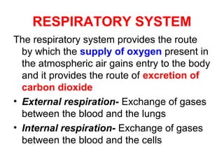
  • 1. RESPIRATORY SYSTEM The respiratory system provides the route by which the supply of oxygen present in the atmospheric air gains entry to the body and it provides the route of excretion of carbon dioxide • External respiration- Exchange of gases between the blood and the lungs • Internal respiration- Exchange of gases between the blood and the cells
  • 3. Organs of Respiratory System • Nose • Pharynx Upper Respiratory tract • Larynx • Trachea • Two bronchi • Bronchioles Lower respiratory tract • Two lungs • Coverings of Lungs-Pleura • Muscles of Respiration- Intercostal muscles & Diaphragm
  • 4. Functional Zones of Respiratory System
  • 5. NOSE • Made up of external nose & an inner chamber-Nasal Cavity Anterior nare Posterior nare Opening of Auditory tube
  • 6. Nasal Cavity: • Consists of a large irregular cavity divided into two equal passages by a septum • The posterior bony part of the septum - perpendicular plate of the ethmoid bone and the vomer. Anteriorly - hyaline cartilage. • roof -cribriform plate of the ethmoid bone, the sphenoid bone, frontal bone and nasal bones. • floor- hard palate in front and the soft palate behind. The hard palate is composed of the maxilla and palatine bones and the soft palate consists of involuntary muscle. • medial wall - septum. • lateral walls - maxilla, the ethmoid bone and the inferior conchae. • posterior wall - posterior wall of the pharynx.
  • 7. Openings into the nasal cavity • The anterior nares, or nostrils, are the openings from the exterior into the nasal cavity. Hairs are present in this area. • The posterior nares are the openings from the nasal cavity into the pharynx. • The paranasal sinuses are cavities in the bones of the face and the cranium which contain air. There are tiny openings between the paranasal sinuses and the nasal cavity. They are lined with mucous membrane, continuous with that of the nasal cavity. The main sinuses are: • maxillary sinuses in the lateral walls • frontal and sphenoidal sinuses in the roof • ethmoidal sinuses in the upper part of the lateral walls
  • 8. Respiratory function of the nose • The function of the nose is to begin the process by which the air is warmed, moistened and 'filtered'. • The projecting conchae increase the surface area and cause turbulence, spreading inspired air over the whole nasal surface. The large surface area maximises warming, humidification and filtering.
  • 9. Warming: This is due to the immense vascularity of the mucosa. This explains the large blood loss when a nosebleed (epistaxis) occurs. Filtering and cleaning of air: This occurs as hairs at the anterior nares trap larger particles. Smaller particles such as dust and microbes settle and adhere to the mucus. Mucus protects the underlying epithelium from irritation and prevents drying. Synchronous beating of the cilia wafts the mucus towards the throat where it is swallowed or expectorated. Humidification: This occurs as air travels over the moist mucosa and becomes saturated with water vapour. Irritation of the nasal mucosa results in sneezing, a reflex action that forcibly expels an irritant.
  • 11. PHARYNX-ANATOMY • Pharynx is a tube 12 to 14 cm long that extends from the base of the skull to the level of the 6th cervical vertebra. • Anatomical regions of pharynx: Name Region Nasopharynx Behind nasal cavity-internal nare to soft palate Oropharynx Behind roof of tongue-soft palate to epiglottis & hyoid Laryngopharynx Behind larynx-epiglottis to cricoid cartilage of larynx
  • 12. The pharynx is composed of three layers of tissue: 1. Mucous membrane lining: The mucosa varies slightly in the different parts. In the nasopharynx it is continuous with the lining of the nose and consists of ciliated columnar epithelium; in the oropharynx and laryngopharynx it is formed by tougher stratified squamous epithelium which is continuous with the lining of the mouth and oesophagus. 2. Fibrous tissue: This forms the intermediate layer. It is thicker in the nasopharynx, where there is little muscle, and becomes thinner towards the lower end, where the muscle layer is thicker. 3. Muscle tissue: This consists of several involuntary constrictor muscles that play an important part in the mechanism of swallowing (deglutition) which, in the pharynx, is not under voluntary control. The upper end of the oesophagus is closed by the lower constrictor muscle, except during swallowing.
  • 13. Functions: Passageway for air and food: The pharynx is an organ involved in both the respiratory and the digestive systems: air passes through the nasal and oral parts, and food through the oral and laryngeal parts. Warming and humidifying: By the same methods as in the nose, the air is further warmed and moistened as it passes through the pharynx. Taste: There are olfactory nerve endings of the sense of taste in the epithelium of the oral and pharyngeal parts. Hearing: The auditory tube, extending from the nasal part to each middle ear, allows air to enter the middle ear. Satisfactory hearing depends on the presence of air at atmospheric pressure on each side of the tympanic membrane (ear drum) Protection: The lymphatic tissue of the pharyngeal and laryngeal tonsils produces antibodies in response to antigens, e.g. microbes. The tonsils are larger in children and tend to atrophy in adults. Speech: The pharynx functions in speech; by acting as a resonating chamber for the sound ascending from the larynx, it helps (together with the sinuses) to give the voice its individual characteristics.
  • 15.
  • 16. Anatomy: • The larynx or 'voice box' extends from the root of the tongue and the hyoid bone to the trachea. • It lies in front of the laryngopharynx at the level of the 3rd, 4th, 5th and 6th cervical vertebrae. • In males, it grows larger in size during puberty. • The larynx is composed of several irregularly shaped cartilages attached to each other by ligaments and membranes. The main cartilages are: • Thyroid, Cricoid, 2 Arytenoid, • 2 Corniculate, 2Cuneiform and Epiglottis • Vocal Folds: medially into lumen of larynx 2 sets of vocal folds, extend from back of thyroid cartilage to front of arytenoid cartilage
  • 17. Functions: Production of Speech: This occurs during expiration when the sounds produced by the vocal cords are manipulated by the tongue, cheeks and lips. Protection of the lower respiratory tract: During swallowing (deglutition) the larynx moves upwards, occluding the opening into it from the pharynx and the hinged epiglottis closes over the larynx. This ensures that food passes into the esophagus and not into the lower respiratory passages. Passageway for air: between the pharynx and trachea. Humidifying, filtering and warming: These continue as inspired air travels through the larynx.
  • 18. TRACHEA / WINDPIPE • The trachea or windpipe is a continuation of the larynx & extends downwards to about the level of the 5th thoracic vertebra where it bifurcates at the carina into the right and left bronchi, one bronchus going to each lung. • It is approximately 10 to 12 cm long and lies in front of the esophagus. • It is composed of from 16 to 20 posteriorly incomplete (C-shaped) rings of hyaline cartilages lying one above the other.
  • 19.
  • 20. •Connective tissue and involuntary muscle join the cartilages and form the posterior wall where they are incomplete. •Trachea composed of three layers of tissue which 'clothe' the cartilages. The outer layer: consists of fibrous and elastic tissue and encloses the cartilages. The middle layer: consists of cartilages and bands of smooth muscle that wind round the trachea in a helical arrangement. There is some areolar tissue, containing blood and lymph vessels and autonomic nerves. The inner lining: consists of ciliated columnar epithelium, containing mucus-secreting goblet cells.
  • 21. Functions: Support and patency: The arrangement of cartilage and elastic tissue prevents kinking and obstruction of the airway as the head and neck move. The absence of cartilage posteriorly allows the trachea to dilate and constrict in response to nerve stimulation, and for indentation as the esophagus distends during swallowing. The cartilages prevent collapse of the tube when the internal pressure is less than intrathoracic pressure, i.e. at the end of forced expiration. Mucociliary escalator: cilia wafts mucus with adherent articles upwards towards the larynx where it is swallowed or expectorated Cough reflex: Nerve endings in the larynx, trachea and bronchi are sensitive to irritation that generates nerve impulses which are conducted by the vagus nerves to the respiratory centre in the brain stem. The reflex motor response is deep inspiration followed by closure of the glottis. The abdominal and respiratory muscles then contract and suddenly the air is released under pressure expelling mucus and/or foreign material from the mouth. Warming, humidifying and filtering of air: These continue as in the nose, although air is normally saturated and at body temperature when it reaches the trachea.
  • 22. BRONCHI & BRONCHIOLES • At the level of 5th thoracic vertebra trachea bifurcates into 2 primary(1o ) bronchus: • Right & Left Primary bronchus • The right bronchus: is wider, shorter and more vertical than the left bronchus. It is approximately 2.5 cm long. After entering the right lung at the hilum it divides into three branches one to each lobe [lobar bronchus(2o )] • The left bronchus: This is about 5 cm long and is narrower than the right. After entering the lung at the hilum it divides into two branches, one to each lobe. [lobar bronchus (2o )]
  • 23.
  • 24. • Within a lobe, tertiary (3o ) bronchi branch from secondary bronchi. Each tertiary bronchus conducts air to & from a bronchopulmonary segment. • There are 10 bronchopulmonary segments in right lung & 8 in left lung. • The bronchi are composed of the same tissues as the trachea. They are lined with ciliated columnar epithelium. • The bronchi progressively subdivide into bronchioles, terminal bronchioles, respiratory bronchioles, alveolar ducts and finally, alveoli.
  • 26. Functions of air passages not involved in gaseous exchange • Control of air entry. The diameter of the respiratory passages may be altered by contraction or relaxation of the involuntary muscles in their walls, thus regulating the volume of air entering the lungs. These changes are controlled by the autonomic nerve supply: parasympathetic stimulation causes constriction and sympathetic stimulation causes dilatation • Following functions continue as in the upper airways: warming and humidifying; support and patency removal of particulate matter; cough reflex.
  • 27. Respiratory bronchioles and alveoli • Each bronchopulmonary segment is partitioned by connective tissue into lobules. Each lobule measure about 3.5 mm in diameter. • Lobule consisting of: respiratory bronchioles, alveolar ducts and alveoli • Walls gradually become thinner until muscle and connective tissue fade out leaving a single layer of simple squamous epithelial cells in the alveolar ducts and alveoli. • These respiratory passages are supported by a loose network of elastic connective tissue in which macrophages, fibroblasts, nerves and blood and lymph vessels are embedded.
  • 28. Functions of respiratory bronchioles and alveoli • External respiration: • Defense against microbes: protective cells present within the lung tissue- lymphocytes and plasma cells, which produce antibodies in the presence of antigens, and macrophages and polymorphonuclear lymphocytes, which are phagocytic.. • Warming and humidifying. These continue as in the upper airways.
  • 29. LUNGS • Two lungs, one lying on each side of the midline in the thoracic cavity • cone-shaped and are described as having an apex, a base, costal surface and medial surface • Structures enter and leave at the hilum include the primary bronchus, the pulmonary artery supplying the lung and the two pulmonary veins draining it, the bronchial artery and veins, and the lymphatic and nerve supply
  • 30. Pulmonary veins Right bronchus Pulmonary artery Apex Superior lobe Middle lobe Inferior lobe Base
  • 31. Pulmonary veins Left bronchus Pulmonary artery Apex Superior lobe Inferior lobe Base
  • 32. Organisation of Lungs: • The right lung -3 lobes & left lung -2 lobes • In right lung, superior lobe and middle lobe are separated by depression –horizontal fissure. Inferiorly, an oblique fissure separates middle & inferior lobe. • In left lung oblique fissure separates superior & inferior lobe. • The lungs are composed of the bronchi and smaller air passages, alveoli, connective tissue, blood vessels, lymph vessels and nerves.
  • 33. Pleura & Pleural cavity • The pleura: sac of serous membrane (one for each lung) which contains a small amount of serous fluid. The lung is invaginated into this sac so that it forms two layers: The visceral pleura: adherent to the lung, covering each lobe and passing into the fissures which separate them. • The parietal pleura: adherent to the inside of the chest wall and the thoracic surface of the diaphragm. It is continuous with the visceral pleura round the edges of the hilum. • The pleural cavity: This is only a potential space. In health, the two layers of pleura are separated by only a thin film of serous fluid which allows them to glide over each other, preventing friction between them during breathing. The serous fluid is secreted by the epithelial cells of the membrane.
  • 34.
  • 35. RESPIRATION • Inflation and deflation of the lungs occurring with each breath ensures that regular exchange of gases takes place between the alveoli and the external air. • Muscles of respiration: Intercostal muscles -11 pairs of external & internal Intercostal muscles supplied by Intercostal nerves Diaphragm-Dome-shaped muscle supplied by Phrenic nerve • The Intercostal muscles and the diaphragm contract simultaneously ensuring the enlargement of the thoracic cavity in all directions, that is from back to front, side to side and top to bottom
  • 36.
  • 37.
  • 38. Cycle of Respiration: • It occurs 12 -15 per minute & consists of 3 phases: inspiration, expiration & pause • Inspiration: • Simultaneous contraction of the intercostal muscles and the diaphragm capacity of the thoracic cavity is increased parietal pleura moves with the walls of the thorax and the diaphragm pressure in the pleural cavity to a level considerably lower than atmospheric pressure. • The visceral pleura follows the parietal pleura pulling the lung with it. This stretches the lungs and the pressure within the alveoli and in the air passages falls, drawing air into the lungs in an attempt to equalize the atmospheric and alveolar air pressures.
  • 39. •Expiration: • Relaxation of the intercostal muscles and the diaphragm in downward and inward movement of the rib cage and elastic recoil of the lungs. • As this occurs, pressure inside the lungs exceeds that in the atmosphere and therefore air is expelled from the respiratory tract. • The lungs still contain some air and are prevented from complete collapse by the intact pleura. • This process is passive as it does not require the expenditure of energy. • After expiration, there is a pause before the next cycle begins.
  • 40. Physiological variables affecting respiration: • Elasticity: Ability of the lung to return to its normal shape after each breath. • Loss of elasticity of the connective tissue in the lungs necessitates forced expiration and increased effort on inspiration. • Compliance: a measure of the distensibility of the lungs, i.e. the effort required to inflate the alveoli. When compliance is low the effort needed to inflate the lungs is greater than normal, e.g. in some diseases where elasticity is reduced or when insufficient surfactant is present. Compliance and Elasticity are opposing forces. • Airflow resistance: When this is increased, e.g. in bronchoconstriction, more respiratory effort is required to inflate the lungs.
  • 41. Lung Volumes & Capacities
  • 42. Lung Volumes: • Tidal volume (TV): amount of air which passes into and out of the lungs during each cycle of quiet breathing ( 500 ml). • Inspiratory reserve volume (IRV): This is the extra volume of air that can be inhaled into the lungs during maximal inspiration (3100 ml). • Expiratory reserve volume (ERV): This is the largest volume of air which can be expelled from the lungs during maximal expiration (1200 ml). • Residual volume (RV): This is the volume of air remaining in the lungs after forced expiration.
  • 43. Lung Capacities: • Inspiratory capacity (IC): This is the amount of air that can be inspired with maximum effort. It consists of the tidal volume plus the inspiratory reserve volume.(3600 ml) • Functional residual capacity (FRC): This is the amount of air remaining in the air passages and alveoli at the end of quiet expiration.(2400 ml) • Vital capacity (VC): This is the maximum volume of air which can be moved into and out of the lungs (4800 ml) VC = Tidal volume + IRV + ERV • Total Lung Capacity ???
  • 44. External Respiration • This is exchange of gases by diffusion between the alveoli and the blood. Each alveolar wall is one cell thick and is surrounded by a network of tiny capillaries. The total area for gas exchange in the lungs is 70 to 80 sq.m. • Venous blood arriving at the lungs has traveled from all the active tissues of the body, and contains high levels of CO2 and low levels of O2. • Carbon dioxide diffuses from venous blood down its concentration gradient into the alveoli until equilibrium with alveolar air is reached. By the same process, oxygen diffuses from the alveoli into the blood. The slow flow of blood through the capillaries increases the time available for diffusion to occur. When blood leaves the alveolar capillaries, the oxygen and carbon dioxide concentrations are in equilibrium with those of alveolar air.
  • 45. External respiration: exchange of gases between alveolar air and capillary blood.
  • 46. Internal Respiration • This is exchange of gases by diffusion between blood in the capillaries and the body cells. • Blood arriving at the tissues has a higher PO2 and a lower PCO2 than the tissues. This creates concentration gradients between the blood and the tissues, and gaseous exchange therefore occurs by diffusion. • O2 diffuses from the bloodstream through the capillary wall into the tissues. CO2 diffuses from the cells into the extracellular fluid then into the bloodstream towards the venous end of the capillary.
  • 47. Internal respiration: exchange of gases between capillary blood and tissue cells
  • 48. Disorders of Respiratory System • Sinusitis This is usually caused by spread of microbes from the nose and pharynx to the mucous membrane lining the paranasal sinuses in maxillary, sphenoidal, ethmoidal and frontal bones. The primary viral infection is usually followed by bacterial infection, e.g. Streptococcus pyogenes, Streptococcus pneumoniae, Staphylococcus aureus. The congested mucosa may block the openings between the nose and the sinuses, preventing drainage of mucopurulent discharge.
  • 49. Bronchitis • This is usually a secondary bacterial infection of the bronchi. It is usually preceded by a common cold or influenza Asthma • Asthma is an inflammatory disease of the airways in which the mucous membrane and muscle layers of the bronchi become thickened and the mucous glands enlarge, reducing airflow in the lower respiratory tract.