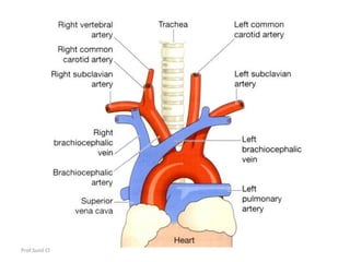
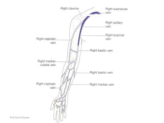
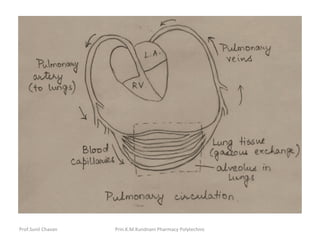
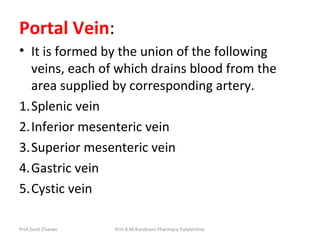
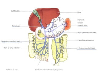
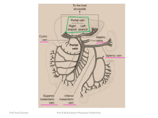
Prof.Sunil Chavan Prin.K.M.Kundnani Pharmacy Polytechnic
Systemic or general circulation
• The blood pumped out from the left
ventricle is carried by the branches of the
aorta around the body and is returned to
the right atrium of the heart by the
superior and inferior venae cavae.
Prof.Sunil Chavan Prin.K.M.Kundnani Pharmacy Polytechnic
Aorta
• The aorta begins at the upper part of the left
ventricle and, after passing upwards for a
short way, it arches backwards and to the left.
It then descends behind the heart through the
thoracic cavity a little to the left of the
thoracic vertebrae.
• At the level of the 12th thoracic vertebra it
passes behind the diaphragm then
downwards in the abdominal cavity to the
level of the 4th lumbar vertebra, where it
divides into the right and left common iliac
arteries.
Prof.Sunil Chavan Prin.K.M.Kundnani Pharmacy Polytechnic
Thoracic aorta
• This part of the aorta is above the
diaphragm and is described in three
parts:
• ascending aorta
• arch of the aorta
• descending aorta in the thorax.
Prof.Sunil Chavan Prin.K.M.Kundnani Pharmacy Polytechnic
Ascending aorta
• This is about 5 cm long and lies behind the
sternum.
• The right and left coronary arteries are its only
branches and they arise from the aorta just
above the level of the aortic valve
Prof.Sunil Chavan Prin.K.M.Kundnani Pharmacy Polytechnic
Arch of the aorta
• The arch of the aorta is a continuation of the
ascending aorta. It begins behind the manubrium
of the sternum and runs upwards, backwards and
to the left in front of the trachea. It then passes
downwards to the left of the trachea and is
continuous with the descending aorta. Three
branches are given off from its upper aspect:
• brachiocephalic artery or trunk
• left common carotid artery
• left subclavian artery
Prof.Sunil Chavan Prin.K.M.Kundnani Pharmacy Polytechnic
The brachiocephalic artery is about 4 to 5
cm long and passes obliquely upwards,
backwards and to the right.
At the level of the sternoclavicular joint it
divides into the right common carotid
artery and the right subclavian artery.
Prof.Sunil Chavan Prin.K.M.Kundnani Pharmacy Polytechnic
Circulation of blood to the head and
neck
Prof.Sunil Chavan Prin.K.M.Kundnani Pharmacy Polytechnic
Arterial supply
• The paired arteries supplying the head and
neck are the common carotid arteries and
the vertebral arteries
• Carotid arteries: The right common carotid
artery is a branch of the brachiocephalic
artery. The left common carotid artery arises
directly from the arch of the aorta. They pass
upwards on either side of the neck and have
the same distribution on each side.
Prof.Sunil Chavan Prin.K.M.Kundnani Pharmacy Polytechnic
The common carotid arteries at the level of the upper border of
the thyroid cartilage they divide into: External carotid artery &
Internal carotid artery
External carotid artery :This artery supplies the superficial
tissues of the head and neck, via a number of branches.
•The superior thyroid artery -thyroid gland and adjacent muscles.
• The lingual artery -tongue, the lining membrane of the mouth,
the structures in the floor of the mouth, the tonsil and the
epiglottis.
• The facial artery -muscles of facial expression and structures in
the mouth.
• The occipital artery -the posterior part of the scalp.
• The temporal artery -frontal, temporal and parietal parts of the
scalp.
• The maxillary artery -muscles of mastication and a branch of
this artery, the middle meningeal artery, runs deeply to supply
structures in the interior of the skull.
Prof.Sunil Chavan Prin.K.M.Kundnani Pharmacy Polytechnic
Internal carotid artery
• The internal carotid artery is a major
contributor to the circulus arteriosus (circle
of Willis) which supplies the greater part of
the brain.
• It also has branches that supply the eyes,
forehead and nose.
• It ascends to the base of the skull and passes
through the carotid foramen in the temporal
bone.
Prof.Sunil Chavan Prin.K.M.Kundnani Pharmacy Polytechnic
Circulus arteriosus (circle of Willis)
Prof.Sunil Chavan Prin.K.M.Kundnani Pharmacy Polytechnic
The greater part of the brain is supplied with arterial blood
by an arrangement of arteries called the circulus arteriosus
or the circle of Willis. two internal carotid arteries and two
vertebral arteries contribute to its formation:
The circulus arteriosus is therefore formed by:
• 2 anterior cerebral arteries
• 2 internal carotid arteries
• 1 anterior communicating artery
• 2 posterior communicating arteries
• 2 posterior cerebral arteries
• 1 basilar artery
Prof.Sunil Chavan Prin.K.M.Kundnani Pharmacy Polytechnic
The arrangement in the circulus arteriosus is
such that the brain as a whole receives an
adequate blood supply when a contributing
artery is damaged and during extreme
movements of the head and neck.
Prof.Sunil Chavan Prin.K.M.Kundnani Pharmacy Polytechnic
Venous return from the head and
neck
• The venous blood from the head and neck is
returned by deep and superficial veins.
• Superficial veins return venous blood from the
superficial structures of the face and scalp and
unite to form the external jugular vein.
• External jugular vein begins in the neck
entering the subclavian vein
Prof.Sunil Chavan Prin.K.M.Kundnani Pharmacy Polytechnic
The venous blood from the deep areas of the brain is
collected into channels called the dural venous sinuses
The main venous sinuses are:
• 1 superior sagittal sinus
• 1 inferior sagittal sinus
• 1 straight sinus
• 2 transverse or lateral sinuses
• 2 sigmoid sinuses
Each internal jugular vein is the continuation of a sigmoid
sinus.
They run downwards in the neck behind the
sternocleidomastoid muscles. Behind the clavicle they
unite with the subclavian veins, carrying blood from the
upper limbs, to form the brachiocephalic veins.
Prof.Sunil Chavan Prin.K.M.Kundnani Pharmacy Polytechnic
The left brachiocephalic vein is longer than the
right and passes obliquely behind the
manubrium of the sternum, where it joins the
right brachiocephalic vein to form the
SUPERIOR VENA CAVA.
The superior vena cava, which drains all the
venous blood from the head, neck and upper
limbs, is about 7 cm long. It passes downwards
along the right border of the sternum and ends
in the right atrium of the heart.
Prof.Sunil Chavan Prin.K.M.Kundnani Pharmacy Polytechnic
Circulation of blood to the upper limb
Prof.Sunil Chavan Prin.K.M.Kundnani Pharmacy Polytechnic
Systemic Circulation
• Systemic circulation is the part of the
cardiovascular system which
carries oxygenated blood away from the
heart to the body, and returns deoxygenated
blood back to the heart.
Prof.Sunil Chavan Prin.K.M.Kundnani Pharmacy Polytechnic
• From the left ventricle, blood is pumped
through the aortic valve and into the aorta,
the body's largest artery. The aorta arches and
branches into major arteries to the upper
body before passing through the diaphragm,
where it branches further into arteries which
supply the lower parts of the body. The
arteries branch into smaller arteries,
arterioles, and finally capillaries. It provides
oxygen and nutrients to cells of all body
tissues.
Prof.Sunil Chavan Prin.K.M.Kundnani Pharmacy Polytechnic
• Waste and carbon dioxide diffuse out of the
cell into the blood. The deoxygenated blood
continues through the capillaries which merge
into venules, then veins, and finally the
superior and inferior venae cavae, which drain
into the right atrium of the heart. From the
right atrium, the blood will travel through the
pulmonary circulation to be oxygenated
before returning gain to the system
circulation.
Prof.Sunil Chavan Prin.K.M.Kundnani Pharmacy Polytechnic
Pulmonary Circulation
• The deoxygenated blood from the right
ventricle is forced into the pulmonary artery.
The pulmonary artery divides into 2 branches
carrying blood to right and left lungs.
• In the lung tissue, the artery divides and re-
divides to form a net of blood capillaries
surrounding the alveoli. The blood in the blood
capillaries and oxygen in the alveoli are
separated by double layer of flat epithelium.
This membrane is called alveolar capillary
membrane.
Prof.Sunil Chavan Prin.K.M.Kundnani Pharmacy Polytechnic
• The oxygen from alveoli diffuses through this
membrane and passes to the blood in the
capillary. The carbon dioxide from blood
capillaries diffuses into alveoli. Thus the
exchange of gases occurs in lungs causing
oxygenation of blood.
• The oxygenated blood is transported by 4
pulmonary veins from right and left lung and
poured into left atrium.
• This course of blood from right ventricle to
the left atrium via lungs is called
pulmonary/lesser circulation.
Prof.Sunil Chavan Prin.K.M.Kundnani Pharmacy Polytechnic
Portal Circulation
• In portal circulation, venous blood passes
from the capillary beds of abdominal part of
the digestive system, spleen and pancreas to
liver.
• It passes through a second capillary bed, the
hepatic sinusoids, in the liver before entering
the general circulation.
Prof.Sunil Chavan Prin.K.M.Kundnani Pharmacy Polytechnic
Portal Vein:
• It is formed by the union of the following
veins, each of which drains blood from the
area supplied by corresponding artery.
1.Splenic vein
2.Inferior mesenteric vein
3.Superior mesenteric vein
4.Gastric vein
5.Cystic vein
Prof.Sunil Chavan Prin.K.M.Kundnani Pharmacy Polytechnic
• The splenic vein drains blood from the spleen,
the pancreas and part of stomach.
• The inferior mesenteric vein returns venous
blood from the rectum, pelvic and descending
colon. It joins the splenic vein.
• The superior mesenteric vein returns blood
from the small intestine and proximal part of
large intestine. It unites the splenic vein to form
portal vein.
• The gastric veins drain blood from stomach and
distal end of esophagus, then join the portal
vein.
Prof.Sunil Chavan Prin.K.M.Kundnani Pharmacy Polytechnic
• The cystic vein which drains venous blood
from the gall bladder joins the portal vein.
Importance:
• Blood with the high concentration of nutrients
absorbed from the stomach & intestine goes
to liver first. In the liver certain modifications
takes place including the blood nutrient level.
The venous blood then leaves liver via hepatic
vein & joins the inferior vena cava.
Prof.Sunil Chavan Prin.K.M.Kundnani Pharmacy Polytechnic

07 general circulation

  • 1.
    Prof.Sunil Chavan Prin.K.M.KundnaniPharmacy Polytechnic Systemic or general circulation • The blood pumped out from the left ventricle is carried by the branches of the aorta around the body and is returned to the right atrium of the heart by the superior and inferior venae cavae.
  • 2.
    Prof.Sunil Chavan Prin.K.M.KundnaniPharmacy Polytechnic Aorta • The aorta begins at the upper part of the left ventricle and, after passing upwards for a short way, it arches backwards and to the left. It then descends behind the heart through the thoracic cavity a little to the left of the thoracic vertebrae. • At the level of the 12th thoracic vertebra it passes behind the diaphragm then downwards in the abdominal cavity to the level of the 4th lumbar vertebra, where it divides into the right and left common iliac arteries.
  • 3.
    Prof.Sunil Chavan Prin.K.M.KundnaniPharmacy Polytechnic Thoracic aorta • This part of the aorta is above the diaphragm and is described in three parts: • ascending aorta • arch of the aorta • descending aorta in the thorax.
  • 4.
    Prof.Sunil Chavan Prin.K.M.KundnaniPharmacy Polytechnic Ascending aorta • This is about 5 cm long and lies behind the sternum. • The right and left coronary arteries are its only branches and they arise from the aorta just above the level of the aortic valve
  • 5.
  • 6.
  • 7.
    Prof.Sunil Chavan Prin.K.M.KundnaniPharmacy Polytechnic Arch of the aorta • The arch of the aorta is a continuation of the ascending aorta. It begins behind the manubrium of the sternum and runs upwards, backwards and to the left in front of the trachea. It then passes downwards to the left of the trachea and is continuous with the descending aorta. Three branches are given off from its upper aspect: • brachiocephalic artery or trunk • left common carotid artery • left subclavian artery
  • 8.
    Prof.Sunil Chavan Prin.K.M.KundnaniPharmacy Polytechnic The brachiocephalic artery is about 4 to 5 cm long and passes obliquely upwards, backwards and to the right. At the level of the sternoclavicular joint it divides into the right common carotid artery and the right subclavian artery.
  • 9.
    Prof.Sunil Chavan Prin.K.M.KundnaniPharmacy Polytechnic Circulation of blood to the head and neck
  • 10.
    Prof.Sunil Chavan Prin.K.M.KundnaniPharmacy Polytechnic Arterial supply • The paired arteries supplying the head and neck are the common carotid arteries and the vertebral arteries • Carotid arteries: The right common carotid artery is a branch of the brachiocephalic artery. The left common carotid artery arises directly from the arch of the aorta. They pass upwards on either side of the neck and have the same distribution on each side.
  • 11.
  • 12.
    Prof.Sunil Chavan Prin.K.M.KundnaniPharmacy Polytechnic The common carotid arteries at the level of the upper border of the thyroid cartilage they divide into: External carotid artery & Internal carotid artery External carotid artery :This artery supplies the superficial tissues of the head and neck, via a number of branches. •The superior thyroid artery -thyroid gland and adjacent muscles. • The lingual artery -tongue, the lining membrane of the mouth, the structures in the floor of the mouth, the tonsil and the epiglottis. • The facial artery -muscles of facial expression and structures in the mouth. • The occipital artery -the posterior part of the scalp. • The temporal artery -frontal, temporal and parietal parts of the scalp. • The maxillary artery -muscles of mastication and a branch of this artery, the middle meningeal artery, runs deeply to supply structures in the interior of the skull.
  • 13.
    Prof.Sunil Chavan Prin.K.M.KundnaniPharmacy Polytechnic Internal carotid artery • The internal carotid artery is a major contributor to the circulus arteriosus (circle of Willis) which supplies the greater part of the brain. • It also has branches that supply the eyes, forehead and nose. • It ascends to the base of the skull and passes through the carotid foramen in the temporal bone.
  • 14.
    Prof.Sunil Chavan Prin.K.M.KundnaniPharmacy Polytechnic Circulus arteriosus (circle of Willis)
  • 15.
    Prof.Sunil Chavan Prin.K.M.KundnaniPharmacy Polytechnic The greater part of the brain is supplied with arterial blood by an arrangement of arteries called the circulus arteriosus or the circle of Willis. two internal carotid arteries and two vertebral arteries contribute to its formation: The circulus arteriosus is therefore formed by: • 2 anterior cerebral arteries • 2 internal carotid arteries • 1 anterior communicating artery • 2 posterior communicating arteries • 2 posterior cerebral arteries • 1 basilar artery
  • 16.
    Prof.Sunil Chavan Prin.K.M.KundnaniPharmacy Polytechnic The arrangement in the circulus arteriosus is such that the brain as a whole receives an adequate blood supply when a contributing artery is damaged and during extreme movements of the head and neck.
  • 17.
    Prof.Sunil Chavan Prin.K.M.KundnaniPharmacy Polytechnic Venous return from the head and neck • The venous blood from the head and neck is returned by deep and superficial veins. • Superficial veins return venous blood from the superficial structures of the face and scalp and unite to form the external jugular vein. • External jugular vein begins in the neck entering the subclavian vein
  • 18.
    Prof.Sunil Chavan Prin.K.M.KundnaniPharmacy Polytechnic The venous blood from the deep areas of the brain is collected into channels called the dural venous sinuses The main venous sinuses are: • 1 superior sagittal sinus • 1 inferior sagittal sinus • 1 straight sinus • 2 transverse or lateral sinuses • 2 sigmoid sinuses Each internal jugular vein is the continuation of a sigmoid sinus. They run downwards in the neck behind the sternocleidomastoid muscles. Behind the clavicle they unite with the subclavian veins, carrying blood from the upper limbs, to form the brachiocephalic veins.
  • 19.
    Prof.Sunil Chavan Prin.K.M.KundnaniPharmacy Polytechnic The left brachiocephalic vein is longer than the right and passes obliquely behind the manubrium of the sternum, where it joins the right brachiocephalic vein to form the SUPERIOR VENA CAVA. The superior vena cava, which drains all the venous blood from the head, neck and upper limbs, is about 7 cm long. It passes downwards along the right border of the sternum and ends in the right atrium of the heart.
  • 20.
    Prof.Sunil Chavan Prin.K.M.KundnaniPharmacy Polytechnic Circulation of blood to the upper limb
  • 21.
  • 22.
    Systemic Circulation • Systemiccirculation is the part of the cardiovascular system which carries oxygenated blood away from the heart to the body, and returns deoxygenated blood back to the heart. Prof.Sunil Chavan Prin.K.M.Kundnani Pharmacy Polytechnic
  • 23.
    • From theleft ventricle, blood is pumped through the aortic valve and into the aorta, the body's largest artery. The aorta arches and branches into major arteries to the upper body before passing through the diaphragm, where it branches further into arteries which supply the lower parts of the body. The arteries branch into smaller arteries, arterioles, and finally capillaries. It provides oxygen and nutrients to cells of all body tissues. Prof.Sunil Chavan Prin.K.M.Kundnani Pharmacy Polytechnic
  • 24.
    • Waste andcarbon dioxide diffuse out of the cell into the blood. The deoxygenated blood continues through the capillaries which merge into venules, then veins, and finally the superior and inferior venae cavae, which drain into the right atrium of the heart. From the right atrium, the blood will travel through the pulmonary circulation to be oxygenated before returning gain to the system circulation. Prof.Sunil Chavan Prin.K.M.Kundnani Pharmacy Polytechnic
  • 25.
    Pulmonary Circulation • Thedeoxygenated blood from the right ventricle is forced into the pulmonary artery. The pulmonary artery divides into 2 branches carrying blood to right and left lungs. • In the lung tissue, the artery divides and re- divides to form a net of blood capillaries surrounding the alveoli. The blood in the blood capillaries and oxygen in the alveoli are separated by double layer of flat epithelium. This membrane is called alveolar capillary membrane. Prof.Sunil Chavan Prin.K.M.Kundnani Pharmacy Polytechnic
  • 26.
    • The oxygenfrom alveoli diffuses through this membrane and passes to the blood in the capillary. The carbon dioxide from blood capillaries diffuses into alveoli. Thus the exchange of gases occurs in lungs causing oxygenation of blood. • The oxygenated blood is transported by 4 pulmonary veins from right and left lung and poured into left atrium. • This course of blood from right ventricle to the left atrium via lungs is called pulmonary/lesser circulation. Prof.Sunil Chavan Prin.K.M.Kundnani Pharmacy Polytechnic
  • 27.
  • 28.
    Portal Circulation • Inportal circulation, venous blood passes from the capillary beds of abdominal part of the digestive system, spleen and pancreas to liver. • It passes through a second capillary bed, the hepatic sinusoids, in the liver before entering the general circulation. Prof.Sunil Chavan Prin.K.M.Kundnani Pharmacy Polytechnic
  • 29.
    Portal Vein: • Itis formed by the union of the following veins, each of which drains blood from the area supplied by corresponding artery. 1.Splenic vein 2.Inferior mesenteric vein 3.Superior mesenteric vein 4.Gastric vein 5.Cystic vein Prof.Sunil Chavan Prin.K.M.Kundnani Pharmacy Polytechnic
  • 30.
  • 31.
  • 32.
    • The splenicvein drains blood from the spleen, the pancreas and part of stomach. • The inferior mesenteric vein returns venous blood from the rectum, pelvic and descending colon. It joins the splenic vein. • The superior mesenteric vein returns blood from the small intestine and proximal part of large intestine. It unites the splenic vein to form portal vein. • The gastric veins drain blood from stomach and distal end of esophagus, then join the portal vein. Prof.Sunil Chavan Prin.K.M.Kundnani Pharmacy Polytechnic
  • 33.
    • The cysticvein which drains venous blood from the gall bladder joins the portal vein. Importance: • Blood with the high concentration of nutrients absorbed from the stomach & intestine goes to liver first. In the liver certain modifications takes place including the blood nutrient level. The venous blood then leaves liver via hepatic vein & joins the inferior vena cava. Prof.Sunil Chavan Prin.K.M.Kundnani Pharmacy Polytechnic