06 lymphatic system
•Download as PPT, PDF•
3 likes•276 views

The document summarizes the key components and functions of the lymphatic system. It describes the lymph, lymphatic vessels, lymph nodes, spleen, and thymus. The lymphatic system is responsible for immunity and drains interstitial fluid via a network of lymph capillaries, vessels, nodes, and ducts. Lymph nodes filter foreign substances and allow immune cell proliferation. The spleen and thymus also play important roles in immune functions.

Recommended
More Related Content
What's hot
What's hot (20)
Similar to 06 lymphatic system
Similar to 06 lymphatic system (20)
More from Prin.K.M.Kundnani Pharmacy Polytechnic
More from Prin.K.M.Kundnani Pharmacy Polytechnic (18)
Recently uploaded
Recently uploaded (20)
06 lymphatic system
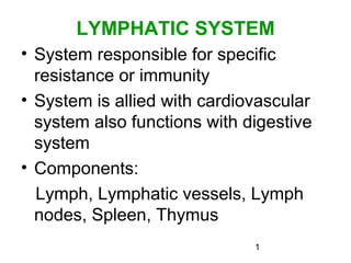
- 1. 1 LYMPHATIC SYSTEM • System responsible for specific resistance or immunity • System is allied with cardiovascular system also functions with digestive system • Components: Lymph, Lymphatic vessels, Lymph nodes, Spleen, Thymus
- 2. 2 LYMPH • Blood plasma filter through blood capillary walls and form interstitial fluid. • Interstitial fluid passes into lymph capillaries, is called lymph. • It is pale yellow colored liquid. • Composition of lymph similar to that of plasma but concentrations of constituents are different. • Also contains substances that are large to pass through blood capillary walls. • Lymph passes through vessels of increasing size and no. of lymph nodes and return to blood.
- 3. 3
- 4. 4 LYMPHATIC VESSELS • Lymphatic vessels begin as lymphatic capillaries. • Lymphatic capillary originate as blind-end tube in the interstitial spaces, made of single-layer endothelial cells. • When pressure is greater in interstitial fluid than in lymph capillary the endothelial cells separate slightly and interstitial fluid enters capillary • When pressure inside capillary is more cells adhere closely and prevent back flow of lymph. • Lymph moves further.
- 5. 5 Lymph Vessels: • Lymph capillaries carry lymph into lymph vessels. • Lymph vessels has outer fibrous covering, middle layer of smooth muscle and elastic tissue and inner lining of endothelium. • It has numerous cup shaped valves that prevent back flow of lymph. • Lymph vessels unite to form Lymph trunks.
- 6. 6
- 7. 7 Lymph Trunks Name of trunk Organs drained Lumbar trunks (L ,R) Lower limbs, Pelvis region, kidneys, adrenal glands, abdominal walls Intestinal trunk Stomach, Intestines, Pancreas, part of liver Bronchomediastinal trunks (L,R) Thoracic wall, Lungs, Heart Subclavian trunks (L,R) Upper limbs Jugular trunks (L,R) Head, Neck
- 8. 8 Lymphatic Ducts • Lymph passes from lymph trunks into 2 ducts: Thoracic (Left Lymphatic) duct & Right Lymphatic duct • Thoracic duct:38-45cm long • Begin as dilation called Cisterna chyli at anterior to second lumbar vertebra • It receives lymph from Lumbar trunks (L ,R), Intestinal trunk, Bronchomediastinal trunks (L), Subclavian trunks (L), Jugular trunks (L) i.e. 75% of body • Right lymphatic duct:1cm long, receives lymph from Bronchomediastinal trunks (L), Subclavian trunks (R), Jugular trunks (R) • Thoracic & Right lymphatic duct pour lymph at the junction of left internal jugular vein & left subclavian vein and right internal jugular vein & right subclavian vein
- 9. 9 Drained by Thoracic Duct Drained by Right Lymphatic Duct
- 10. 10 LYMPH NODES • About 600, bean-shaped, located along lymph vessels, scattered throughout body • Vary In size from 1-25mm long • Lymph node is covered by fibrous tissue capsule. It extends into node, these extensions are called trabecule. • Internal to capsule is supporting network of reticular fibers and fibroblasts • Capsule, trabecule, reticular fibers and fibroblasts constitute stroma (supporting tissue) of lymph node
- 11. 11
- 12. 12 • Parenchyma (functioning part) of lymph node is divided into superficial cortex and deep medulla • Cortex consists of an outer cortex and inner cortex. • Outer cortex composed of B-cells, follicular dendritic cells and macrophages. • Inner cortex consists of T-cells and dendritic cells • Medulla of lymph node contains B-cells, plasma cells and macrophages. • Afferent lymphatic vessels penetrate node at several points. Lymph flows through trabecular sinuses into medullary sinuses.
- 13. 13
- 14. 14 • Medullary sinuses drain lymph into one or two efferent lymphatic vessels • Efferent lymphatic vessels emerge from one side of lymph node, at a slight depression-hilum • Blood vessels also enter and leave node at hilum Functions: 1.Filteration:Foreign substances are trapped by reticular fibers. Macrophages and lymphocytes destroy these . Filtered lymph then leaves other end of lymph node. 2.Proliferation:T-lymphocytes and b-lymphocytes undergo multiplication in lymph node.
- 15. 15 SPLEEN • Slightly oval in shape, 12cm in length, 7cm in width, 2.5cm thick • Located in left hypochondriac region between stomach and diaphragm. • Surrounded by fibroelastic capsule & Trabecule extend inwards from capsule forms stroma • Parenchyma of spleen consists of: White pulp & Red pulp • White pulp- consists of lymphocytes & macrophages • Red pulp -consists of blood filled venous sinuses and sple 15nic cords. It consists of RBCs, lymphocytes, macrophages and granulocytes. • Functions: 1.White pulp carry out immune functions. 2.Red pulp performs 3 functions: -Removal by macrophages of ruptured, worn out or defective blood cells -Storage of platelets, up to 1/3 of body supply -Production of blood cells during fetal life
- 16. 16 SPLEEN
- 18. 18 THYMUS • A bilobed organ, located in upper part of mediastinum between sternum & arota • Weighs about 70g at birth, at puberty between 30-40g and in old age 3-5g • Both lobes are covered in envelope of areolar tissue. Each lobe has capsule of fibroelastic tissue. Trabecule form lobules. • Each lobules consists of outer cortex and central medulla. • Cortex consists of large no.of T-cells, dendritic cells & macrophages • Medulla consists of T-cells, dendritic cells & macrophages. Function:Maturation of T-cells & acts as source of future generations of T-cells
- 19. 19
- 20. 20 FUNCTIONS OF LYMPHATIC SYSTEM 1. Draining excess interstitial fluid: Lymphatic vessels drain excess of interstitial fluid from tissue spaces & return to blood. 2. Transporting dietary lipids: Lymphatic vessels transport lipid & lipid-soluble vitamins absorbed by g.i.t to blood. 3. Carrying out immune responses: T-cells & B-cells assisted by macrophages recognize foreign cells, microbes, toxins and cancer cells and destroy them by releasing cytotoxic substances or by producing antibodies.
Editor's Notes
- Page 140 fig 6.1