Downloaded 171 times
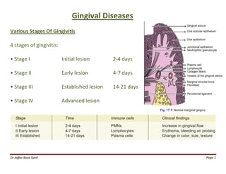
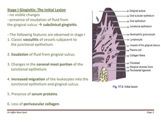


The document discusses the stages of gingivitis. It describes 4 stages: initial lesion, early lesion, established lesion, and advanced lesion. Each stage is characterized by specific clinical and microscopic features that progress as gingivitis advances. The initial lesion shows vasculitis and fluid exudation. The early lesion displays erythema and bleeding on probing. The established lesion has blood stasis and junctional epithelium proliferation. The advanced lesion results in bone loss, pocket formation, and collagen loss. The document also discusses classification of gingivitis, changes in gingival position, causes of recession, and clinical significance of recession.


![Principles of periodontal instrumentation [autosaved]](https://cdn.slidesharecdn.com/ss_thumbnails/principlesofperiodontalinstrumentationautosaved-210220074109-thumbnail.jpg?width=640&height=640&fit=bounds)


