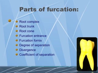
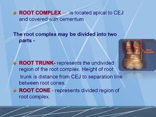
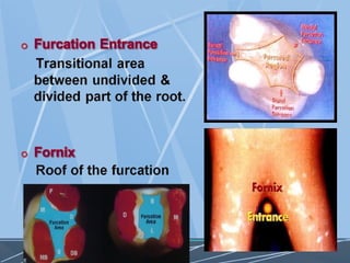
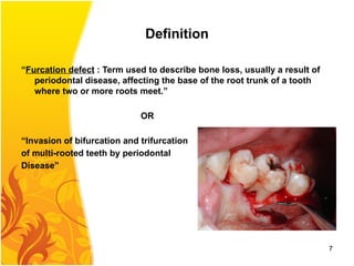
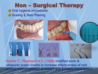
Definition
“Furcation defect :Term used to describe bone loss, usually a result of
periodontal disease, affecting the base of the root trunk of a tooth
where two or more roots meet.”
OR
“Invasion of bifurcation and trifurcation
of multi-rooted teeth by periodontal
Disease”
7
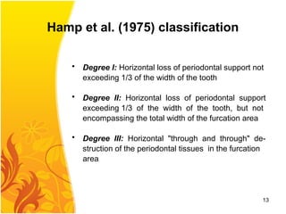
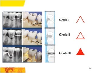
Hamp et al.(1975) classification
• Degree I: Horizontal loss of periodontal support not
exceeding 1/3 of the width of the tooth
• Degree II: Horizontal loss of periodontal support
exceeding 1/3 of the width of the tooth, but not
encompassing the total width of the furcation area
• Degree III: Horizontal "through and through" de-
struction of the periodontal tissues in the furcation
area
13
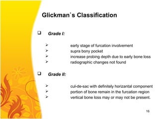
Glickman`s Classification
GradeI:
early stage of furcation involvement
supra bony pocket
increase probing depth due to early bone loss
radiographic changes not found
Grade II:
cul-de-sac with definitely horizantal component
portion of bone remain in the furcation region
vertical bone loss may or may not be present.
16
17.
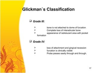
Glickman`s Classification
GradeIII:
bone is not attached to dome of furcation
Complete loss of interadicular bone
appearance of radiolucent area with pocket
formation
Grade IV:
loss of attachment and gingival recession
furcation is clinically visible
Probe passes easily through and through.
17
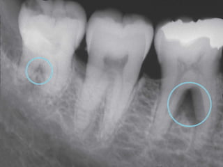
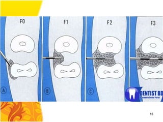
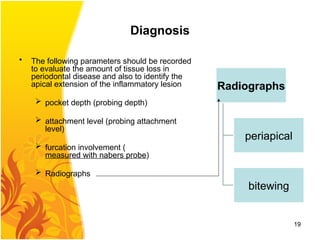
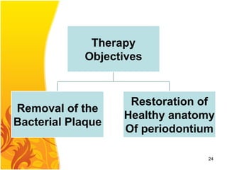
Diagnosis
• The followingparameters should be recorded
to evaluate the amount of tissue loss in
periodontal disease and also to identify the
apical extension of the inflammatory lesion
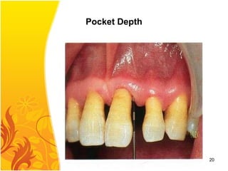
pocket depth (probing depth)
attachment level (probing attachment
level)
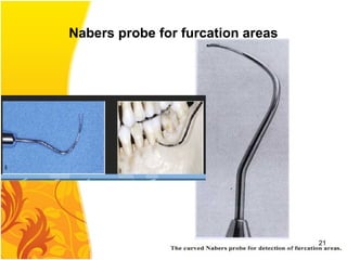
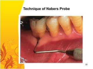
furcation involvement (
measured with nabers probe)
Radiographs
Radiographs
periapical
bitewing
19
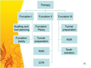
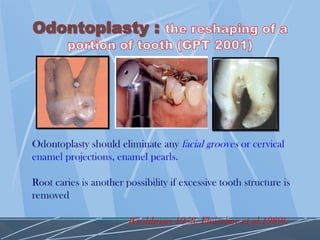
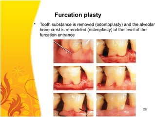
Furcation plasty
• Toothsubstance is removed (odontoplasty) and the alveolar
bone crest is remodeled (osteoplasty) at the level of the
furcation entrance
28
29.
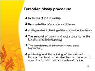
Furcation plasty procedure
Reflection of soft tissue flap.
Removal of the inflammatory soft tissue
scaling and root planning of the exposed root surfaces.
The removal of crown and root substance in the
furcation area (odontoplasty)
The recontouring of the alveolar bone crest
(osteoplasty)
positioning and the suturing of the mucosal
flaps at the level of the alveolar crest in order to
cover the furcation entrance with soft tissue.
29
30.
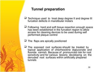
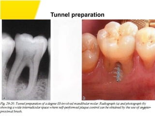
Tunnel preparation
Techniqueused to treat deep degree II and degree III
furcation defects in mandibular molars
Following hard and soft tissue resection enough space
has been established in the furcation region to allow
access for cleaning devices to be used during self
performed plaque control
The flaps are apically positioned
The exposed root surfaces should be treated by
topical application of chlorhexidine digluconate and
fluoride varnish. Because of pronounced risk for root
sensitivity and for carious lesions developing on the
denuded root surfaces within artificially prepared
tunnels
30
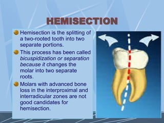
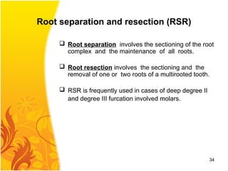
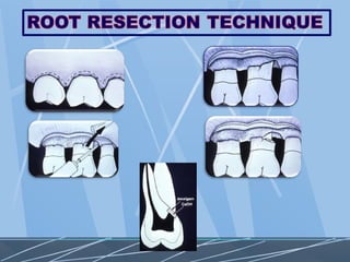
Root separation andresection (RSR)
Root separation involves the sectioning of the root
complex and the maintenance of all roots.
Root resection involves the sectioning and the
removal of one or two roots of a multirooted tooth.
RSR is frequently used in cases of deep degree II
and degree III furcation involved molars.
34
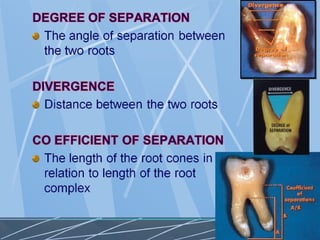
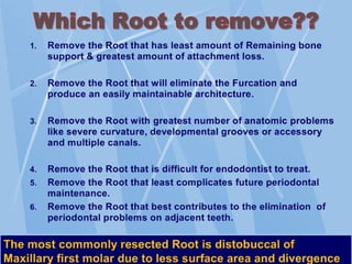
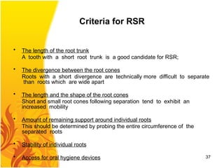
Criteria for RSR
•The length of the root trunk
A tooth with a short root trunk is a good candidate for RSR;
• The divergence between the root cones
Roots with a short divergence are technically more difficult to separate
than roots which are wide apart
• The length and the shape of the root cones
Short and small root cones following separation tend to exhibit an
increased mobility
• Amount of remaining support around individual roots
This should be determined by probing the entire circumference of the
separated roots
• Stability of individual roots
• Access for oral hygiene devices 37
38.
Regeneration of furcationdefects
• "guided tissue regeneration" (GTR) therapy is provided
• GTR is more successful in degree II furcation involvements then in
degree III involvements
38
GTR limits
• Themorphology of the periodontal defect
Horizantal bone loss
• The anatomy of the Furcation with complex
morphology more in maxillary than mandibular
tooth
• The varying and changing location of the soft tissue
margins during the early phase of healing with a
possible recession of the flap margin and early
exposure of both the membrane material and the
fornix of the Furcation
40
41.
GTR feasibility improvesif
• Adequate debridement area of exposed root
surface
• The membrane material is properly placed
• A plaque control program is put in place.
This should include daily rinsing with a
chlorhexidine solution and professional
toothcleaning once a week for the first month, and
once every 2-3 weeks for at least another 6
months of healing following the surgical
procedure
41
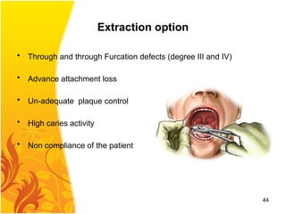
Extraction option
• Throughand through Furcation defects (degree III and IV)
• Advance attachment loss
• Un-adequate plaque control
• High caries activity
• Non compliance of the patient
44