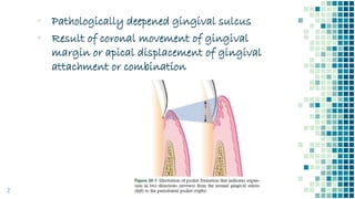
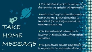
PERIODONTAL
POCKET
DR. SHRADDHA KODE
â–Ş Pathologically deepened gingival sulcus
â–Ş Result of coronal movement of gingival
margin or apical displacement of gingival
attachment or combination
2
“
â–Ş Gingival pocket: Due to gingival enlargement
without destruction of the underlying
periodontal tissues. The sulcus is deepened
because of the increased bulk of the gingiva.
â–Ş Periodontal pocket: It leads to destruction of the
supporting periodontal tissues, thereby leading
to the loosening and exfoliation of the teeth.
▪ Two types of periodontal pocket –
â–Ş Suprabony/Supraalveolar/Supracrestal: bottom
of the pocket is coronal to the underlying alveolar
bone
â–Ş Infrabony /Intrabony/Subcrestal/Intraalveolar:
bottom of the pocket is apical to the level of the
adjacent alveolar bone3
4
5
6
“
â–Ş Simple pocket: Involves one tooth surface
â–Ş Compound pocket: Involves two or more tooth
surfaces
â–Ş Spiral pocket: Originating on one tooth surface
and twisting around the tooth to involve one or
more additional surfaces. Most common in
furcation areas
7
CLINICAL FEATURES
8
â–Ş bluish-red thickened marginal gingiva
â–Ş a bluish-red vertical zone from the gingival margin to the
alveolar mucosa
â–Ş gingival bleeding and suppuration
â–Ş tooth mobility
â–Ş diastema formation
▪ symptoms such as localized pain or pain “deep in the bone.”
Initial lesion is the inflammation of the gingiva
in response to a bacterial challenge
Gingival sulcus deepens due to edematous
enlargement of the gingiva – Gingival pocket
Anaerobic micro-organisms tend to colonise the
subgingival plaque (spirochetes and motile rods)
Large number of PMN leukocytes and
macrophages migrate to the gingival tissue in
response to bacterial challenge
2 mechanisms of collagen loss: lysosomal
enzymes (collagenase) released by PMN
leukocytes and fibroblasts phagocytose collagen
fibres
When the collagen fibres apical to the junctional
epithelium get destroyed, the epithelial cells
proliferate along the root surface in an apical
direction until they come in contact with
healthy collagen fibres
At the same time, coronal portion of the
junctional epithelium gets detached from the
tooth surface
PMN cells migrates towards the coronal portion
of the junctional epithelium
When volume of PMN leukocytes at the coronal
portion of junctional epithelium exceeds 60% the
epithelium separates from the tooth surface
POCKET FORMATION
Plaque removal is difficult or impossible from deep
pocket thus favouring growth of pathogenic organism
Further attachment loss and bone loss
12
MICROTOPOGRAPHY OF
PERIODONTAL POCKET WALL
13
â–Ş The soft tissue wall of periodontal pocket under scanning
electron microscope show following areas from superficial surface
towards the connective tissue:
▪ Areas of relative quiescence – Boundaries of epithelial cells with
occasional shedding of cells
▪ Areas of bacterial accumulation – Bacterial plaque penetrates
into the enlarged intercellular spaces of the pocket epithelium
▪ Areas of emergence of leukocytes – Due to leukocyte epithelial
interaction
14
▪ Areas of epithelial desquamation – Areas of semi-loosened and
folded epithelial squames with one end attached to the pocket
wall surface and other free end towards the pocket surface
▪ Areas of ulceration – Areas with exposed connective tissue
▪ Areas of hemorrhage – With numerous erythrocytes
SURFACE TOPOGRAPHY OF
TOOTH WALL OF
PERIODONTAL POCKET
15
â–Ş Zones from the tooth surface outwards:
▪ Cementum covered by calculus – Demonstrates various
pathological surface changes
▪ Zone of attached plaque – Plaque covers calculus and extends
apically upto 100 to 500um
▪ Zone of unattached plaque – Zone of unattached plaque is
covered by this zone
16
â–Ş Zone where the junctional epithelium is
attached to the tooth – This zone is
reduced to 100um in a periodontal pocket
▪ Zone of connective tissue destruction –
It lies apical to the junctional epithelium
and demonstrates the connective tissue
destruction
SITE SPECIFICITY
17
The pocket formation may only be
on a few sites around the tooth.
Therefore, periodontal destruction is
site specific, not tooth specific
POCKET CONTENTS
Debris consisting of micro-organisms and their products
like enzymes, endotoxins and other metabolic products
Gingival fluid
Food remnants
Salivary mucin
Desquamated epithelial cells
Leukocytes
Plaque covered calculus
Purulent exudate may be present consisting of degenerated
and necrotic leukocytes, dead bacteria, serum and fibrin
PERIODONTAL POCKETS AS
HEALING LESIONS
19
â–Ş Periodontal pocket are chronic inflammatory lesions and thus
are constantly undergoing repair
â–Ş Complete healing does not occur due to persistence of bacterial
attack
â–Ş Condition of the soft tissue wall of periodontal pocket results
from interplay of destructive and constructive tissue changes
â–Ş If the inflammatory fluid and cellular exudate predominate,
pocket wall is bluish red, soft, spongy and friable with smooth
shiny surface, it is referred to as edematous pocket
20
â–Ş If there is a relative predominance of newly formed connective
tissues cells and fibres the pocket wall is more firm and pink
and referred to as the fibrotic pocket
â–Ş Depending on the predominance of exudative and constructive
changes, they can be either edematous or fibrotic pockets
â–Ş Fibrotic pocket walls may be misleading because they do not
reflect what is taking place throughout the pocket wall.
21
â–Ş In some cases inflammation and
ulceration on the inside of the pocket is
walled off by the fibrous tissue on the
outer aspect
â–Ş Externally the pocket appears pink and
fibrotic despite the inflammatory changes
occurring internally
CHANGES IN CEMENTUM FACING
PERIODONTAL POCKET
Deposition of plaque onto the root surface causes degradation
of collagen fibres embedded in cementum
Acidic environment in this area may thus soften the
cementum surface and micro organisms get embedded into
the cementum
Areas of variable calcification are present. Hypercalcified
areas where saliva is a constant source of minerals whereas
hypocalcified areas where plaque is constantly present
Endotoxins produced by plaque bacteria can be detected on
the root surface. These endotoxins cause structural changes in
the cementum and may interfere with healing during
periodontal treatment
Thus, the main aim of periodontal therapy is to produce
smooth root surface which provides a compatible surface for
repair and regeneration
AREA BETWEEN BASE OF
THE POCKET AND ALVEOLAR
BONE
24
â–Ş The distance between apical end of junctional epithelium and the
alveolar bone is relatively constant
â–Ş The distance between the apical extent of the calculus and the
alveolar crest is most constant – a mean of 1.97mm
â–Ş The distance from the attached plaque to the bone is never less
than 0.5mm and never more than 2.7mm. This shows that the
bone resorbing activity induced by the bacteria is within these
distances
PULP CHANGES ASSOCIATED
WITH PERIODONTAL POCKET
Spread of infection from the periodontal pocket may lead to
pathologic changes in the pulp giving rise to painful
symptoms
Involvement of the pulp occurs through apical foramen or
lateral canals after pocket infection reaches them
PERIODONTAL ABSCESS
26
â–Ş Localized purulent inflammation in the periodontal tissues
â–Ş Also known as a lateral abscess or a parietal abscess
â–Ş Abscesses that are localized in the gingiva, that are caused by
injury to the outer surface of the gingiva, and that do not
involve the supporting structures are called gingival abscesses
â–Ş Gingival abscesses may occur in the presence or absence of a
periodontal pocket
CAUSES:
1. Extension of infection from periodontal pocket
deeply into the supporting periodontal tissues
and along the lateral aspect of the root.
2. Formation of the abscess results when drainage
into the pocket space is impaired.
3. Formation in a pocket with a tortuous course
around the root. A periodontal abscess may
form in the cul-de-sac, the deep end of which is
shut off from the surface.
CAUSES:
4. Incomplete removal of calculus during
treatment of a periodontal pocket. The gingival
wall shrinks, thereby occluding the pocket
orifice, and a periodontal abscess occurs in the
sealed-off portion of the pocket.
5. After trauma to the tooth or with perforation of
the lateral wall of the root in endodontic
therapy. In these situations, a periodontal
abscess may occur in the absence of
periodontal disease.
LATERAL PERIODONTAL CYST
29
â–Ş The lateral periodontal cyst also called as the periodontal cyst
produces localised destruction of the periodontal tissues on the
lateral aspect of the root most commonly in the mandibular
canine-premolar area
â–Ş It is considered to be derived from the cell rests of Malassez or
other proliferating odontogenic rests
â–Ş Usually asymptomatic without grossly detectable changes but
may present as localised tender swelling
▪ Radiographically – radiolucent area bordered by radiopaque line
on the side of the root
TAKE
HOME
MESSAGE
30
The periodontal pocket formation is the
first step in the periodontal destruction
Understanding the etiopathogenesis of
the periodontal pocket formation is
important for the diagnosis and the
treatment planning
The host-microbial interaction is
involved in the initiation of the pocket
formation
The periodontal disease progression rate
is responsible for periodontal destruction
THANK YOU!
31

Periodontal Pocket

  • 1.
  • 2.
    â–Ş Pathologically deepenedgingival sulcus â–Ş Result of coronal movement of gingival margin or apical displacement of gingival attachment or combination 2
  • 3.
    “ ▪ Gingival pocket:Due to gingival enlargement without destruction of the underlying periodontal tissues. The sulcus is deepened because of the increased bulk of the gingiva. ▪ Periodontal pocket: It leads to destruction of the supporting periodontal tissues, thereby leading to the loosening and exfoliation of the teeth. ▪ Two types of periodontal pocket – ▪ Suprabony/Supraalveolar/Supracrestal: bottom of the pocket is coronal to the underlying alveolar bone ▪ Infrabony /Intrabony/Subcrestal/Intraalveolar: bottom of the pocket is apical to the level of the adjacent alveolar bone3
  • 4.
  • 5.
  • 6.
  • 7.
    “ ▪ Simple pocket:Involves one tooth surface ▪ Compound pocket: Involves two or more tooth surfaces ▪ Spiral pocket: Originating on one tooth surface and twisting around the tooth to involve one or more additional surfaces. Most common in furcation areas 7
  • 8.
    CLINICAL FEATURES 8 ▪ bluish-redthickened marginal gingiva ▪ a bluish-red vertical zone from the gingival margin to the alveolar mucosa ▪ gingival bleeding and suppuration ▪ tooth mobility ▪ diastema formation ▪ symptoms such as localized pain or pain “deep in the bone.”
  • 9.
    Initial lesion isthe inflammation of the gingiva in response to a bacterial challenge Gingival sulcus deepens due to edematous enlargement of the gingiva – Gingival pocket Anaerobic micro-organisms tend to colonise the subgingival plaque (spirochetes and motile rods) Large number of PMN leukocytes and macrophages migrate to the gingival tissue in response to bacterial challenge
  • 10.
    2 mechanisms ofcollagen loss: lysosomal enzymes (collagenase) released by PMN leukocytes and fibroblasts phagocytose collagen fibres When the collagen fibres apical to the junctional epithelium get destroyed, the epithelial cells proliferate along the root surface in an apical direction until they come in contact with healthy collagen fibres At the same time, coronal portion of the junctional epithelium gets detached from the tooth surface
  • 11.
    PMN cells migratestowards the coronal portion of the junctional epithelium When volume of PMN leukocytes at the coronal portion of junctional epithelium exceeds 60% the epithelium separates from the tooth surface POCKET FORMATION Plaque removal is difficult or impossible from deep pocket thus favouring growth of pathogenic organism Further attachment loss and bone loss
  • 12.
  • 13.
    MICROTOPOGRAPHY OF PERIODONTAL POCKETWALL 13 ▪ The soft tissue wall of periodontal pocket under scanning electron microscope show following areas from superficial surface towards the connective tissue: ▪ Areas of relative quiescence – Boundaries of epithelial cells with occasional shedding of cells ▪ Areas of bacterial accumulation – Bacterial plaque penetrates into the enlarged intercellular spaces of the pocket epithelium ▪ Areas of emergence of leukocytes – Due to leukocyte epithelial interaction
  • 14.
    14 ▪ Areas ofepithelial desquamation – Areas of semi-loosened and folded epithelial squames with one end attached to the pocket wall surface and other free end towards the pocket surface ▪ Areas of ulceration – Areas with exposed connective tissue ▪ Areas of hemorrhage – With numerous erythrocytes
  • 15.
    SURFACE TOPOGRAPHY OF TOOTHWALL OF PERIODONTAL POCKET 15 ▪ Zones from the tooth surface outwards: ▪ Cementum covered by calculus – Demonstrates various pathological surface changes ▪ Zone of attached plaque – Plaque covers calculus and extends apically upto 100 to 500um ▪ Zone of unattached plaque – Zone of unattached plaque is covered by this zone
  • 16.
    16 ▪ Zone wherethe junctional epithelium is attached to the tooth – This zone is reduced to 100um in a periodontal pocket ▪ Zone of connective tissue destruction – It lies apical to the junctional epithelium and demonstrates the connective tissue destruction
  • 17.
    SITE SPECIFICITY 17 The pocketformation may only be on a few sites around the tooth. Therefore, periodontal destruction is site specific, not tooth specific
  • 18.
    POCKET CONTENTS Debris consistingof micro-organisms and their products like enzymes, endotoxins and other metabolic products Gingival fluid Food remnants Salivary mucin Desquamated epithelial cells Leukocytes Plaque covered calculus Purulent exudate may be present consisting of degenerated and necrotic leukocytes, dead bacteria, serum and fibrin
  • 19.
    PERIODONTAL POCKETS AS HEALINGLESIONS 19 â–Ş Periodontal pocket are chronic inflammatory lesions and thus are constantly undergoing repair â–Ş Complete healing does not occur due to persistence of bacterial attack â–Ş Condition of the soft tissue wall of periodontal pocket results from interplay of destructive and constructive tissue changes â–Ş If the inflammatory fluid and cellular exudate predominate, pocket wall is bluish red, soft, spongy and friable with smooth shiny surface, it is referred to as edematous pocket
  • 20.
    20 â–Ş If thereis a relative predominance of newly formed connective tissues cells and fibres the pocket wall is more firm and pink and referred to as the fibrotic pocket â–Ş Depending on the predominance of exudative and constructive changes, they can be either edematous or fibrotic pockets â–Ş Fibrotic pocket walls may be misleading because they do not reflect what is taking place throughout the pocket wall.
  • 21.
    21 â–Ş In somecases inflammation and ulceration on the inside of the pocket is walled off by the fibrous tissue on the outer aspect â–Ş Externally the pocket appears pink and fibrotic despite the inflammatory changes occurring internally
  • 22.
    CHANGES IN CEMENTUMFACING PERIODONTAL POCKET Deposition of plaque onto the root surface causes degradation of collagen fibres embedded in cementum Acidic environment in this area may thus soften the cementum surface and micro organisms get embedded into the cementum Areas of variable calcification are present. Hypercalcified areas where saliva is a constant source of minerals whereas hypocalcified areas where plaque is constantly present
  • 23.
    Endotoxins produced byplaque bacteria can be detected on the root surface. These endotoxins cause structural changes in the cementum and may interfere with healing during periodontal treatment Thus, the main aim of periodontal therapy is to produce smooth root surface which provides a compatible surface for repair and regeneration
  • 24.
    AREA BETWEEN BASEOF THE POCKET AND ALVEOLAR BONE 24 ▪ The distance between apical end of junctional epithelium and the alveolar bone is relatively constant ▪ The distance between the apical extent of the calculus and the alveolar crest is most constant – a mean of 1.97mm ▪ The distance from the attached plaque to the bone is never less than 0.5mm and never more than 2.7mm. This shows that the bone resorbing activity induced by the bacteria is within these distances
  • 25.
    PULP CHANGES ASSOCIATED WITHPERIODONTAL POCKET Spread of infection from the periodontal pocket may lead to pathologic changes in the pulp giving rise to painful symptoms Involvement of the pulp occurs through apical foramen or lateral canals after pocket infection reaches them
  • 26.
    PERIODONTAL ABSCESS 26 â–Ş Localizedpurulent inflammation in the periodontal tissues â–Ş Also known as a lateral abscess or a parietal abscess â–Ş Abscesses that are localized in the gingiva, that are caused by injury to the outer surface of the gingiva, and that do not involve the supporting structures are called gingival abscesses â–Ş Gingival abscesses may occur in the presence or absence of a periodontal pocket
  • 27.
    CAUSES: 1. Extension ofinfection from periodontal pocket deeply into the supporting periodontal tissues and along the lateral aspect of the root. 2. Formation of the abscess results when drainage into the pocket space is impaired. 3. Formation in a pocket with a tortuous course around the root. A periodontal abscess may form in the cul-de-sac, the deep end of which is shut off from the surface.
  • 28.
    CAUSES: 4. Incomplete removalof calculus during treatment of a periodontal pocket. The gingival wall shrinks, thereby occluding the pocket orifice, and a periodontal abscess occurs in the sealed-off portion of the pocket. 5. After trauma to the tooth or with perforation of the lateral wall of the root in endodontic therapy. In these situations, a periodontal abscess may occur in the absence of periodontal disease.
  • 29.
    LATERAL PERIODONTAL CYST 29 ▪The lateral periodontal cyst also called as the periodontal cyst produces localised destruction of the periodontal tissues on the lateral aspect of the root most commonly in the mandibular canine-premolar area ▪ It is considered to be derived from the cell rests of Malassez or other proliferating odontogenic rests ▪ Usually asymptomatic without grossly detectable changes but may present as localised tender swelling ▪ Radiographically – radiolucent area bordered by radiopaque line on the side of the root
  • 30.
    TAKE HOME MESSAGE 30 The periodontal pocketformation is the first step in the periodontal destruction Understanding the etiopathogenesis of the periodontal pocket formation is important for the diagnosis and the treatment planning The host-microbial interaction is involved in the initiation of the pocket formation The periodontal disease progression rate is responsible for periodontal destruction
  • 31.