Pathologic changes ingingivitis are associated with
the presence of microorganisms in gingival sulcus
3.
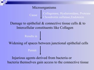
Microorganisms
Collagenase, Hyaluronidase, Protease
Chondroitinsulfatase,Endotoxin
Cause
Damage to epithelial & connective tissue cells & to
Intercellular constituents like Collagen
Results in
Widening of spaces between junctional epithelial cells
Permit
Injurious agents derived from bacteria or
bacteria themselves gain access to the connective tissue
4.
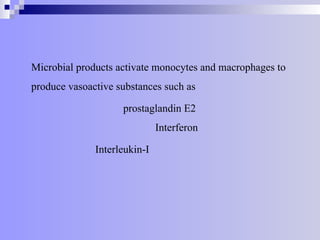
Microbial products activatemonocytes and macrophages to
produce vasoactive substances such as
prostaglandin E2
Interferon
Interleukin-I
5.
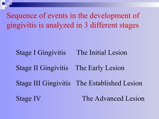
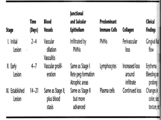
Sequence of eventsin the development of
gingivitis is analyzed in 3 different stages
Stage I Gingivitis The Initial Lesion
Stage II Gingivitis The Early Lesion
Stage III Gingivitis The Established Lesion
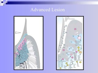
Stage IV The Advanced Lesion
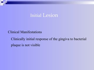
Initial Lesion
Microscopic changes
Features of acute inflammation are seen in connective
tissue beneath junctional epithelium.
Changes in blood vessel morphologic features
(Widening)
Adherence of neutrophils to vessels (Margination)
Leukocytes mainly PMN’s leave the capillaries by
migrating through walls (Diapedisis, Emigration)
Exudation of fluid from the gingival sulcus
8.
Initial Lesion
The characterand intensity of the host response
determines whether the initial lesion resolves
rapidly or evolves into a chronic inflammatory
lesion
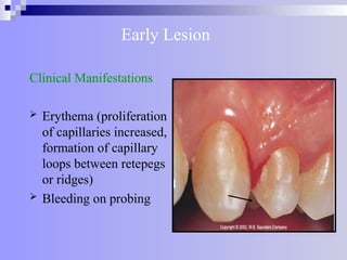
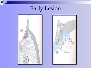
Early Lesion
Clinical Manifestations
Erythema (proliferation
of capillaries increased,
formation of capillary
loops between retepegs
or ridges)
Bleeding on probing
11.
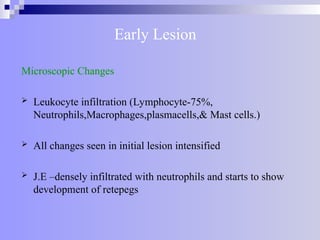
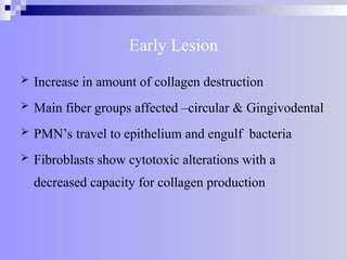
Early Lesion
Microscopic Changes
Leukocyte infiltration (Lymphocyte-75%,
Neutrophils,Macrophages,plasmacells,& Mast cells.)
All changes seen in initial lesion intensified
J.E –densely infiltrated with neutrophils and starts to show
development of retepegs
12.
Early Lesion
Increasein amount of collagen destruction
Main fiber groups affected –circular & Gingivodental
PMN’s travel to epithelium and engulf bacteria
Fibroblasts show cytotoxic alterations with a
decreased capacity for collagen production
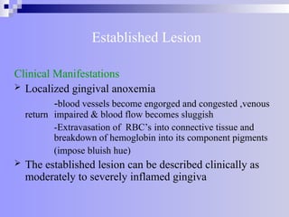
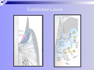
Established Lesion
Clinical Manifestations
Localized gingival anoxemia
-blood vessels become engorged and congested ,venous
return impaired & blood flow becomes sluggish
-Extravasation of RBC’s into connective tissue and
breakdown of hemoglobin into its component pigments
(impose bluish hue)
The established lesion can be described clinically as
moderately to severely inflamed gingiva
15.
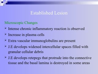
Established Lesion
Microscopic Changes
Intense chronic inflammatory reaction is observed
Increase in plasma cells
Extra vascular immunoglobulins are present
J.E develops widened intercellular spaces filled with
granular cellular debris
J.E develops retepegs that protrude into the connective
tissue and the basal lamina is destroyed in some areas
16.
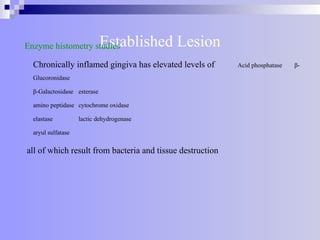
Established Lesion
Enzyme histometrystudies
Chronically inflamed gingiva has elevated levels of Acid phosphatase β-
Glucoronidase
β-Galactosidase esterase
amino peptidase cytochrome oxidase
elastase lactic dehydrogenase
aryul sulfatase
all of which result from bacteria and tissue destruction
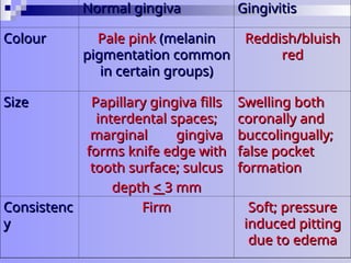
Normal gingiva
Normal gingivaGingivitis
Gingivitis
Colour
Colour Pale pink
Pale pink (melanin
(melanin
pigmentation common
pigmentation common
in certain groups)
in certain groups)
Reddish/bluish
Reddish/bluish
red
red
Size
Size Papillary gingiva fills
Papillary gingiva fills
interdental spaces;
interdental spaces;
marginal gingiva
marginal gingiva
forms knife edge with
forms knife edge with
tooth surface; sulcus
tooth surface; sulcus
depth
depth <
< 3 mm
3 mm
Swelling both
Swelling both
coronally and
coronally and
buccolingually;
buccolingually;
false pocket
false pocket
formation
formation
Consistenc
Consistenc
y
y
Firm
Firm Soft; pressure
Soft; pressure
induced pitting
induced pitting
due to edema
due to edema
22.
Normal Gingiva
Normal GingivaGingivitis
Gingivitis
Contour
Contour Scalloped
Scalloped
troughs in
troughs in
marginal areas
marginal areas
rise to peaks in
rise to peaks in
interdental
interdental
areas
areas
Edema which
Edema which
blunts the
blunts the
marginal and
marginal and
papillary tissues
papillary tissues
leads to loss of
leads to loss of
knife edge
knife edge
adaptation.
adaptation.
Marginal
Marginal
swelling leads to
swelling leads to
less accentuated
less accentuated
scalloping
scalloping
23.
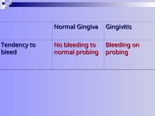
Normal Gingiva
Normal GingivaGingivitis
Gingivitis
Tendency to
Tendency to
bleed
bleed
No bleeding to
No bleeding to
normal probing
normal probing
Bleeding on
Bleeding on
probing
probing


![CTEV [ clubfoot] DR ARUN LAL ,DR MOHAMED ASHRAF travancore medical college k...](https://cdn.slidesharecdn.com/ss_thumbnails/ctevclubfootdrarunlaldrmohamedashraftravancoremedicalcollegekollamkeralaindia-260208063247-18fc466c-thumbnail.jpg?width=640&height=640&fit=bounds)


![ONFH[AVN HIP] -TRIPLE REGIME -A NOVAL SURGICAL CONCEPT .pptx](https://cdn.slidesharecdn.com/ss_thumbnails/onfhavnhip2026koaconcalicutdrgokuldevdrmashraf-260210064517-213ec005-thumbnail.jpg?width=640&height=640&fit=bounds)

