2. DSA (Digital Subtraction
Angiography)
Pemeriksaan baku dan standar dari pembuluh darah
otak untuk melihat aliran darah di pembuluh darah
arteri sampai ke jaringan lalu ke pembuluh vena
secara langsung dan terus menerus menggunakan
alat angiografi atau keterisasi
3.
4.
5. DSA (Digital Subtraction
Angiography)
Angiografi yang dilakukan pada pembuluh
darah otak
Memasukkan kateter ke dalam arteri pada
lengan maupun tungkai.
Injeksikan kontras ke dalam pembuluh
darah menuju otak
Cerebral angiogram lebih akurat
dibanding karotid Doppler
6. Menolong deteksi dan diagnosa stroke
akut
Mendeteksi kelainan pembuluh darah
yang menuju otak (misalnya, aneurisma,
malformasi pembuluh darah, trombosis,
penyempitan atau penyumbatan)
7. Pelajari PD otak yang letaknya abnormal
(tumor, gumpalan darah, spasme, tekanan
otak meningkat, atau hidrosefalus)
Bantu pembedahan
8. Angiografi tidak boleh dilakukan
pada penderita penyakit hati,
ginjal, atau tiroid, atau alergi
kontras
9. Hasil abnormal
Spasme, plak, fistula, malformasi
arteriovenosus, atau arteriosclerosis.
Penurunan suplai aliran darah ke otak.
Pembuluh darah otak yang letaknya tidak
lazim menunjukkan adanya tumor, daerah
pembengkakan, atau penyumbatan aliran
cairan spinal.
10. Transcranial Doppler
Teknik ultrasonografi non invasif yang
mempunyai kemampuan untuk mengukur
kecepatan dan arah aliran pembuluh
darah di otak.
11. Kelebihan Transcranial Doppler (TCD) :
1. Menggunakan teknik sonografi yang non invasif
sehingga menghindarkan pasien dari rasa tidak
nyaman selama pemeriksaan
2. Aman, karena teknik ini bebas dari bahaya
radiasi
3. Tidak memerlukan ruangan khusus dalam
pelaksanaan
4. Dapat dilakukan berulang kali untuk monitoring
tanpa adanya efek samping
12. 5. Tidak memerlukan penggunaan zat
kontras yang mempunyai resiko terjadinya
efek samping seperti alergi
6. Biaya yang lebih murah dibandingkan
dengan teknik lain seperti arteriografi.
13. Peran TCD di bidang medis:
- Mendeteksi adanya gangguan aliran
pembuluh darah otak
- Menilai faktor resiko terjadinya stroke pada
pasien beresiko
- Mendeteksi adanya emboli
- Menilai respon hasil terapi post stroke
- Mendeteksi adanya vasospasme (spasme
pembuluh darah) misalnya setelah
terjadinya perdarahan sub arrachnoid
14. - Sebagai penunjang diagnosis bersama
dengan pemeriksaan lain seperti CT scan,
MRI, MRA.
- Sebagai penunjang terapi
(Sonotrombolisis)
- Untuk mendeteksi kematian otak (Brain
Death)
15. TCD dapat diaplikasikan pada kasus-
kasus seperti :
• Resiko terjadinya stroke pada anak-anak
penderita sickle cell anemia
• TIA (Transient Ischemic Attack) dan Stroke
untuk menilai stenosis pembuluh darah dan
aliran kolateral.
• Menilai adanya trombosis atau emboli pada
TIA atau Stroke
• Post Trauma kepala atau perdarahan dari
aneurysma sub arachnoid yang beresiko
terjadi vasospasme pembuluh darah
16. • Mengkonfirmasi diagnosis klinik dari
kematian otak (Brain Death)
• Vascular Headache ( seperti pada kasus
migrain) dan beberapa kasus lain yang
masih dalam tahap pengembangan dan
penelitian
17.
18. Cath Lab
Mendeteksi penyakit artherosclerosis
pada arteri carotis di leher, yang
menggangu aliran darah ke otak dan
bahkan dapat menyebabkan stroke.
Kateter angiography dapat menampilkan
gambar pembuluh darah secara detil, jelas dan
akurat. Sangat membantu dalam tindakan
prosedur operasi atau Percutaneous
Transluminal Coronary Angioplasty (PTCA).
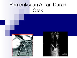
20. What Are They?
Computed Tomography Angiography
(CTA)
Imaging of the vasculature using CT techniques
Can be 3-D
Requires contrast
Magnetic Resonance Angiography (MRA)
Imaging of the vasculature system using MRI
techniques
Can be 3-D
No contrast required
21. How Does
CTA Work?
Uses X-rays
Tube rotates around patient at high speed
Detector picks up attenuated beam
Computer generates the image
Collects image in axial plane but can be
converted into sagittal, or coronal views
Computer can generate 3-d images
22. How Does MRA work?
Uses a large magnet align hydrogen atom in the
body
Pulsed radio-waves cause hydrogen atoms to
flip out of alignment
When the radio-waves are turned off the
hydrogen atom flip back and give off their own
signal
Signal is detected by the computer and used to
generate an image
Different body tissues
have different amounts
of Hydrogen
23. What is CTA used for?
Imaging of calcified atherosclerotic plaque
Areas that are scan for plaque include:
Carotid Bulb
Iliac arteries
Coronary arteries
Used to detect legs clots before they
break and cause pulmonary emboli
25. What is MRA used for?
MRA is used to image many peripheral vessels
Areas that are imaged include:
Circle of Willis
Cerebral Arteries
Renal Arteries
If there is a contrast allergy then MRA is used
26. Benefits of CTA
Detecting narrowing vessels in time for
intervention
Better anatomical detail than with MRA or
ultrasound
Can be used for screening for arterial disease
Less costly and safer than
conventional angiography
Contrast reactions are
less severe
27. Risks of CTA
Allergic reactions to contrast media
Avoided in patients with kidney disease
due to contrast
Ionizing radiation is used
Pregnant women should not have a CT
due to radiation
28. Benefits of MRA
Detailed images without damaging the
artery with a catheter
Shorter procedure and recovery times
than with conventional angiography
Less costly than catheter angiography
No exposure to ionizing radiation
Use of contrast is
not necessary to
obtain good images
29. Risks of MRA
Metal implants may be affected by the
magnetic field
Claustrophobic patients may need ot be
sedated
It is unknown how the magnetic field
affects the fetus so first trimester patients
should not have an MRI
30. Limitations of CTA
Fuzzy images if there is patient movement
Heart beat can blur images
Blocked vessels are harder to interpret
Not reliable for imaging small twisted
vessels in rapidly moving organs
Faster gantry times will solve this problem
31. Limitations of MRA
Any metal object in the patient is
contraindicative
Image clarity is not as good as
conventional angiography
Cannot image calcified plaque
Hard to image very small vessels
32. Accuracy of CTA
16-slice multidetector machine
Sensitive and specific for 2mm diameter or
greater 92-93%
Ultra fast 16-slice multidetector machine
Sensitive and specific for 2mm diameter or
greater 95-98%
64-slice multidetector machine
Sensitive and specific for 2mm diameter or
greater 92-93%
33. Accuracy of MRA
Detecting Cerebral aneurysms with an average
accuracy of about 70% without contrast
False positives averaged approx. 27
With contrast 100% sensitive for aneurysmal
and stenotic lesions
Specificity of completely occluded lesions also
100%
Drops to 83.3% when only partially occluded
34. What’s to come?
Improved spatial resolution
Resolutions due to software advancements are
improving
Decreasing costs
Screening
CTA screening for CAD
Joint Modalities
Combining of the two for better images
Better accuracy
New contrast medias for MR and faster CT machines
35. Bibliography
Dargan, R., Volkin, L., New Application Enhances Capabilities of
Angiographic CT, retrieved February 5, from:
www.asrt.org/content/News/IndustryNews
Briefs/CT/NewApplica050318.aspx
Sheth, T. et. al., (2005), Coronary Computed Tomography
Angiography: Emerging Technique for Coronary Artery Imaging,
JACR 2005:56(1):15-24
Tripathi, RP, et. al., (2002), Three-Dimensional Contrast-Enhanced
Magnetic Resonance Angiography- Our Preliminary Experience, Ind.
J. Raiol. Imag. 2002, 12:2:179-188
www.cir.uc.edu/ research.html
www.medscape.com/viewarticle/521872?rss
www.radiologyinfo.org/content/ct-angiography.htm
www.radiologyinfo.org/content/mr-angiography.htm