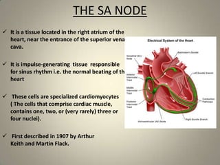
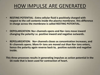
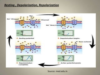
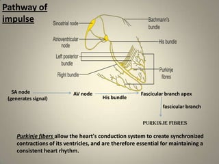
HEART’S PACE MAKER,
THE SINOATRIAL NODE

PRESENTED BY:
MINHAZ AHMED
BBI11014
INTEGRATED M.SC
3RD SEMESTER,MBBT
Tezpur University,Assam
THE SA NODE
 It is a tissue located in the right atrium of the
heart, near the entrance of the superior vena
cava.
 It is impulse-generating tissue responsible
for sinus rhythm i.e. the normal beating of the
heart

 These cells are specialized cardiomyocytes
( The cells that comprise cardiac muscle,
contains one, two, or (very rarely) three or
four nuclei).
 First described in 1907 by Arthur
Keith and Martin Flack.
HOW IMPULSE ARE GENERATED
• RESTING POTENTIAL : Extra cellular fluid is positively charged with
respect to the cell contents inside the plasma membrane. this difference
in charge across the membrane is called RESTING POTENTIAL.
• DEPOLARIZATION: Na+ channels opens and Na+ ions move inward
changing the polarity i.e. positive inward and negative outwards.
• REPOLARIZATION: Na+ channels closes as concentration increases, and
K+ channels opens. More K+ ions are moved out than Na+ ions enters,
hence the polarity again reverse back to, positive outside and negative
inside.
This three processes results in generating impulses as action potential in the
SA node that is been used for contraction of heart.
Resting , Depolarization, Repolarization

Source: med.edu.in
Pathway of
impulse

SA node
(generates signal)

AV node

His bundle

Fascicular branch apex
fascicular branch
Purkinje fibres

Purkinje fibers allow the heart's conduction system to create synchronized
contractions of its ventricles, and are therefore essential for maintaining a
consistent heart rhythm.
Cardiac pacemaker
 Primary (SA node):
main generator of electrical impulse.
 Secondary (AV junction & Bundle of His)
If the SA node does not function, a group of cells further
down the heart will become the heart's pacemaker.

AV node normally discharge at about 40-60 beats per
minute.

Bundle of His along with the Purkinje fibres, can
produce a spontaneous action potential at a rate of 30-40
beats per minute
ARTIFICIAL PACEMAKER
• pacemaker is a medical device that uses electrical impulses,
delivered by electrodes contacting the heart muscles, to
regulate the beating of the heart.
• The primary purpose of a pacemaker is to maintain an
adequate heart rate, either because the heart's native
pacemaker is not fast enough, or there is a block in
the heart's electrical conduction system.
Heart’s pace maker, the sinoatrial node

Heart’s pace maker, the sinoatrial node

  • 1.
    HEART’S PACE MAKER, THESINOATRIAL NODE PRESENTED BY: MINHAZ AHMED BBI11014 INTEGRATED M.SC 3RD SEMESTER,MBBT Tezpur University,Assam
  • 2.
    THE SA NODE It is a tissue located in the right atrium of the heart, near the entrance of the superior vena cava.  It is impulse-generating tissue responsible for sinus rhythm i.e. the normal beating of the heart  These cells are specialized cardiomyocytes ( The cells that comprise cardiac muscle, contains one, two, or (very rarely) three or four nuclei).  First described in 1907 by Arthur Keith and Martin Flack.
  • 3.
    HOW IMPULSE AREGENERATED • RESTING POTENTIAL : Extra cellular fluid is positively charged with respect to the cell contents inside the plasma membrane. this difference in charge across the membrane is called RESTING POTENTIAL. • DEPOLARIZATION: Na+ channels opens and Na+ ions move inward changing the polarity i.e. positive inward and negative outwards. • REPOLARIZATION: Na+ channels closes as concentration increases, and K+ channels opens. More K+ ions are moved out than Na+ ions enters, hence the polarity again reverse back to, positive outside and negative inside. This three processes results in generating impulses as action potential in the SA node that is been used for contraction of heart.
  • 4.
    Resting , Depolarization,Repolarization Source: med.edu.in
  • 5.
    Pathway of impulse SA node (generatessignal) AV node His bundle Fascicular branch apex fascicular branch Purkinje fibres Purkinje fibers allow the heart's conduction system to create synchronized contractions of its ventricles, and are therefore essential for maintaining a consistent heart rhythm.
  • 6.
    Cardiac pacemaker  Primary(SA node): main generator of electrical impulse.  Secondary (AV junction & Bundle of His) If the SA node does not function, a group of cells further down the heart will become the heart's pacemaker.  AV node normally discharge at about 40-60 beats per minute.  Bundle of His along with the Purkinje fibres, can produce a spontaneous action potential at a rate of 30-40 beats per minute
  • 7.
    ARTIFICIAL PACEMAKER • pacemakeris a medical device that uses electrical impulses, delivered by electrodes contacting the heart muscles, to regulate the beating of the heart. • The primary purpose of a pacemaker is to maintain an adequate heart rate, either because the heart's native pacemaker is not fast enough, or there is a block in the heart's electrical conduction system.