SlideShare a Scribd company logo
1 of 6
Download to read offline
International Research Journal of Engineering and Technology (IRJET) e-ISSN: 2395-0056
Volume: 07 Issue: 03 | Mar 2020 www.irjet.net p-ISSN: 2395-0072
© 2020, IRJET | Impact Factor value: 7.34 | ISO 9001:2008 Certified Journal | Page 4615
Diabetic Retinopathy
Vaishnavi Kukutkar1, Rehan Pathan2, Shubham Mansute3, Madan Bhendare4, Jyoti Kubade5
1,2,3,4,5Student, Dept. of Computer Science & Engineering, Datta Meghe Institute of Engineering, Technology &
Research, Wardha, Maharashtra, India
---------------------------------------------------------------------***----------------------------------------------------------------------
Abstract - Here we address the detection of Hemorrhages
and microaneurysms in color fundus images. Inpre-Processing
we separate red, green, blue color channel from the retinal
images. The green channel will pass tothefurtherprocess. The
green color plane was utilized in theanalysissinceitshowsthe
simplest contrast between the vessels and therefore the
background retina. Then we extract the GLCM (Gray Level Co-
Occurrence Matrix) feature. Within the GLCMs, several
statistics information are derived using the various formulas.
These statistics provide informationaboutthefeelofapicture,
like Energy, Entropy, Dissimilarity, Contrast, Inverse
difference, correlation Homogeneity, Auto correlation,Cluster
Shade Cluster Prominence, Maximum probability, Sum of
Squares are going to be calculated for texture image. After
feature Extraction, we offer this feature to classifier. Finally
it'll predict about the retinal whether it's hemorrhages or
microaneurysms. After predicting problems in the retinal
image we'll localize the affected place. For segmenting the
localized place we'll use adaptive thresholding segmentation.
Key Words: Image representation, dissimilarity space,
class learning.
1. INTRODUCTION
1.1 Overview
Diabetic retinopathy (DR) is a vascular disease of the
retina which affects patients with diabetes mellitus. Itis
the number one cause of blindness in people between
the ages of 20-64 in the United States. It is, therefore, a
worthwhile topic for all medical students to review.
Diabetes mellitus is extremely common, so it is not
surprising that DR affects 3.4 percent of the population
(4.1 million individuals). Of the millions of people with
DR, nearly one-fourth have vision-threatening disease.
The likelihood of developing diabetic retinopathy is
related to the duration of the disease. Type 2 diabetes
has an insidious onset and cangounnoticedforyears.As
a result, patients may already have DR at the time of
diagnosis. Type 1 diabetics, on the other hand, are
diagnosed early in the course of their disease, and they
typically do not develop retinopathy until yearsafterthe
diagnosis is made. The risk of developing retinopathy
increases after puberty. Twenty years after the
diagnosis of diabetes, 80% of type2diabeticsandnearly
all type 1 diabetics show some signs of retinopathy.
While these numbers are eye-opening, diabetics can
decrease their risk of retinopathy and slow the
progression of the disease after it has begun with tight
glucose control.
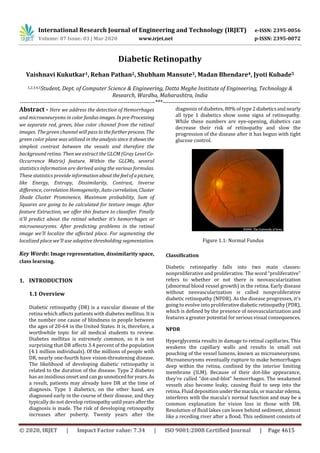
Figure 1.1: Normal Fundus
Classification
Diabetic retinopathy falls into two main classes:
nonproliferative and proliferative. The word “proliferative”
refers to whether or not there is neovascularization
(abnormal blood vessel growth) in the retina. Early disease
without neovascularization is called nonproliferative
diabetic retinopathy (NPDR). As the disease progresses, it's
going to evolve into proliferativediabeticretinopathy(PDR),
which is defined by the presence of neovascularization and
features a greater potential for serious visual consequences.
NPDR
Hyperglycemia results in damage to retinal capillaries. This
weakens the capillary walls and results in small out
pouching of the vessel lumens, known as microaneurysms.
Microaneurysms eventually rupture to make hemorrhages
deep within the retina, confined by the interior limiting
membrane (ILM). Because of their dot-like appearance,
they're called “dot-and-blot” hemorrhages. The weakened
vessels also become leaky, causing fluid to seep into the
retina. Fluid depositionunderthemacula,ormacularedema,
interferes with the macula’s normal function and may be a
common explanation for vision loss in those with DR.
Resolution of fluid lakes can leave behind sediment, almost
like a receding river after a flood. This sediment consists of
International Research Journal of Engineering and Technology (IRJET) e-ISSN: 2395-0056
Volume: 07 Issue: 03 | Mar 2020 www.irjet.net p-ISSN: 2395-0072
© 2020, IRJET | Impact Factor value: 7.34 | ISO 9001:2008 Certified Journal | Page 4616
lipid byproducts and appears aswaxy,yellowdepositscalled
hard exudates. As NPDR progresses, the affected vessels
eventually become obstructed. This obstruction may cause
infarction of the nerve fiber layer, leading to fluffy, white
patches called cotton spots (CWS).
Figure 1.2: NPDR
Morphological Processing
Morphological processing is the most common methodused
for detection of microaneurysm.Morphological processingis
a collection of techniques that can be used for image
component extraction. In 1996, Spencer et al. [11] used
morphological processing which detects microaneurysms
present in fluorescein angiograms. After preprocessing
stage, a bilinear top-hat transformation and matched
filtering are used to provide an initial segmentation of the
images. Then Thresholding is used to produce a binary
image that contains candidate microaneurysms. Then a
novel region-growing algorithm results in the final
segmentation of microaneurysms.
Then Frame et al. [12] produced a list of features like shape
features and pixel-intensityfeatures oneachcandidate.After
that the classifier is used to classify each candidate as
microaneurysm and non-microaneurysm by using these
features.
Niemeijer et al. [6] used a hybrid approach to detect the red
lesions by combining the prior works by Spencer et al. [11]
and Frame et al. [12] with two important new contributions.
Their first contribution is the useofnewredlesioncandidate
detection system whichisbasedonpixel classification.Blood
vessels and red lesions are separated from the background
by using this technique. Remaining objectsareconsideredas
possible red lesions after removal of vasculature. Their
second contribution is the addition of a large numberofnew
features to those proposed by Spencer Frame. Then k-
nearest neighbor classifier is used to classify the detected
candidate objects. The images for the dataset are takenfrom
the hospital. This method has achieved 100%sensitivity and
87% specificity but the time required is more for initial
detection of red lesions.
Then Zhang and Fan [13] proposed a spot lesion detection
algorithm that uses multiscale morphological processing.
Using scale-based lesion validation, blood vessels are
removed. This algorithm was testedon30retinal images and
it has achieved the sensitivity of84.10%andpredictivevalue
of 89.20%.
Karnowski et al. [14] proposed a morphological
reconstruction method for the segmentation of retinal
lesions. In this method, the segmentation is performed by
using ground-truth data at a variety of scales determined, to
separate nuisance blobs from true lesions. The ground truth
data is used to design post-processing classifiers toseparate
the machine segmented results into nuisance and actual
lesion classes. This segmentationresultisusedtoclassifythe
images as “normal” or “abnormal”. This method was tested
on 86 retinal images and these images are taken from the
hospital. This method has achieved a sensitivity of 90% and
specificity of 90%. The weakness of this method is that
classification features for the post-processing steps is
inaccurate so this method can be improved onmorerealistic
dataset.
Kande et al. [15] proposed an approach that takes into
account the advantages of pixel-based classification and
morphological based detection. Local entropy thresholding
algorithm is used to distinguish between enhanced red
lesion segments and the background. Then morphological
top-hat transformation is used to suppress thebloodvessels
and then SVM (Support Vector Machine) is used to classify
the red lesions from other dark regions. This approachtakes
the same time as proposed in [11] and [12] and performs
better. The author has selected 80 retinal images randomly
from the Clemson, DIARETDB0 and DIARETDB1 databases.
The sensitivity was 96.22% and specificity was 99.53% of
this approach.
1.2 Motivation
The prolonged diabetes leads to the formation of micro-
aneurysms and subsequently it leads to exudates as well as
hemorrhages. These are thefeaturesofDR andthatthey may
cause severe vision loss or maybe blindness. In order to
avoid these complications, it is very important to detect DR
early. This can be done by an accurate detection of
microaneurysms. It is very difficult to detect the exudates
clearly, because they are tiny spots on the retina. Also, the
detection of hemorrhages is very challenging. The texture of
hemorrhages and macula is almost the same. So, we'd liketo
possess robust algorithms which detect these features.
1.3 Objectives
The primary objective of evaluating and managing diabetic
retinopathy is to prevent, retard, or reverse visual loss,
thereby maintaining or improving vision-related quality of
life. In computer based retinal image analysis system, image
processing techniques are used in order to facilitate and
International Research Journal of Engineering and Technology (IRJET) e-ISSN: 2395-0056
Volume: 07 Issue: 03 | Mar 2020 www.irjet.net p-ISSN: 2395-0072
© 2020, IRJET | Impact Factor value: 7.34 | ISO 9001:2008 Certified Journal | Page 4617
improve diagnosis. Manual analysis of the images can be
improved and problem of detection of diabetic retinopathy
in the late stage for optimal treatment may be resolved.
Based on these the main objectives of the project are as
follows:-
i. Detection of macular region.
ii. Detection of retinal blood vessels.
iii. Early detection of diabetic retinopathy.
iv. To predict intensity of infection in eye usingfeature
extraction.
v. Detection of Hemorrhages and Microaneurysms in
color fundus image.
2. METHODOLOGY
2.1 Problem Definition
 Diabetic Retinopathy (DR) is oneoftheleadingcausesof
blindness in the industrialized world.
 Early detection is the key in providing effective
treatment.
 However, the current number of trained eye care
specialists is inadequate to screen the increasing
number of diabetic patients.
 In recent years,automatedandsemi-automatedsystems
to detect DR with color fundus images have been
developed with encouraging, but not fully satisfactory
results.
2.2 Proposed Approach
Microaneurysm Detection
Automated microaneurysm detection is very useful in
diagnosing the diabetic retinopathy for the prevention of
blindness. With the help of automated system, the work of
ophthalmologists can be reduced and the costofdetectionof
diabetic retinopathy canalsobereduced.Mostoftheexisting
methods of microaneurysms detection work in two stages:
microaneurysm candidate extractionandclassification.First
stage requires image preprocessing for the reduction of
noise and contrast enhancement. Image preprocessing is
performed on the green color plane of RGB image becausein
green color plane microaneurysms have the higher contrast
with the background. After that candidate regions for
microaneurysms are detected. Then blood vessel
segmentation algorithms are applied to extract blood vessel
from the candidates for the reduction in false positives
because many of the blood vessels may appear as false
positives in the preprocessed image. Thenfeatureanalysisis
used in which feature extraction and feature selection is
performed to detect the microaneurysms. In second stage,
the classification algorithm is applied to categorize these
features intomicroaneurysmcandidate(abnormal)andnon-
microaneurysm candidate (normal). The probability is
estimated for each candidate using a classifieranda large set
of specifically designed features to represent a
microaneurysm. In general the process for the detection of
microaneurysms is concluded in Figure given below.
Figure 2.2.1: Microaneurysm Detection
Algorithm Description
Phase 1
Image Pre-processing
Images are enhancedbysharpeningandremovingunwanted
outliers.
Figure 2.2.2: Image Pre-processing
Phase 2
Segmentation
Image will be segmented to fetch out the image edges and
then detected all required parameters.
Retinal fundus image
Image preprocessing
Candidate extraction
Vessel segmentation
Determination of candidate lesion
features
Microaneurysm
classification
Performance measure
International Research Journal of Engineering and Technology (IRJET) e-ISSN: 2395-0056
Volume: 07 Issue: 03 | Mar 2020 www.irjet.net p-ISSN: 2395-0072
© 2020, IRJET | Impact Factor value: 7.34 | ISO 9001:2008 Certified Journal | Page 4618
Figure 2.2.3: Segmentation
Phase 3
Recognition and Classification
Ones the image is segmented it can be tested to recognize it
first and then classify it using SVM algorithm.
Figure 2.2.4: Recognition and Classification
3. IMPLEMENTATION DETAILS
3.1 Test Environment
Processor Intel® Core™ i3
Processor 2.40 GHz
RAM 4 GB
Operating
System
64 bit
Programming
Tool
MATLAB R 2015 a
3.2 MATLAB
Millions of engineers and scientists worldwide use
MATLAB® to analyze and design the systems and products.
MATLAB is in automobile active safety systems,
interplanetary spacecraft, health monitoring devices, smart
power grids. It is used for machine learning, signal
processing, image processing, computer vision,
computational finance, control design, robotics, and much
more. MATLAB is stands for Matrix Laboratory. Clave Molar
is a mathematician and a computer programmer has
invented MATLAB in mid 1970s. MATLAB is a high
performance language for technical computing. It takes part
in computation, visualization, and programming in an easy-
to-use environment where problems and solutions are
expressed in familiar mathematical code. Typical uses
include: Math and computation. MATLAB is programmable
software for multi-paradigm numerical environment. This
software is developed by the Math Works.
4. RESULT AND DISCUSSION
In figure 4.1, the main GUI is shown which has sevenbuttons
to perform different operations on the fundus image.
Figure 4.1: Main GUI Window
Figure 4.2 shows the windowafterclickingthe“InputImage”
button to fetch dataset.
Figure 4.2: Fetching Dataset
Figure 4.3 shows the working of Neural Network algorithm
on a fundus image to train the dataset forfurtherprocessing.
International Research Journal of Engineering and Technology (IRJET) e-ISSN: 2395-0056
Volume: 07 Issue: 03 | Mar 2020 www.irjet.net p-ISSN: 2395-0072
© 2020, IRJET | Impact Factor value: 7.34 | ISO 9001:2008 Certified Journal | Page 4619
Figure 4.3: Working of Neural Network on a Fundus Image
In Figure 4.4, RGB channels are extracted and stored by
clicking on the “Preprocessing” tab.
Figure 4.4: RGB Channel Extraction of an Input Image
By clicking on “Feature Extraction” button,differentfeatures
such as contrast, energy, etc. are extractedanddisplayedina
table as shown in figure 4.5.
Figure 4.5: Feature Extraction of Fundus Image
The “Classification” tab will navigate the user to actual
disease, for example, Hemorrhages, in the fundus image
which is shown in figure 4.6.
Figure 4.6: Detection of Actual Disease
Which site or vessel of fundus image contains the problemis
detected and it is shown by separating the background and
foreground, as shown in figure 4.7.
International Research Journal of Engineering and Technology (IRJET) e-ISSN: 2395-0056
Volume: 07 Issue: 03 | Mar 2020 www.irjet.net p-ISSN: 2395-0072
© 2020, IRJET | Impact Factor value: 7.34 | ISO 9001:2008 Certified Journal | Page 4620
Figure 4.7: Detecting actual sites of problem
5. CONCLUSIONS
Prolonged diabetes leads to DR, where the retina is
damaged due to fluid leaking fromthebloodvessels.Usually,
the stage of DR is judged based on blood vessels, exudes,
hemorrhages, microaneurysmsandtexture.Inthispaper, we
have discussed different methodsforfeaturesextraction and
automatic DR stage detection. An ophthalmologist uses an
ophthalmoscope to visualize the blood vesselsandhisorher
brain to detect the DR stages. Recently digital imaging
became available as a tool for DR screening. It provides high
quality permanent records of the retinal appearance, which
can be used for monitoring of progression or response to
treatment, and which can be reviewed by an
ophthalmologist, digital images have the potential to be
processed by automatic analysis systems. A combination of
both accurate and early diagnosis as well as correct
application of treatment canpreventblindnesscausedby DR
in more than 50% of all cases. Automatic detection of
microaneurysm presents many of the challenges. The size
and color of microaneurysm is very similar to the blood
vessels. Its size is variable and often very small so it can be
easily confused with noise present in the image. In human
retina, there is a pigmentation variation, texture, size and
location of human features from person to person. Themore
false positives occur when the blood vessels areoverlapping
or adjacent with microaneurysms. So there is a need of an
effective automated microaneurysm detection method so
that diabetic retinopathy can be treated atanearlystageand
the blindness due to diabetic retinopathy can be prevented.
REFERENCES
[1] S. Wild, G. Roglic, A Green, “Global prevalenceofdiabetes:
estimates for the year 2000 and projections for 2030,”
Diabetes Care, 27, pp.l047- 1053, 2004.
[2] S. R. Nirmala, M. K. Nath, and S. Dandapat, “Retinal Image
Analysis: A Review,” International Journal of Computer &
Communication Technology (IJCCT), vol-2, pp. 11-15, 2011.
[3] National Eye Institute, National Institutes of Health,
“Diabetic Retinopathy: Whatyoushouldknow,”Booklet,NIH
Publication, no: 06-2171, 2003.
[4] A. D. Fleming, K. A. Goatman, and J. A. Olson, “The role of
haemorrhage and exudate detectioninautomatedgrading of
diabetic retinopathy,” British Journal of Ophthalmology, vol
94, no. 6, pp. 706- 711, 2010.
[5] A. D. Fleming, S. Philip, K. A. Goatman, J. A. Olson, andP. F.
Sharp, “Automated microaneurysm detection using local
contrast normalization and local vessel detection,” IEEE
Trans. Med. Imag., vol. 25, no. 9, pp. 1223–1232, Sep. 2006.
[6] M.Niemeijer,B. V. Ginneken, J. Staal, M. S. A. Suttorp-
Schulten, and M. D. Abramoff, “Automatic detection of red
lesions in digital color fundus photographs,” IEEE Trans.
Med. Imag., vol. 24, no. 5, pp. 584– 592, May 2005.
[7] M. J. Cree, J. A. Olsoni, K. C. McHardyt, J. V. Forresters and
P. F. Sharp, “Automated microaneurysms detection,” IEEE
conference, pp. 699-702, 1996
[8] A. Mizutani, C. Muramatsu, Y. Hatanaka, S. Suemori, T.
Hara, and H. Fujita, “Automated microaneurysm detection
method based on double ring filter in retinal fundusimages,”
in Proc. SPIE, Med. Imag, Imag,Computer-Aided Diagnosis,
vol. 7260, 2009.
[9] K. Ram, G. D. Joshi and J. Sivaswamy, “A successiveclutter
rejectionbased-approach for early detection of diabetic
retinopathy,” IEEE Transaction on Biomediacl Engineering,
vol. 58, no. 3, pp. 664-673, Mar. 2011
[10] M. Esmaeili, H. Rabbani, A. M. Dehnavi, and A.Dehghani,
“A new curvlet transform based method forextractionofred
lesions in digital color retinal images,”in Proc.ICIP,pp.4093-
4096, 2010.
[11] T. Spencer, J. Olson, K. McHardy, P. Sharp, and J.
Forrester, “An image processing strategy for the
segmentation and quantification in fluorescein angiograms
of the ocular fundus,” Comput. Biomed.Res.,vol.29,pp.284–
302, 1996.
[12] A. Frame, P. Undrill, M. Cree, J. Olson and K. McHardy, P.
Sharp, and J. Forrester, “A comparison of computer based
classification methods applied to the detection of
microaneurysms in ophthalmic fluorescein angiograms,”
Comput. Biol. Med., vol. 28, pp. 225–238, 1998.
[13] X. Zhang and G. Fan, “Retinal Spot Lesion Detection
Using Adaptive Multiscale Morphological Processing,” in
Proc. ISVC (2), pp.490-501, 2006.
[14] T. P. Karnowski, V. P. Govindasamy, K. W. Tobin, E.
Chaum, M. D. Abramoff, “Retina Lesion and Microaneurysm
Segmentation using Morphological ReconstructionMethods
with Ground-Truth Data,” Conf Proc IEEE Eng Med BioI Soc
1, pp. 5433-5436, 2008.
[15] G.B. Kande, S.S. Tirumala, P.V. Subbaiah, and M.R.
Tagore, “Detection of Red Lesions in Digital Fundus Images,”
in Proc. ISBI, pp.558-561, 2009.

More Related Content

What's hot

IRJET- Automatic Detection of Blood Vessels and Classification of Retinal...
IRJET-  	  Automatic Detection of Blood Vessels and Classification of Retinal...IRJET-  	  Automatic Detection of Blood Vessels and Classification of Retinal...
IRJET- Automatic Detection of Blood Vessels and Classification of Retinal...IRJET Journal
 
IRJET- Approach for Diabetic Retinopathy Analysis using Artificial Neural Net...
IRJET- Approach for Diabetic Retinopathy Analysis using Artificial Neural Net...IRJET- Approach for Diabetic Retinopathy Analysis using Artificial Neural Net...
IRJET- Approach for Diabetic Retinopathy Analysis using Artificial Neural Net...IRJET Journal
 
Detection and Grading of Diabetic Maculopathy Automatically in Digital Retina...
Detection and Grading of Diabetic Maculopathy Automatically in Digital Retina...Detection and Grading of Diabetic Maculopathy Automatically in Digital Retina...
Detection and Grading of Diabetic Maculopathy Automatically in Digital Retina...paperpublications3
 
Diagnosis of Diabetic Retinopathy by Detection of Microneurysm And Exudates
Diagnosis of Diabetic Retinopathy by Detection of Microneurysm And ExudatesDiagnosis of Diabetic Retinopathy by Detection of Microneurysm And Exudates
Diagnosis of Diabetic Retinopathy by Detection of Microneurysm And ExudatesIRJET Journal
 
IRJET- Study on Segmentation and Classification Techniques for Automatic ...
IRJET-  	  Study on Segmentation and Classification Techniques for Automatic ...IRJET-  	  Study on Segmentation and Classification Techniques for Automatic ...
IRJET- Study on Segmentation and Classification Techniques for Automatic ...IRJET Journal
 
IRJET- Automatic Detection of Diabetic Retinopathy Lesions
IRJET- Automatic Detection of Diabetic Retinopathy LesionsIRJET- Automatic Detection of Diabetic Retinopathy Lesions
IRJET- Automatic Detection of Diabetic Retinopathy LesionsIRJET Journal
 
An Approach for the Detection of Vascular Abnormalities in Diabetic Retinopathy
An Approach for the Detection of Vascular Abnormalities in Diabetic RetinopathyAn Approach for the Detection of Vascular Abnormalities in Diabetic Retinopathy
An Approach for the Detection of Vascular Abnormalities in Diabetic Retinopathyijdmtaiir
 
Detection of Macular Edema by using Various Techniques of Feature Extraction ...
Detection of Macular Edema by using Various Techniques of Feature Extraction ...Detection of Macular Edema by using Various Techniques of Feature Extraction ...
Detection of Macular Edema by using Various Techniques of Feature Extraction ...IRJET Journal
 
C LASSIFICATION O F D IABETES R ETINA I MAGES U SING B LOOD V ESSEL A REAS
C LASSIFICATION  O F D IABETES  R ETINA I MAGES  U SING B LOOD V ESSEL A REASC LASSIFICATION  O F D IABETES  R ETINA I MAGES  U SING B LOOD V ESSEL A REAS
C LASSIFICATION O F D IABETES R ETINA I MAGES U SING B LOOD V ESSEL A REASIJCI JOURNAL
 
Diabetic Retinopathy Detection System from Retinal Images
Diabetic Retinopathy Detection System from Retinal ImagesDiabetic Retinopathy Detection System from Retinal Images
Diabetic Retinopathy Detection System from Retinal Imagesijtsrd
 
IRJET - Automatic Detection of Diabetic Retinopathy in Retinal Image
IRJET -  	  Automatic Detection of Diabetic Retinopathy in Retinal ImageIRJET -  	  Automatic Detection of Diabetic Retinopathy in Retinal Image
IRJET - Automatic Detection of Diabetic Retinopathy in Retinal ImageIRJET Journal
 
Enhancements to a Computer : Assisted Screening Technology for Diabetic Retin...
Enhancements to a Computer : Assisted Screening Technology for Diabetic Retin...Enhancements to a Computer : Assisted Screening Technology for Diabetic Retin...
Enhancements to a Computer : Assisted Screening Technology for Diabetic Retin...Apollo Hospitals Group and ATNF
 
IRJET -An Automatated Learning Approach for Detection of Diabetic Retinopathy...
IRJET -An Automatated Learning Approach for Detection of Diabetic Retinopathy...IRJET -An Automatated Learning Approach for Detection of Diabetic Retinopathy...
IRJET -An Automatated Learning Approach for Detection of Diabetic Retinopathy...IRJET Journal
 
Blood vessel segmentation in fundus images
Blood vessel segmentation in fundus imagesBlood vessel segmentation in fundus images
Blood vessel segmentation in fundus imagesIRJET Journal
 
AUTOMATED DETECTION OF HARD EXUDATES IN FUNDUS IMAGES USING IMPROVED OTSU THR...
AUTOMATED DETECTION OF HARD EXUDATES IN FUNDUS IMAGES USING IMPROVED OTSU THR...AUTOMATED DETECTION OF HARD EXUDATES IN FUNDUS IMAGES USING IMPROVED OTSU THR...
AUTOMATED DETECTION OF HARD EXUDATES IN FUNDUS IMAGES USING IMPROVED OTSU THR...IJCSES Journal
 
Efficient Diabetic Retinopathy Space Subjective Fuzzy Clustering In Digital F...
Efficient Diabetic Retinopathy Space Subjective Fuzzy Clustering In Digital F...Efficient Diabetic Retinopathy Space Subjective Fuzzy Clustering In Digital F...
Efficient Diabetic Retinopathy Space Subjective Fuzzy Clustering In Digital F...IOSR Journals
 

What's hot (18)

IRJET- Automatic Detection of Blood Vessels and Classification of Retinal...
IRJET-  	  Automatic Detection of Blood Vessels and Classification of Retinal...IRJET-  	  Automatic Detection of Blood Vessels and Classification of Retinal...
IRJET- Automatic Detection of Blood Vessels and Classification of Retinal...
 
IRJET- Approach for Diabetic Retinopathy Analysis using Artificial Neural Net...
IRJET- Approach for Diabetic Retinopathy Analysis using Artificial Neural Net...IRJET- Approach for Diabetic Retinopathy Analysis using Artificial Neural Net...
IRJET- Approach for Diabetic Retinopathy Analysis using Artificial Neural Net...
 
Detection and Grading of Diabetic Maculopathy Automatically in Digital Retina...
Detection and Grading of Diabetic Maculopathy Automatically in Digital Retina...Detection and Grading of Diabetic Maculopathy Automatically in Digital Retina...
Detection and Grading of Diabetic Maculopathy Automatically in Digital Retina...
 
Diagnosis of Diabetic Retinopathy by Detection of Microneurysm And Exudates
Diagnosis of Diabetic Retinopathy by Detection of Microneurysm And ExudatesDiagnosis of Diabetic Retinopathy by Detection of Microneurysm And Exudates
Diagnosis of Diabetic Retinopathy by Detection of Microneurysm And Exudates
 
IRJET- Study on Segmentation and Classification Techniques for Automatic ...
IRJET-  	  Study on Segmentation and Classification Techniques for Automatic ...IRJET-  	  Study on Segmentation and Classification Techniques for Automatic ...
IRJET- Study on Segmentation and Classification Techniques for Automatic ...
 
IRJET- Automatic Detection of Diabetic Retinopathy Lesions
IRJET- Automatic Detection of Diabetic Retinopathy LesionsIRJET- Automatic Detection of Diabetic Retinopathy Lesions
IRJET- Automatic Detection of Diabetic Retinopathy Lesions
 
SYSTEM BASED ON THE NEURAL NETWORK FOR THE DIAGNOSIS OF DIABETIC RETINOPATHY
SYSTEM BASED ON THE NEURAL NETWORK FOR THE DIAGNOSIS OF DIABETIC RETINOPATHYSYSTEM BASED ON THE NEURAL NETWORK FOR THE DIAGNOSIS OF DIABETIC RETINOPATHY
SYSTEM BASED ON THE NEURAL NETWORK FOR THE DIAGNOSIS OF DIABETIC RETINOPATHY
 
An Approach for the Detection of Vascular Abnormalities in Diabetic Retinopathy
An Approach for the Detection of Vascular Abnormalities in Diabetic RetinopathyAn Approach for the Detection of Vascular Abnormalities in Diabetic Retinopathy
An Approach for the Detection of Vascular Abnormalities in Diabetic Retinopathy
 
Detection of Macular Edema by using Various Techniques of Feature Extraction ...
Detection of Macular Edema by using Various Techniques of Feature Extraction ...Detection of Macular Edema by using Various Techniques of Feature Extraction ...
Detection of Macular Edema by using Various Techniques of Feature Extraction ...
 
C LASSIFICATION O F D IABETES R ETINA I MAGES U SING B LOOD V ESSEL A REAS
C LASSIFICATION  O F D IABETES  R ETINA I MAGES  U SING B LOOD V ESSEL A REASC LASSIFICATION  O F D IABETES  R ETINA I MAGES  U SING B LOOD V ESSEL A REAS
C LASSIFICATION O F D IABETES R ETINA I MAGES U SING B LOOD V ESSEL A REAS
 
Diabetic Retinopathy Detection System from Retinal Images
Diabetic Retinopathy Detection System from Retinal ImagesDiabetic Retinopathy Detection System from Retinal Images
Diabetic Retinopathy Detection System from Retinal Images
 
IRJET - Automatic Detection of Diabetic Retinopathy in Retinal Image
IRJET -  	  Automatic Detection of Diabetic Retinopathy in Retinal ImageIRJET -  	  Automatic Detection of Diabetic Retinopathy in Retinal Image
IRJET - Automatic Detection of Diabetic Retinopathy in Retinal Image
 
Enhancements to a Computer : Assisted Screening Technology for Diabetic Retin...
Enhancements to a Computer : Assisted Screening Technology for Diabetic Retin...Enhancements to a Computer : Assisted Screening Technology for Diabetic Retin...
Enhancements to a Computer : Assisted Screening Technology for Diabetic Retin...
 
IRJET -An Automatated Learning Approach for Detection of Diabetic Retinopathy...
IRJET -An Automatated Learning Approach for Detection of Diabetic Retinopathy...IRJET -An Automatated Learning Approach for Detection of Diabetic Retinopathy...
IRJET -An Automatated Learning Approach for Detection of Diabetic Retinopathy...
 
Blood vessel segmentation in fundus images
Blood vessel segmentation in fundus imagesBlood vessel segmentation in fundus images
Blood vessel segmentation in fundus images
 
AUTOMATED DETECTION OF HARD EXUDATES IN FUNDUS IMAGES USING IMPROVED OTSU THR...
AUTOMATED DETECTION OF HARD EXUDATES IN FUNDUS IMAGES USING IMPROVED OTSU THR...AUTOMATED DETECTION OF HARD EXUDATES IN FUNDUS IMAGES USING IMPROVED OTSU THR...
AUTOMATED DETECTION OF HARD EXUDATES IN FUNDUS IMAGES USING IMPROVED OTSU THR...
 
Efficient Diabetic Retinopathy Space Subjective Fuzzy Clustering In Digital F...
Efficient Diabetic Retinopathy Space Subjective Fuzzy Clustering In Digital F...Efficient Diabetic Retinopathy Space Subjective Fuzzy Clustering In Digital F...
Efficient Diabetic Retinopathy Space Subjective Fuzzy Clustering In Digital F...
 
Ijecet 06 09_007
Ijecet 06 09_007Ijecet 06 09_007
Ijecet 06 09_007
 

Similar to IRJET - Diabetic Retinopathy

A Survey on early stage detection of Microaneurysms
A Survey on early stage detection of MicroaneurysmsA Survey on early stage detection of Microaneurysms
A Survey on early stage detection of MicroaneurysmsIRJET Journal
 
IRJET- Detection of Microaneurysm in Diabetic Retinopathy: A Review
IRJET- Detection of Microaneurysm in Diabetic Retinopathy: A ReviewIRJET- Detection of Microaneurysm in Diabetic Retinopathy: A Review
IRJET- Detection of Microaneurysm in Diabetic Retinopathy: A ReviewIRJET Journal
 
Automatic identification and classification of microaneurysms for detection o...
Automatic identification and classification of microaneurysms for detection o...Automatic identification and classification of microaneurysms for detection o...
Automatic identification and classification of microaneurysms for detection o...eSAT Journals
 
A Review on Computer Vision based Classification of Diabetic Retinopathy usin...
A Review on Computer Vision based Classification of Diabetic Retinopathy usin...A Review on Computer Vision based Classification of Diabetic Retinopathy usin...
A Review on Computer Vision based Classification of Diabetic Retinopathy usin...IRJET Journal
 
IRJET- Automatic Detection of Blood Vessels and Classification of Retinal Ima...
IRJET- Automatic Detection of Blood Vessels and Classification of Retinal Ima...IRJET- Automatic Detection of Blood Vessels and Classification of Retinal Ima...
IRJET- Automatic Detection of Blood Vessels and Classification of Retinal Ima...IRJET Journal
 
Diabetic Retinopathy Detection Design and Implementation on Retinal Images
Diabetic Retinopathy Detection Design and Implementation on Retinal ImagesDiabetic Retinopathy Detection Design and Implementation on Retinal Images
Diabetic Retinopathy Detection Design and Implementation on Retinal ImagesIRJET Journal
 
DIABETIC RETINOPATHY DETECTION USING MACHINE LEARNING TECHNIQUE
DIABETIC RETINOPATHY DETECTION USING MACHINE LEARNING TECHNIQUEDIABETIC RETINOPATHY DETECTION USING MACHINE LEARNING TECHNIQUE
DIABETIC RETINOPATHY DETECTION USING MACHINE LEARNING TECHNIQUEIRJET Journal
 
IRJET- Detection of Diabetic Retinopathy using Convolutional Neural Network
IRJET- 	  Detection of Diabetic Retinopathy using Convolutional Neural NetworkIRJET- 	  Detection of Diabetic Retinopathy using Convolutional Neural Network
IRJET- Detection of Diabetic Retinopathy using Convolutional Neural NetworkIRJET Journal
 
Automatic detection of microaneurysms and hemorrhages in color eye fundus images
Automatic detection of microaneurysms and hemorrhages in color eye fundus imagesAutomatic detection of microaneurysms and hemorrhages in color eye fundus images
Automatic detection of microaneurysms and hemorrhages in color eye fundus imagesijcsit
 
An Efficient Integrated Approach for the Detection of Exudates and Diabetic M...
An Efficient Integrated Approach for the Detection of Exudates and Diabetic M...An Efficient Integrated Approach for the Detection of Exudates and Diabetic M...
An Efficient Integrated Approach for the Detection of Exudates and Diabetic M...acijjournal
 
A Novel Advanced Approach Using Morphological Image Processing Technique for ...
A Novel Advanced Approach Using Morphological Image Processing Technique for ...A Novel Advanced Approach Using Morphological Image Processing Technique for ...
A Novel Advanced Approach Using Morphological Image Processing Technique for ...CSCJournals
 
Rapid detection of diabetic retinopathy in retinal images: a new approach usi...
Rapid detection of diabetic retinopathy in retinal images: a new approach usi...Rapid detection of diabetic retinopathy in retinal images: a new approach usi...
Rapid detection of diabetic retinopathy in retinal images: a new approach usi...IJECEIAES
 
IRJET- Eye Diabetic Retinopathy by using Deep Learning
IRJET- Eye Diabetic Retinopathy by using Deep LearningIRJET- Eye Diabetic Retinopathy by using Deep Learning
IRJET- Eye Diabetic Retinopathy by using Deep LearningIRJET Journal
 
Diabetic Retinopathy detection using Machine learning
Diabetic Retinopathy detection using Machine learningDiabetic Retinopathy detection using Machine learning
Diabetic Retinopathy detection using Machine learningIRJET Journal
 
Discovering Abnormal Patches and Transformations of Diabetics Retinopathy in ...
Discovering Abnormal Patches and Transformations of Diabetics Retinopathy in ...Discovering Abnormal Patches and Transformations of Diabetics Retinopathy in ...
Discovering Abnormal Patches and Transformations of Diabetics Retinopathy in ...cscpconf
 
DISCOVERING ABNORMAL PATCHES AND TRANSFORMATIONS OF DIABETICS RETINOPATHY IN ...
DISCOVERING ABNORMAL PATCHES AND TRANSFORMATIONS OF DIABETICS RETINOPATHY IN ...DISCOVERING ABNORMAL PATCHES AND TRANSFORMATIONS OF DIABETICS RETINOPATHY IN ...
DISCOVERING ABNORMAL PATCHES AND TRANSFORMATIONS OF DIABETICS RETINOPATHY IN ...csandit
 
Optical Disc Detection, Localization and Glaucoma Identification of Retinal F...
Optical Disc Detection, Localization and Glaucoma Identification of Retinal F...Optical Disc Detection, Localization and Glaucoma Identification of Retinal F...
Optical Disc Detection, Localization and Glaucoma Identification of Retinal F...IRJET Journal
 
FEATURE EXTRACTION TO DETECT AND CLASSIFY DIABETIC RETINOPATHY USING FUNDAL I...
FEATURE EXTRACTION TO DETECT AND CLASSIFY DIABETIC RETINOPATHY USING FUNDAL I...FEATURE EXTRACTION TO DETECT AND CLASSIFY DIABETIC RETINOPATHY USING FUNDAL I...
FEATURE EXTRACTION TO DETECT AND CLASSIFY DIABETIC RETINOPATHY USING FUNDAL I...IRJET Journal
 
IRJET- Blood Vessel Segmentation in Retinal Images using Matlab
IRJET- Blood Vessel Segmentation in Retinal Images using MatlabIRJET- Blood Vessel Segmentation in Retinal Images using Matlab
IRJET- Blood Vessel Segmentation in Retinal Images using MatlabIRJET Journal
 

Similar to IRJET - Diabetic Retinopathy (20)

A Survey on early stage detection of Microaneurysms
A Survey on early stage detection of MicroaneurysmsA Survey on early stage detection of Microaneurysms
A Survey on early stage detection of Microaneurysms
 
IRJET- Detection of Microaneurysm in Diabetic Retinopathy: A Review
IRJET- Detection of Microaneurysm in Diabetic Retinopathy: A ReviewIRJET- Detection of Microaneurysm in Diabetic Retinopathy: A Review
IRJET- Detection of Microaneurysm in Diabetic Retinopathy: A Review
 
Automatic identification and classification of microaneurysms for detection o...
Automatic identification and classification of microaneurysms for detection o...Automatic identification and classification of microaneurysms for detection o...
Automatic identification and classification of microaneurysms for detection o...
 
A Review on Computer Vision based Classification of Diabetic Retinopathy usin...
A Review on Computer Vision based Classification of Diabetic Retinopathy usin...A Review on Computer Vision based Classification of Diabetic Retinopathy usin...
A Review on Computer Vision based Classification of Diabetic Retinopathy usin...
 
V01761152156
V01761152156V01761152156
V01761152156
 
IRJET- Automatic Detection of Blood Vessels and Classification of Retinal Ima...
IRJET- Automatic Detection of Blood Vessels and Classification of Retinal Ima...IRJET- Automatic Detection of Blood Vessels and Classification of Retinal Ima...
IRJET- Automatic Detection of Blood Vessels and Classification of Retinal Ima...
 
Diabetic Retinopathy Detection Design and Implementation on Retinal Images
Diabetic Retinopathy Detection Design and Implementation on Retinal ImagesDiabetic Retinopathy Detection Design and Implementation on Retinal Images
Diabetic Retinopathy Detection Design and Implementation on Retinal Images
 
DIABETIC RETINOPATHY DETECTION USING MACHINE LEARNING TECHNIQUE
DIABETIC RETINOPATHY DETECTION USING MACHINE LEARNING TECHNIQUEDIABETIC RETINOPATHY DETECTION USING MACHINE LEARNING TECHNIQUE
DIABETIC RETINOPATHY DETECTION USING MACHINE LEARNING TECHNIQUE
 
IRJET- Detection of Diabetic Retinopathy using Convolutional Neural Network
IRJET- 	  Detection of Diabetic Retinopathy using Convolutional Neural NetworkIRJET- 	  Detection of Diabetic Retinopathy using Convolutional Neural Network
IRJET- Detection of Diabetic Retinopathy using Convolutional Neural Network
 
Automatic detection of microaneurysms and hemorrhages in color eye fundus images
Automatic detection of microaneurysms and hemorrhages in color eye fundus imagesAutomatic detection of microaneurysms and hemorrhages in color eye fundus images
Automatic detection of microaneurysms and hemorrhages in color eye fundus images
 
An Efficient Integrated Approach for the Detection of Exudates and Diabetic M...
An Efficient Integrated Approach for the Detection of Exudates and Diabetic M...An Efficient Integrated Approach for the Detection of Exudates and Diabetic M...
An Efficient Integrated Approach for the Detection of Exudates and Diabetic M...
 
A Novel Advanced Approach Using Morphological Image Processing Technique for ...
A Novel Advanced Approach Using Morphological Image Processing Technique for ...A Novel Advanced Approach Using Morphological Image Processing Technique for ...
A Novel Advanced Approach Using Morphological Image Processing Technique for ...
 
Rapid detection of diabetic retinopathy in retinal images: a new approach usi...
Rapid detection of diabetic retinopathy in retinal images: a new approach usi...Rapid detection of diabetic retinopathy in retinal images: a new approach usi...
Rapid detection of diabetic retinopathy in retinal images: a new approach usi...
 
IRJET- Eye Diabetic Retinopathy by using Deep Learning
IRJET- Eye Diabetic Retinopathy by using Deep LearningIRJET- Eye Diabetic Retinopathy by using Deep Learning
IRJET- Eye Diabetic Retinopathy by using Deep Learning
 
Diabetic Retinopathy detection using Machine learning
Diabetic Retinopathy detection using Machine learningDiabetic Retinopathy detection using Machine learning
Diabetic Retinopathy detection using Machine learning
 
Discovering Abnormal Patches and Transformations of Diabetics Retinopathy in ...
Discovering Abnormal Patches and Transformations of Diabetics Retinopathy in ...Discovering Abnormal Patches and Transformations of Diabetics Retinopathy in ...
Discovering Abnormal Patches and Transformations of Diabetics Retinopathy in ...
 
DISCOVERING ABNORMAL PATCHES AND TRANSFORMATIONS OF DIABETICS RETINOPATHY IN ...
DISCOVERING ABNORMAL PATCHES AND TRANSFORMATIONS OF DIABETICS RETINOPATHY IN ...DISCOVERING ABNORMAL PATCHES AND TRANSFORMATIONS OF DIABETICS RETINOPATHY IN ...
DISCOVERING ABNORMAL PATCHES AND TRANSFORMATIONS OF DIABETICS RETINOPATHY IN ...
 
Optical Disc Detection, Localization and Glaucoma Identification of Retinal F...
Optical Disc Detection, Localization and Glaucoma Identification of Retinal F...Optical Disc Detection, Localization and Glaucoma Identification of Retinal F...
Optical Disc Detection, Localization and Glaucoma Identification of Retinal F...
 
FEATURE EXTRACTION TO DETECT AND CLASSIFY DIABETIC RETINOPATHY USING FUNDAL I...
FEATURE EXTRACTION TO DETECT AND CLASSIFY DIABETIC RETINOPATHY USING FUNDAL I...FEATURE EXTRACTION TO DETECT AND CLASSIFY DIABETIC RETINOPATHY USING FUNDAL I...
FEATURE EXTRACTION TO DETECT AND CLASSIFY DIABETIC RETINOPATHY USING FUNDAL I...
 
IRJET- Blood Vessel Segmentation in Retinal Images using Matlab
IRJET- Blood Vessel Segmentation in Retinal Images using MatlabIRJET- Blood Vessel Segmentation in Retinal Images using Matlab
IRJET- Blood Vessel Segmentation in Retinal Images using Matlab
 

More from IRJET Journal

TUNNELING IN HIMALAYAS WITH NATM METHOD: A SPECIAL REFERENCES TO SUNGAL TUNNE...
TUNNELING IN HIMALAYAS WITH NATM METHOD: A SPECIAL REFERENCES TO SUNGAL TUNNE...TUNNELING IN HIMALAYAS WITH NATM METHOD: A SPECIAL REFERENCES TO SUNGAL TUNNE...
TUNNELING IN HIMALAYAS WITH NATM METHOD: A SPECIAL REFERENCES TO SUNGAL TUNNE...IRJET Journal
 
STUDY THE EFFECT OF RESPONSE REDUCTION FACTOR ON RC FRAMED STRUCTURE
STUDY THE EFFECT OF RESPONSE REDUCTION FACTOR ON RC FRAMED STRUCTURESTUDY THE EFFECT OF RESPONSE REDUCTION FACTOR ON RC FRAMED STRUCTURE
STUDY THE EFFECT OF RESPONSE REDUCTION FACTOR ON RC FRAMED STRUCTUREIRJET Journal
 
A COMPARATIVE ANALYSIS OF RCC ELEMENT OF SLAB WITH STARK STEEL (HYSD STEEL) A...
A COMPARATIVE ANALYSIS OF RCC ELEMENT OF SLAB WITH STARK STEEL (HYSD STEEL) A...A COMPARATIVE ANALYSIS OF RCC ELEMENT OF SLAB WITH STARK STEEL (HYSD STEEL) A...
A COMPARATIVE ANALYSIS OF RCC ELEMENT OF SLAB WITH STARK STEEL (HYSD STEEL) A...IRJET Journal
 
Effect of Camber and Angles of Attack on Airfoil Characteristics
Effect of Camber and Angles of Attack on Airfoil CharacteristicsEffect of Camber and Angles of Attack on Airfoil Characteristics
Effect of Camber and Angles of Attack on Airfoil CharacteristicsIRJET Journal
 
A Review on the Progress and Challenges of Aluminum-Based Metal Matrix Compos...
A Review on the Progress and Challenges of Aluminum-Based Metal Matrix Compos...A Review on the Progress and Challenges of Aluminum-Based Metal Matrix Compos...
A Review on the Progress and Challenges of Aluminum-Based Metal Matrix Compos...IRJET Journal
 
Dynamic Urban Transit Optimization: A Graph Neural Network Approach for Real-...
Dynamic Urban Transit Optimization: A Graph Neural Network Approach for Real-...Dynamic Urban Transit Optimization: A Graph Neural Network Approach for Real-...
Dynamic Urban Transit Optimization: A Graph Neural Network Approach for Real-...IRJET Journal
 
Structural Analysis and Design of Multi-Storey Symmetric and Asymmetric Shape...
Structural Analysis and Design of Multi-Storey Symmetric and Asymmetric Shape...Structural Analysis and Design of Multi-Storey Symmetric and Asymmetric Shape...
Structural Analysis and Design of Multi-Storey Symmetric and Asymmetric Shape...IRJET Journal
 
A Review of “Seismic Response of RC Structures Having Plan and Vertical Irreg...
A Review of “Seismic Response of RC Structures Having Plan and Vertical Irreg...A Review of “Seismic Response of RC Structures Having Plan and Vertical Irreg...
A Review of “Seismic Response of RC Structures Having Plan and Vertical Irreg...IRJET Journal
 
A REVIEW ON MACHINE LEARNING IN ADAS
A REVIEW ON MACHINE LEARNING IN ADASA REVIEW ON MACHINE LEARNING IN ADAS
A REVIEW ON MACHINE LEARNING IN ADASIRJET Journal
 
Long Term Trend Analysis of Precipitation and Temperature for Asosa district,...
Long Term Trend Analysis of Precipitation and Temperature for Asosa district,...Long Term Trend Analysis of Precipitation and Temperature for Asosa district,...
Long Term Trend Analysis of Precipitation and Temperature for Asosa district,...IRJET Journal
 
P.E.B. Framed Structure Design and Analysis Using STAAD Pro
P.E.B. Framed Structure Design and Analysis Using STAAD ProP.E.B. Framed Structure Design and Analysis Using STAAD Pro
P.E.B. Framed Structure Design and Analysis Using STAAD ProIRJET Journal
 
A Review on Innovative Fiber Integration for Enhanced Reinforcement of Concre...
A Review on Innovative Fiber Integration for Enhanced Reinforcement of Concre...A Review on Innovative Fiber Integration for Enhanced Reinforcement of Concre...
A Review on Innovative Fiber Integration for Enhanced Reinforcement of Concre...IRJET Journal
 
Survey Paper on Cloud-Based Secured Healthcare System
Survey Paper on Cloud-Based Secured Healthcare SystemSurvey Paper on Cloud-Based Secured Healthcare System
Survey Paper on Cloud-Based Secured Healthcare SystemIRJET Journal
 
Review on studies and research on widening of existing concrete bridges
Review on studies and research on widening of existing concrete bridgesReview on studies and research on widening of existing concrete bridges
Review on studies and research on widening of existing concrete bridgesIRJET Journal
 
React based fullstack edtech web application
React based fullstack edtech web applicationReact based fullstack edtech web application
React based fullstack edtech web applicationIRJET Journal
 
A Comprehensive Review of Integrating IoT and Blockchain Technologies in the ...
A Comprehensive Review of Integrating IoT and Blockchain Technologies in the ...A Comprehensive Review of Integrating IoT and Blockchain Technologies in the ...
A Comprehensive Review of Integrating IoT and Blockchain Technologies in the ...IRJET Journal
 
A REVIEW ON THE PERFORMANCE OF COCONUT FIBRE REINFORCED CONCRETE.
A REVIEW ON THE PERFORMANCE OF COCONUT FIBRE REINFORCED CONCRETE.A REVIEW ON THE PERFORMANCE OF COCONUT FIBRE REINFORCED CONCRETE.
A REVIEW ON THE PERFORMANCE OF COCONUT FIBRE REINFORCED CONCRETE.IRJET Journal
 
Optimizing Business Management Process Workflows: The Dynamic Influence of Mi...
Optimizing Business Management Process Workflows: The Dynamic Influence of Mi...Optimizing Business Management Process Workflows: The Dynamic Influence of Mi...
Optimizing Business Management Process Workflows: The Dynamic Influence of Mi...IRJET Journal
 
Multistoried and Multi Bay Steel Building Frame by using Seismic Design
Multistoried and Multi Bay Steel Building Frame by using Seismic DesignMultistoried and Multi Bay Steel Building Frame by using Seismic Design
Multistoried and Multi Bay Steel Building Frame by using Seismic DesignIRJET Journal
 
Cost Optimization of Construction Using Plastic Waste as a Sustainable Constr...
Cost Optimization of Construction Using Plastic Waste as a Sustainable Constr...Cost Optimization of Construction Using Plastic Waste as a Sustainable Constr...
Cost Optimization of Construction Using Plastic Waste as a Sustainable Constr...IRJET Journal
 

More from IRJET Journal (20)

TUNNELING IN HIMALAYAS WITH NATM METHOD: A SPECIAL REFERENCES TO SUNGAL TUNNE...
TUNNELING IN HIMALAYAS WITH NATM METHOD: A SPECIAL REFERENCES TO SUNGAL TUNNE...TUNNELING IN HIMALAYAS WITH NATM METHOD: A SPECIAL REFERENCES TO SUNGAL TUNNE...
TUNNELING IN HIMALAYAS WITH NATM METHOD: A SPECIAL REFERENCES TO SUNGAL TUNNE...
 
STUDY THE EFFECT OF RESPONSE REDUCTION FACTOR ON RC FRAMED STRUCTURE
STUDY THE EFFECT OF RESPONSE REDUCTION FACTOR ON RC FRAMED STRUCTURESTUDY THE EFFECT OF RESPONSE REDUCTION FACTOR ON RC FRAMED STRUCTURE
STUDY THE EFFECT OF RESPONSE REDUCTION FACTOR ON RC FRAMED STRUCTURE
 
A COMPARATIVE ANALYSIS OF RCC ELEMENT OF SLAB WITH STARK STEEL (HYSD STEEL) A...
A COMPARATIVE ANALYSIS OF RCC ELEMENT OF SLAB WITH STARK STEEL (HYSD STEEL) A...A COMPARATIVE ANALYSIS OF RCC ELEMENT OF SLAB WITH STARK STEEL (HYSD STEEL) A...
A COMPARATIVE ANALYSIS OF RCC ELEMENT OF SLAB WITH STARK STEEL (HYSD STEEL) A...
 
Effect of Camber and Angles of Attack on Airfoil Characteristics
Effect of Camber and Angles of Attack on Airfoil CharacteristicsEffect of Camber and Angles of Attack on Airfoil Characteristics
Effect of Camber and Angles of Attack on Airfoil Characteristics
 
A Review on the Progress and Challenges of Aluminum-Based Metal Matrix Compos...
A Review on the Progress and Challenges of Aluminum-Based Metal Matrix Compos...A Review on the Progress and Challenges of Aluminum-Based Metal Matrix Compos...
A Review on the Progress and Challenges of Aluminum-Based Metal Matrix Compos...
 
Dynamic Urban Transit Optimization: A Graph Neural Network Approach for Real-...
Dynamic Urban Transit Optimization: A Graph Neural Network Approach for Real-...Dynamic Urban Transit Optimization: A Graph Neural Network Approach for Real-...
Dynamic Urban Transit Optimization: A Graph Neural Network Approach for Real-...
 
Structural Analysis and Design of Multi-Storey Symmetric and Asymmetric Shape...
Structural Analysis and Design of Multi-Storey Symmetric and Asymmetric Shape...Structural Analysis and Design of Multi-Storey Symmetric and Asymmetric Shape...
Structural Analysis and Design of Multi-Storey Symmetric and Asymmetric Shape...
 
A Review of “Seismic Response of RC Structures Having Plan and Vertical Irreg...
A Review of “Seismic Response of RC Structures Having Plan and Vertical Irreg...A Review of “Seismic Response of RC Structures Having Plan and Vertical Irreg...
A Review of “Seismic Response of RC Structures Having Plan and Vertical Irreg...
 
A REVIEW ON MACHINE LEARNING IN ADAS
A REVIEW ON MACHINE LEARNING IN ADASA REVIEW ON MACHINE LEARNING IN ADAS
A REVIEW ON MACHINE LEARNING IN ADAS
 
Long Term Trend Analysis of Precipitation and Temperature for Asosa district,...
Long Term Trend Analysis of Precipitation and Temperature for Asosa district,...Long Term Trend Analysis of Precipitation and Temperature for Asosa district,...
Long Term Trend Analysis of Precipitation and Temperature for Asosa district,...
 
P.E.B. Framed Structure Design and Analysis Using STAAD Pro
P.E.B. Framed Structure Design and Analysis Using STAAD ProP.E.B. Framed Structure Design and Analysis Using STAAD Pro
P.E.B. Framed Structure Design and Analysis Using STAAD Pro
 
A Review on Innovative Fiber Integration for Enhanced Reinforcement of Concre...
A Review on Innovative Fiber Integration for Enhanced Reinforcement of Concre...A Review on Innovative Fiber Integration for Enhanced Reinforcement of Concre...
A Review on Innovative Fiber Integration for Enhanced Reinforcement of Concre...
 
Survey Paper on Cloud-Based Secured Healthcare System
Survey Paper on Cloud-Based Secured Healthcare SystemSurvey Paper on Cloud-Based Secured Healthcare System
Survey Paper on Cloud-Based Secured Healthcare System
 
Review on studies and research on widening of existing concrete bridges
Review on studies and research on widening of existing concrete bridgesReview on studies and research on widening of existing concrete bridges
Review on studies and research on widening of existing concrete bridges
 
React based fullstack edtech web application
React based fullstack edtech web applicationReact based fullstack edtech web application
React based fullstack edtech web application
 
A Comprehensive Review of Integrating IoT and Blockchain Technologies in the ...
A Comprehensive Review of Integrating IoT and Blockchain Technologies in the ...A Comprehensive Review of Integrating IoT and Blockchain Technologies in the ...
A Comprehensive Review of Integrating IoT and Blockchain Technologies in the ...
 
A REVIEW ON THE PERFORMANCE OF COCONUT FIBRE REINFORCED CONCRETE.
A REVIEW ON THE PERFORMANCE OF COCONUT FIBRE REINFORCED CONCRETE.A REVIEW ON THE PERFORMANCE OF COCONUT FIBRE REINFORCED CONCRETE.
A REVIEW ON THE PERFORMANCE OF COCONUT FIBRE REINFORCED CONCRETE.
 
Optimizing Business Management Process Workflows: The Dynamic Influence of Mi...
Optimizing Business Management Process Workflows: The Dynamic Influence of Mi...Optimizing Business Management Process Workflows: The Dynamic Influence of Mi...
Optimizing Business Management Process Workflows: The Dynamic Influence of Mi...
 
Multistoried and Multi Bay Steel Building Frame by using Seismic Design
Multistoried and Multi Bay Steel Building Frame by using Seismic DesignMultistoried and Multi Bay Steel Building Frame by using Seismic Design
Multistoried and Multi Bay Steel Building Frame by using Seismic Design
 
Cost Optimization of Construction Using Plastic Waste as a Sustainable Constr...
Cost Optimization of Construction Using Plastic Waste as a Sustainable Constr...Cost Optimization of Construction Using Plastic Waste as a Sustainable Constr...
Cost Optimization of Construction Using Plastic Waste as a Sustainable Constr...
 

Recently uploaded

VIP Call Girls Service Hitech City Hyderabad Call +91-8250192130
VIP Call Girls Service Hitech City Hyderabad Call +91-8250192130VIP Call Girls Service Hitech City Hyderabad Call +91-8250192130
VIP Call Girls Service Hitech City Hyderabad Call +91-8250192130Suhani Kapoor
 
Sheet Pile Wall Design and Construction: A Practical Guide for Civil Engineer...
Sheet Pile Wall Design and Construction: A Practical Guide for Civil Engineer...Sheet Pile Wall Design and Construction: A Practical Guide for Civil Engineer...
Sheet Pile Wall Design and Construction: A Practical Guide for Civil Engineer...Dr.Costas Sachpazis
 
Extrusion Processes and Their Limitations
Extrusion Processes and Their LimitationsExtrusion Processes and Their Limitations
Extrusion Processes and Their Limitations120cr0395
 
CCS335 _ Neural Networks and Deep Learning Laboratory_Lab Complete Record
CCS335 _ Neural Networks and Deep Learning Laboratory_Lab Complete RecordCCS335 _ Neural Networks and Deep Learning Laboratory_Lab Complete Record
CCS335 _ Neural Networks and Deep Learning Laboratory_Lab Complete RecordAsst.prof M.Gokilavani
 
UNIT-V FMM.HYDRAULIC TURBINE - Construction and working
UNIT-V FMM.HYDRAULIC TURBINE - Construction and workingUNIT-V FMM.HYDRAULIC TURBINE - Construction and working
UNIT-V FMM.HYDRAULIC TURBINE - Construction and workingrknatarajan
 
Top Rated Pune Call Girls Budhwar Peth ⟟ 6297143586 ⟟ Call Me For Genuine Se...
Top Rated  Pune Call Girls Budhwar Peth ⟟ 6297143586 ⟟ Call Me For Genuine Se...Top Rated  Pune Call Girls Budhwar Peth ⟟ 6297143586 ⟟ Call Me For Genuine Se...
Top Rated Pune Call Girls Budhwar Peth ⟟ 6297143586 ⟟ Call Me For Genuine Se...Call Girls in Nagpur High Profile
 
UNIT-III FMM. DIMENSIONAL ANALYSIS
UNIT-III FMM.        DIMENSIONAL ANALYSISUNIT-III FMM.        DIMENSIONAL ANALYSIS
UNIT-III FMM. DIMENSIONAL ANALYSISrknatarajan
 
Call for Papers - Educational Administration: Theory and Practice, E-ISSN: 21...
Call for Papers - Educational Administration: Theory and Practice, E-ISSN: 21...Call for Papers - Educational Administration: Theory and Practice, E-ISSN: 21...
Call for Papers - Educational Administration: Theory and Practice, E-ISSN: 21...Christo Ananth
 
OSVC_Meta-Data based Simulation Automation to overcome Verification Challenge...
OSVC_Meta-Data based Simulation Automation to overcome Verification Challenge...OSVC_Meta-Data based Simulation Automation to overcome Verification Challenge...
OSVC_Meta-Data based Simulation Automation to overcome Verification Challenge...Soham Mondal
 
Call Girls Service Nagpur Tanvi Call 7001035870 Meet With Nagpur Escorts
Call Girls Service Nagpur Tanvi Call 7001035870 Meet With Nagpur EscortsCall Girls Service Nagpur Tanvi Call 7001035870 Meet With Nagpur Escorts
Call Girls Service Nagpur Tanvi Call 7001035870 Meet With Nagpur EscortsCall Girls in Nagpur High Profile
 
College Call Girls Nashik Nehal 7001305949 Independent Escort Service Nashik
College Call Girls Nashik Nehal 7001305949 Independent Escort Service NashikCollege Call Girls Nashik Nehal 7001305949 Independent Escort Service Nashik
College Call Girls Nashik Nehal 7001305949 Independent Escort Service NashikCall Girls in Nagpur High Profile
 
KubeKraft presentation @CloudNativeHooghly
KubeKraft presentation @CloudNativeHooghlyKubeKraft presentation @CloudNativeHooghly
KubeKraft presentation @CloudNativeHooghlysanyuktamishra911
 
High Profile Call Girls Nagpur Meera Call 7001035870 Meet With Nagpur Escorts
High Profile Call Girls Nagpur Meera Call 7001035870 Meet With Nagpur EscortsHigh Profile Call Girls Nagpur Meera Call 7001035870 Meet With Nagpur Escorts
High Profile Call Girls Nagpur Meera Call 7001035870 Meet With Nagpur EscortsCall Girls in Nagpur High Profile
 
(MEERA) Dapodi Call Girls Just Call 7001035870 [ Cash on Delivery ] Pune Escorts
(MEERA) Dapodi Call Girls Just Call 7001035870 [ Cash on Delivery ] Pune Escorts(MEERA) Dapodi Call Girls Just Call 7001035870 [ Cash on Delivery ] Pune Escorts
(MEERA) Dapodi Call Girls Just Call 7001035870 [ Cash on Delivery ] Pune Escortsranjana rawat
 
(ANJALI) Dange Chowk Call Girls Just Call 7001035870 [ Cash on Delivery ] Pun...
(ANJALI) Dange Chowk Call Girls Just Call 7001035870 [ Cash on Delivery ] Pun...(ANJALI) Dange Chowk Call Girls Just Call 7001035870 [ Cash on Delivery ] Pun...
(ANJALI) Dange Chowk Call Girls Just Call 7001035870 [ Cash on Delivery ] Pun...ranjana rawat
 
SPICE PARK APR2024 ( 6,793 SPICE Models )
SPICE PARK APR2024 ( 6,793 SPICE Models )SPICE PARK APR2024 ( 6,793 SPICE Models )
SPICE PARK APR2024 ( 6,793 SPICE Models )Tsuyoshi Horigome
 
High Profile Call Girls Nagpur Isha Call 7001035870 Meet With Nagpur Escorts
High Profile Call Girls Nagpur Isha Call 7001035870 Meet With Nagpur EscortsHigh Profile Call Girls Nagpur Isha Call 7001035870 Meet With Nagpur Escorts
High Profile Call Girls Nagpur Isha Call 7001035870 Meet With Nagpur Escortsranjana rawat
 
APPLICATIONS-AC/DC DRIVES-OPERATING CHARACTERISTICS
APPLICATIONS-AC/DC DRIVES-OPERATING CHARACTERISTICSAPPLICATIONS-AC/DC DRIVES-OPERATING CHARACTERISTICS
APPLICATIONS-AC/DC DRIVES-OPERATING CHARACTERISTICSKurinjimalarL3
 
MANUFACTURING PROCESS-II UNIT-2 LATHE MACHINE
MANUFACTURING PROCESS-II UNIT-2 LATHE MACHINEMANUFACTURING PROCESS-II UNIT-2 LATHE MACHINE
MANUFACTURING PROCESS-II UNIT-2 LATHE MACHINESIVASHANKAR N
 
Software Development Life Cycle By Team Orange (Dept. of Pharmacy)
Software Development Life Cycle By  Team Orange (Dept. of Pharmacy)Software Development Life Cycle By  Team Orange (Dept. of Pharmacy)
Software Development Life Cycle By Team Orange (Dept. of Pharmacy)Suman Mia
 

Recently uploaded (20)

VIP Call Girls Service Hitech City Hyderabad Call +91-8250192130
VIP Call Girls Service Hitech City Hyderabad Call +91-8250192130VIP Call Girls Service Hitech City Hyderabad Call +91-8250192130
VIP Call Girls Service Hitech City Hyderabad Call +91-8250192130
 
Sheet Pile Wall Design and Construction: A Practical Guide for Civil Engineer...
Sheet Pile Wall Design and Construction: A Practical Guide for Civil Engineer...Sheet Pile Wall Design and Construction: A Practical Guide for Civil Engineer...
Sheet Pile Wall Design and Construction: A Practical Guide for Civil Engineer...
 
Extrusion Processes and Their Limitations
Extrusion Processes and Their LimitationsExtrusion Processes and Their Limitations
Extrusion Processes and Their Limitations
 
CCS335 _ Neural Networks and Deep Learning Laboratory_Lab Complete Record
CCS335 _ Neural Networks and Deep Learning Laboratory_Lab Complete RecordCCS335 _ Neural Networks and Deep Learning Laboratory_Lab Complete Record
CCS335 _ Neural Networks and Deep Learning Laboratory_Lab Complete Record
 
UNIT-V FMM.HYDRAULIC TURBINE - Construction and working
UNIT-V FMM.HYDRAULIC TURBINE - Construction and workingUNIT-V FMM.HYDRAULIC TURBINE - Construction and working
UNIT-V FMM.HYDRAULIC TURBINE - Construction and working
 
Top Rated Pune Call Girls Budhwar Peth ⟟ 6297143586 ⟟ Call Me For Genuine Se...
Top Rated  Pune Call Girls Budhwar Peth ⟟ 6297143586 ⟟ Call Me For Genuine Se...Top Rated  Pune Call Girls Budhwar Peth ⟟ 6297143586 ⟟ Call Me For Genuine Se...
Top Rated Pune Call Girls Budhwar Peth ⟟ 6297143586 ⟟ Call Me For Genuine Se...
 
UNIT-III FMM. DIMENSIONAL ANALYSIS
UNIT-III FMM.        DIMENSIONAL ANALYSISUNIT-III FMM.        DIMENSIONAL ANALYSIS
UNIT-III FMM. DIMENSIONAL ANALYSIS
 
Call for Papers - Educational Administration: Theory and Practice, E-ISSN: 21...
Call for Papers - Educational Administration: Theory and Practice, E-ISSN: 21...Call for Papers - Educational Administration: Theory and Practice, E-ISSN: 21...
Call for Papers - Educational Administration: Theory and Practice, E-ISSN: 21...
 
OSVC_Meta-Data based Simulation Automation to overcome Verification Challenge...
OSVC_Meta-Data based Simulation Automation to overcome Verification Challenge...OSVC_Meta-Data based Simulation Automation to overcome Verification Challenge...
OSVC_Meta-Data based Simulation Automation to overcome Verification Challenge...
 
Call Girls Service Nagpur Tanvi Call 7001035870 Meet With Nagpur Escorts
Call Girls Service Nagpur Tanvi Call 7001035870 Meet With Nagpur EscortsCall Girls Service Nagpur Tanvi Call 7001035870 Meet With Nagpur Escorts
Call Girls Service Nagpur Tanvi Call 7001035870 Meet With Nagpur Escorts
 
College Call Girls Nashik Nehal 7001305949 Independent Escort Service Nashik
College Call Girls Nashik Nehal 7001305949 Independent Escort Service NashikCollege Call Girls Nashik Nehal 7001305949 Independent Escort Service Nashik
College Call Girls Nashik Nehal 7001305949 Independent Escort Service Nashik
 
KubeKraft presentation @CloudNativeHooghly
KubeKraft presentation @CloudNativeHooghlyKubeKraft presentation @CloudNativeHooghly
KubeKraft presentation @CloudNativeHooghly
 
High Profile Call Girls Nagpur Meera Call 7001035870 Meet With Nagpur Escorts
High Profile Call Girls Nagpur Meera Call 7001035870 Meet With Nagpur EscortsHigh Profile Call Girls Nagpur Meera Call 7001035870 Meet With Nagpur Escorts
High Profile Call Girls Nagpur Meera Call 7001035870 Meet With Nagpur Escorts
 
(MEERA) Dapodi Call Girls Just Call 7001035870 [ Cash on Delivery ] Pune Escorts
(MEERA) Dapodi Call Girls Just Call 7001035870 [ Cash on Delivery ] Pune Escorts(MEERA) Dapodi Call Girls Just Call 7001035870 [ Cash on Delivery ] Pune Escorts
(MEERA) Dapodi Call Girls Just Call 7001035870 [ Cash on Delivery ] Pune Escorts
 
(ANJALI) Dange Chowk Call Girls Just Call 7001035870 [ Cash on Delivery ] Pun...
(ANJALI) Dange Chowk Call Girls Just Call 7001035870 [ Cash on Delivery ] Pun...(ANJALI) Dange Chowk Call Girls Just Call 7001035870 [ Cash on Delivery ] Pun...
(ANJALI) Dange Chowk Call Girls Just Call 7001035870 [ Cash on Delivery ] Pun...
 
SPICE PARK APR2024 ( 6,793 SPICE Models )
SPICE PARK APR2024 ( 6,793 SPICE Models )SPICE PARK APR2024 ( 6,793 SPICE Models )
SPICE PARK APR2024 ( 6,793 SPICE Models )
 
High Profile Call Girls Nagpur Isha Call 7001035870 Meet With Nagpur Escorts
High Profile Call Girls Nagpur Isha Call 7001035870 Meet With Nagpur EscortsHigh Profile Call Girls Nagpur Isha Call 7001035870 Meet With Nagpur Escorts
High Profile Call Girls Nagpur Isha Call 7001035870 Meet With Nagpur Escorts
 
APPLICATIONS-AC/DC DRIVES-OPERATING CHARACTERISTICS
APPLICATIONS-AC/DC DRIVES-OPERATING CHARACTERISTICSAPPLICATIONS-AC/DC DRIVES-OPERATING CHARACTERISTICS
APPLICATIONS-AC/DC DRIVES-OPERATING CHARACTERISTICS
 
MANUFACTURING PROCESS-II UNIT-2 LATHE MACHINE
MANUFACTURING PROCESS-II UNIT-2 LATHE MACHINEMANUFACTURING PROCESS-II UNIT-2 LATHE MACHINE
MANUFACTURING PROCESS-II UNIT-2 LATHE MACHINE
 
Software Development Life Cycle By Team Orange (Dept. of Pharmacy)
Software Development Life Cycle By  Team Orange (Dept. of Pharmacy)Software Development Life Cycle By  Team Orange (Dept. of Pharmacy)
Software Development Life Cycle By Team Orange (Dept. of Pharmacy)
 

IRJET - Diabetic Retinopathy

  • 1. International Research Journal of Engineering and Technology (IRJET) e-ISSN: 2395-0056 Volume: 07 Issue: 03 | Mar 2020 www.irjet.net p-ISSN: 2395-0072 © 2020, IRJET | Impact Factor value: 7.34 | ISO 9001:2008 Certified Journal | Page 4615 Diabetic Retinopathy Vaishnavi Kukutkar1, Rehan Pathan2, Shubham Mansute3, Madan Bhendare4, Jyoti Kubade5 1,2,3,4,5Student, Dept. of Computer Science & Engineering, Datta Meghe Institute of Engineering, Technology & Research, Wardha, Maharashtra, India ---------------------------------------------------------------------***---------------------------------------------------------------------- Abstract - Here we address the detection of Hemorrhages and microaneurysms in color fundus images. Inpre-Processing we separate red, green, blue color channel from the retinal images. The green channel will pass tothefurtherprocess. The green color plane was utilized in theanalysissinceitshowsthe simplest contrast between the vessels and therefore the background retina. Then we extract the GLCM (Gray Level Co- Occurrence Matrix) feature. Within the GLCMs, several statistics information are derived using the various formulas. These statistics provide informationaboutthefeelofapicture, like Energy, Entropy, Dissimilarity, Contrast, Inverse difference, correlation Homogeneity, Auto correlation,Cluster Shade Cluster Prominence, Maximum probability, Sum of Squares are going to be calculated for texture image. After feature Extraction, we offer this feature to classifier. Finally it'll predict about the retinal whether it's hemorrhages or microaneurysms. After predicting problems in the retinal image we'll localize the affected place. For segmenting the localized place we'll use adaptive thresholding segmentation. Key Words: Image representation, dissimilarity space, class learning. 1. INTRODUCTION 1.1 Overview Diabetic retinopathy (DR) is a vascular disease of the retina which affects patients with diabetes mellitus. Itis the number one cause of blindness in people between the ages of 20-64 in the United States. It is, therefore, a worthwhile topic for all medical students to review. Diabetes mellitus is extremely common, so it is not surprising that DR affects 3.4 percent of the population (4.1 million individuals). Of the millions of people with DR, nearly one-fourth have vision-threatening disease. The likelihood of developing diabetic retinopathy is related to the duration of the disease. Type 2 diabetes has an insidious onset and cangounnoticedforyears.As a result, patients may already have DR at the time of diagnosis. Type 1 diabetics, on the other hand, are diagnosed early in the course of their disease, and they typically do not develop retinopathy until yearsafterthe diagnosis is made. The risk of developing retinopathy increases after puberty. Twenty years after the diagnosis of diabetes, 80% of type2diabeticsandnearly all type 1 diabetics show some signs of retinopathy. While these numbers are eye-opening, diabetics can decrease their risk of retinopathy and slow the progression of the disease after it has begun with tight glucose control. Figure 1.1: Normal Fundus Classification Diabetic retinopathy falls into two main classes: nonproliferative and proliferative. The word “proliferative” refers to whether or not there is neovascularization (abnormal blood vessel growth) in the retina. Early disease without neovascularization is called nonproliferative diabetic retinopathy (NPDR). As the disease progresses, it's going to evolve into proliferativediabeticretinopathy(PDR), which is defined by the presence of neovascularization and features a greater potential for serious visual consequences. NPDR Hyperglycemia results in damage to retinal capillaries. This weakens the capillary walls and results in small out pouching of the vessel lumens, known as microaneurysms. Microaneurysms eventually rupture to make hemorrhages deep within the retina, confined by the interior limiting membrane (ILM). Because of their dot-like appearance, they're called “dot-and-blot” hemorrhages. The weakened vessels also become leaky, causing fluid to seep into the retina. Fluid depositionunderthemacula,ormacularedema, interferes with the macula’s normal function and may be a common explanation for vision loss in those with DR. Resolution of fluid lakes can leave behind sediment, almost like a receding river after a flood. This sediment consists of
  • 2. International Research Journal of Engineering and Technology (IRJET) e-ISSN: 2395-0056 Volume: 07 Issue: 03 | Mar 2020 www.irjet.net p-ISSN: 2395-0072 © 2020, IRJET | Impact Factor value: 7.34 | ISO 9001:2008 Certified Journal | Page 4616 lipid byproducts and appears aswaxy,yellowdepositscalled hard exudates. As NPDR progresses, the affected vessels eventually become obstructed. This obstruction may cause infarction of the nerve fiber layer, leading to fluffy, white patches called cotton spots (CWS). Figure 1.2: NPDR Morphological Processing Morphological processing is the most common methodused for detection of microaneurysm.Morphological processingis a collection of techniques that can be used for image component extraction. In 1996, Spencer et al. [11] used morphological processing which detects microaneurysms present in fluorescein angiograms. After preprocessing stage, a bilinear top-hat transformation and matched filtering are used to provide an initial segmentation of the images. Then Thresholding is used to produce a binary image that contains candidate microaneurysms. Then a novel region-growing algorithm results in the final segmentation of microaneurysms. Then Frame et al. [12] produced a list of features like shape features and pixel-intensityfeatures oneachcandidate.After that the classifier is used to classify each candidate as microaneurysm and non-microaneurysm by using these features. Niemeijer et al. [6] used a hybrid approach to detect the red lesions by combining the prior works by Spencer et al. [11] and Frame et al. [12] with two important new contributions. Their first contribution is the useofnewredlesioncandidate detection system whichisbasedonpixel classification.Blood vessels and red lesions are separated from the background by using this technique. Remaining objectsareconsideredas possible red lesions after removal of vasculature. Their second contribution is the addition of a large numberofnew features to those proposed by Spencer Frame. Then k- nearest neighbor classifier is used to classify the detected candidate objects. The images for the dataset are takenfrom the hospital. This method has achieved 100%sensitivity and 87% specificity but the time required is more for initial detection of red lesions. Then Zhang and Fan [13] proposed a spot lesion detection algorithm that uses multiscale morphological processing. Using scale-based lesion validation, blood vessels are removed. This algorithm was testedon30retinal images and it has achieved the sensitivity of84.10%andpredictivevalue of 89.20%. Karnowski et al. [14] proposed a morphological reconstruction method for the segmentation of retinal lesions. In this method, the segmentation is performed by using ground-truth data at a variety of scales determined, to separate nuisance blobs from true lesions. The ground truth data is used to design post-processing classifiers toseparate the machine segmented results into nuisance and actual lesion classes. This segmentationresultisusedtoclassifythe images as “normal” or “abnormal”. This method was tested on 86 retinal images and these images are taken from the hospital. This method has achieved a sensitivity of 90% and specificity of 90%. The weakness of this method is that classification features for the post-processing steps is inaccurate so this method can be improved onmorerealistic dataset. Kande et al. [15] proposed an approach that takes into account the advantages of pixel-based classification and morphological based detection. Local entropy thresholding algorithm is used to distinguish between enhanced red lesion segments and the background. Then morphological top-hat transformation is used to suppress thebloodvessels and then SVM (Support Vector Machine) is used to classify the red lesions from other dark regions. This approachtakes the same time as proposed in [11] and [12] and performs better. The author has selected 80 retinal images randomly from the Clemson, DIARETDB0 and DIARETDB1 databases. The sensitivity was 96.22% and specificity was 99.53% of this approach. 1.2 Motivation The prolonged diabetes leads to the formation of micro- aneurysms and subsequently it leads to exudates as well as hemorrhages. These are thefeaturesofDR andthatthey may cause severe vision loss or maybe blindness. In order to avoid these complications, it is very important to detect DR early. This can be done by an accurate detection of microaneurysms. It is very difficult to detect the exudates clearly, because they are tiny spots on the retina. Also, the detection of hemorrhages is very challenging. The texture of hemorrhages and macula is almost the same. So, we'd liketo possess robust algorithms which detect these features. 1.3 Objectives The primary objective of evaluating and managing diabetic retinopathy is to prevent, retard, or reverse visual loss, thereby maintaining or improving vision-related quality of life. In computer based retinal image analysis system, image processing techniques are used in order to facilitate and
  • 3. International Research Journal of Engineering and Technology (IRJET) e-ISSN: 2395-0056 Volume: 07 Issue: 03 | Mar 2020 www.irjet.net p-ISSN: 2395-0072 © 2020, IRJET | Impact Factor value: 7.34 | ISO 9001:2008 Certified Journal | Page 4617 improve diagnosis. Manual analysis of the images can be improved and problem of detection of diabetic retinopathy in the late stage for optimal treatment may be resolved. Based on these the main objectives of the project are as follows:- i. Detection of macular region. ii. Detection of retinal blood vessels. iii. Early detection of diabetic retinopathy. iv. To predict intensity of infection in eye usingfeature extraction. v. Detection of Hemorrhages and Microaneurysms in color fundus image. 2. METHODOLOGY 2.1 Problem Definition  Diabetic Retinopathy (DR) is oneoftheleadingcausesof blindness in the industrialized world.  Early detection is the key in providing effective treatment.  However, the current number of trained eye care specialists is inadequate to screen the increasing number of diabetic patients.  In recent years,automatedandsemi-automatedsystems to detect DR with color fundus images have been developed with encouraging, but not fully satisfactory results. 2.2 Proposed Approach Microaneurysm Detection Automated microaneurysm detection is very useful in diagnosing the diabetic retinopathy for the prevention of blindness. With the help of automated system, the work of ophthalmologists can be reduced and the costofdetectionof diabetic retinopathy canalsobereduced.Mostoftheexisting methods of microaneurysms detection work in two stages: microaneurysm candidate extractionandclassification.First stage requires image preprocessing for the reduction of noise and contrast enhancement. Image preprocessing is performed on the green color plane of RGB image becausein green color plane microaneurysms have the higher contrast with the background. After that candidate regions for microaneurysms are detected. Then blood vessel segmentation algorithms are applied to extract blood vessel from the candidates for the reduction in false positives because many of the blood vessels may appear as false positives in the preprocessed image. Thenfeatureanalysisis used in which feature extraction and feature selection is performed to detect the microaneurysms. In second stage, the classification algorithm is applied to categorize these features intomicroaneurysmcandidate(abnormal)andnon- microaneurysm candidate (normal). The probability is estimated for each candidate using a classifieranda large set of specifically designed features to represent a microaneurysm. In general the process for the detection of microaneurysms is concluded in Figure given below. Figure 2.2.1: Microaneurysm Detection Algorithm Description Phase 1 Image Pre-processing Images are enhancedbysharpeningandremovingunwanted outliers. Figure 2.2.2: Image Pre-processing Phase 2 Segmentation Image will be segmented to fetch out the image edges and then detected all required parameters. Retinal fundus image Image preprocessing Candidate extraction Vessel segmentation Determination of candidate lesion features Microaneurysm classification Performance measure
  • 4. International Research Journal of Engineering and Technology (IRJET) e-ISSN: 2395-0056 Volume: 07 Issue: 03 | Mar 2020 www.irjet.net p-ISSN: 2395-0072 © 2020, IRJET | Impact Factor value: 7.34 | ISO 9001:2008 Certified Journal | Page 4618 Figure 2.2.3: Segmentation Phase 3 Recognition and Classification Ones the image is segmented it can be tested to recognize it first and then classify it using SVM algorithm. Figure 2.2.4: Recognition and Classification 3. IMPLEMENTATION DETAILS 3.1 Test Environment Processor Intel® Core™ i3 Processor 2.40 GHz RAM 4 GB Operating System 64 bit Programming Tool MATLAB R 2015 a 3.2 MATLAB Millions of engineers and scientists worldwide use MATLAB® to analyze and design the systems and products. MATLAB is in automobile active safety systems, interplanetary spacecraft, health monitoring devices, smart power grids. It is used for machine learning, signal processing, image processing, computer vision, computational finance, control design, robotics, and much more. MATLAB is stands for Matrix Laboratory. Clave Molar is a mathematician and a computer programmer has invented MATLAB in mid 1970s. MATLAB is a high performance language for technical computing. It takes part in computation, visualization, and programming in an easy- to-use environment where problems and solutions are expressed in familiar mathematical code. Typical uses include: Math and computation. MATLAB is programmable software for multi-paradigm numerical environment. This software is developed by the Math Works. 4. RESULT AND DISCUSSION In figure 4.1, the main GUI is shown which has sevenbuttons to perform different operations on the fundus image. Figure 4.1: Main GUI Window Figure 4.2 shows the windowafterclickingthe“InputImage” button to fetch dataset. Figure 4.2: Fetching Dataset Figure 4.3 shows the working of Neural Network algorithm on a fundus image to train the dataset forfurtherprocessing.
  • 5. International Research Journal of Engineering and Technology (IRJET) e-ISSN: 2395-0056 Volume: 07 Issue: 03 | Mar 2020 www.irjet.net p-ISSN: 2395-0072 © 2020, IRJET | Impact Factor value: 7.34 | ISO 9001:2008 Certified Journal | Page 4619 Figure 4.3: Working of Neural Network on a Fundus Image In Figure 4.4, RGB channels are extracted and stored by clicking on the “Preprocessing” tab. Figure 4.4: RGB Channel Extraction of an Input Image By clicking on “Feature Extraction” button,differentfeatures such as contrast, energy, etc. are extractedanddisplayedina table as shown in figure 4.5. Figure 4.5: Feature Extraction of Fundus Image The “Classification” tab will navigate the user to actual disease, for example, Hemorrhages, in the fundus image which is shown in figure 4.6. Figure 4.6: Detection of Actual Disease Which site or vessel of fundus image contains the problemis detected and it is shown by separating the background and foreground, as shown in figure 4.7.
  • 6. International Research Journal of Engineering and Technology (IRJET) e-ISSN: 2395-0056 Volume: 07 Issue: 03 | Mar 2020 www.irjet.net p-ISSN: 2395-0072 © 2020, IRJET | Impact Factor value: 7.34 | ISO 9001:2008 Certified Journal | Page 4620 Figure 4.7: Detecting actual sites of problem 5. CONCLUSIONS Prolonged diabetes leads to DR, where the retina is damaged due to fluid leaking fromthebloodvessels.Usually, the stage of DR is judged based on blood vessels, exudes, hemorrhages, microaneurysmsandtexture.Inthispaper, we have discussed different methodsforfeaturesextraction and automatic DR stage detection. An ophthalmologist uses an ophthalmoscope to visualize the blood vesselsandhisorher brain to detect the DR stages. Recently digital imaging became available as a tool for DR screening. It provides high quality permanent records of the retinal appearance, which can be used for monitoring of progression or response to treatment, and which can be reviewed by an ophthalmologist, digital images have the potential to be processed by automatic analysis systems. A combination of both accurate and early diagnosis as well as correct application of treatment canpreventblindnesscausedby DR in more than 50% of all cases. Automatic detection of microaneurysm presents many of the challenges. The size and color of microaneurysm is very similar to the blood vessels. Its size is variable and often very small so it can be easily confused with noise present in the image. In human retina, there is a pigmentation variation, texture, size and location of human features from person to person. Themore false positives occur when the blood vessels areoverlapping or adjacent with microaneurysms. So there is a need of an effective automated microaneurysm detection method so that diabetic retinopathy can be treated atanearlystageand the blindness due to diabetic retinopathy can be prevented. REFERENCES [1] S. Wild, G. Roglic, A Green, “Global prevalenceofdiabetes: estimates for the year 2000 and projections for 2030,” Diabetes Care, 27, pp.l047- 1053, 2004. [2] S. R. Nirmala, M. K. Nath, and S. Dandapat, “Retinal Image Analysis: A Review,” International Journal of Computer & Communication Technology (IJCCT), vol-2, pp. 11-15, 2011. [3] National Eye Institute, National Institutes of Health, “Diabetic Retinopathy: Whatyoushouldknow,”Booklet,NIH Publication, no: 06-2171, 2003. [4] A. D. Fleming, K. A. Goatman, and J. A. Olson, “The role of haemorrhage and exudate detectioninautomatedgrading of diabetic retinopathy,” British Journal of Ophthalmology, vol 94, no. 6, pp. 706- 711, 2010. [5] A. D. Fleming, S. Philip, K. A. Goatman, J. A. Olson, andP. F. Sharp, “Automated microaneurysm detection using local contrast normalization and local vessel detection,” IEEE Trans. Med. Imag., vol. 25, no. 9, pp. 1223–1232, Sep. 2006. [6] M.Niemeijer,B. V. Ginneken, J. Staal, M. S. A. Suttorp- Schulten, and M. D. Abramoff, “Automatic detection of red lesions in digital color fundus photographs,” IEEE Trans. Med. Imag., vol. 24, no. 5, pp. 584– 592, May 2005. [7] M. J. Cree, J. A. Olsoni, K. C. McHardyt, J. V. Forresters and P. F. Sharp, “Automated microaneurysms detection,” IEEE conference, pp. 699-702, 1996 [8] A. Mizutani, C. Muramatsu, Y. Hatanaka, S. Suemori, T. Hara, and H. Fujita, “Automated microaneurysm detection method based on double ring filter in retinal fundusimages,” in Proc. SPIE, Med. Imag, Imag,Computer-Aided Diagnosis, vol. 7260, 2009. [9] K. Ram, G. D. Joshi and J. Sivaswamy, “A successiveclutter rejectionbased-approach for early detection of diabetic retinopathy,” IEEE Transaction on Biomediacl Engineering, vol. 58, no. 3, pp. 664-673, Mar. 2011 [10] M. Esmaeili, H. Rabbani, A. M. Dehnavi, and A.Dehghani, “A new curvlet transform based method forextractionofred lesions in digital color retinal images,”in Proc.ICIP,pp.4093- 4096, 2010. [11] T. Spencer, J. Olson, K. McHardy, P. Sharp, and J. Forrester, “An image processing strategy for the segmentation and quantification in fluorescein angiograms of the ocular fundus,” Comput. Biomed.Res.,vol.29,pp.284– 302, 1996. [12] A. Frame, P. Undrill, M. Cree, J. Olson and K. McHardy, P. Sharp, and J. Forrester, “A comparison of computer based classification methods applied to the detection of microaneurysms in ophthalmic fluorescein angiograms,” Comput. Biol. Med., vol. 28, pp. 225–238, 1998. [13] X. Zhang and G. Fan, “Retinal Spot Lesion Detection Using Adaptive Multiscale Morphological Processing,” in Proc. ISVC (2), pp.490-501, 2006. [14] T. P. Karnowski, V. P. Govindasamy, K. W. Tobin, E. Chaum, M. D. Abramoff, “Retina Lesion and Microaneurysm Segmentation using Morphological ReconstructionMethods with Ground-Truth Data,” Conf Proc IEEE Eng Med BioI Soc 1, pp. 5433-5436, 2008. [15] G.B. Kande, S.S. Tirumala, P.V. Subbaiah, and M.R. Tagore, “Detection of Red Lesions in Digital Fundus Images,” in Proc. ISBI, pp.558-561, 2009.