SlideShare a Scribd company logo
1 of 4
Download to read offline
ISSN 2349-7815
International Journal of Recent Research in Electrical and Electronics Engineering (IJRREEE)
Vol. 2, Issue 2, pp: (68-71), Month: April 2015 - June 2015, Available at: www.paperpublications.org
Page | 68
Paper Publications
Detection and Grading of Diabetic
Maculopathy Automatically in Digital Retinal
Images
1
Shruti Bhude, 2
Komal Gade, 3
Pragati Kamthe, 4
Prof. Dr.S.R.Patil
Electronics and Telecommunication Dept., Savitribai Phule University of Pune
Bharati Vidyapeeths’s College of Engineering for Women, Dhankawadi, Katraj, Pune-43, Maharashtra, India
Abstract: Diabetic Retinopathy (DR) is a critical eye disease which can be regarded as manifestation of diabetes on
the retina the symptoms can blur or distort the patient’s vision and are a main cause of blindness. Exudates are
one of the signs of Diabetic Retinopathy. If the disease is detected early and treated promptly many of the visual
loss can be prevented. This paper explains the development of an automatic fundus image processing and analytic
system to facilitate diagnosis of the ophthalmologists. The algorithms to detect the optic disc; blood vessels and
exudates are investigated. The proposed system extracts macula from digital retinal image using the optic disc
location. Many common features such as intensity, geometric and correlations are used to distinguish between
them. The system uses GLCM for feature extraction. The system uses a SVM based classifier to differentiate the
retinal images in different stages of maculopathy by using the macula co-ordinates and exudates feature set.
Keywords: Fundus image, exudates, macula, diabetic retinopathy.
I. INTRODUCTION
Diabetes is one of the common disease all over the world, in which the lack of insulin causes high blood sugar in human.
Diabetic Maculopathy (DM) is a complication from diabetic retinopathy that causes gradual and irreversible loss of
central vision. This condition damages the retinal blood vessels causing them to leak which ultimately leads to blindness.
DR is characterized by the development of retinal micro aneurysms, haemorrhages, cotton wool spots (CWS) and hard
exudates. Micro aneurysms are weak red spots developed on blood vessels that bulge outward. They are the first detec-
table change in the retina due to DR. Haemorrhage is usually round or oval in shape and formed by the rapture of micro
aneurysms. They are also red in colour and can be located within the mid-retina. Hard exudates and CWS are collectively
known as exudates. Hard exudates are yellowish deposits of protein present in the retina. CWS are the soft exudates
which are white and fluffy lesions.
Figure: 1. Main components of the human retina along with exudates
There are several stages of DR such as non-proliferative DR(NPDR), proliferative(PDR), and maculopathy or macular
edema(ME). Diabetic maculopathy occurs if exudates appear on or near the macula affecting central vision. The central
portion of the retina which is usually darkest and rich in cones is called the macula. The centre of macula is called fovea.
ISSN 2349-7815
International Journal of Recent Research in Electrical and Electronics Engineering (IJRREEE)
Vol. 2, Issue 2, pp: (68-71), Month: April 2015 - June 2015, Available at: www.paperpublications.org
Page | 69
Paper Publications
The importance of detecting the macula is that it is used for early detection of various diseases. Figure 1 shows the main
component of human retina and also the exudates present on the surface of the retina.
II. PROPOSED METHODOLOGY
Automated screening of human retina and d-etection of early signs can save patient’s vision so it is important to develop
CAD systems for retinal diseases. This paper proposes a complete system for grading of maculopathy to save sudden
vision loss. The proposed system consists of preprocessing, macula and exudates detection, feature extraction and finally
grading of maculopathy using a SVM based classifier. The algorithm improves the quality of automated system by
eliminating blood vessels and optic disc pixels to ensure the reduction in false positives in detailed feature set for accurate
detection of exudates. Figure 2 shows the complete flow diagram of our system starting from input retinal image to its
final grading.
Figure: 2. Flow diagram of proposed system
Pre-processing:
The acquired retinal image may contains extra background pixels which are not required for further processing and add
more time in over all processing. The purpose of pre-processing is to remove the background by differentiating between
background and foreground pixels. Once the back-ground is detected, the smallest window containing foreground pixels
ISSN 2349-7815
International Journal of Recent Research in Electrical and Electronics Engineering (IJRREEE)
Vol. 2, Issue 2, pp: (68-71), Month: April 2015 - June 2015, Available at: www.paperpublications.org
Page | 70
Paper Publications
is retain by eliminating all other unnecessary pixels. Pre-processing consists RGB to Gray conversion, contrast
enhancement, histogram equalisation.
Exudate's Detection:
Exudates are the bright lesions which appear on the surface of the retina if the leaking blood contains fats and proteins
along with water.The presence of optic disc(OD) makes it difficult for an automated system to detect theexudates with
high accuracy.The proposed system detects and removes OD pixels for accurate detection of exudates. The following
steps are used for the exudates detection.
1. Take pre-processed image as an input and apply morphological closing to remove the effect of blood vessel and dark
lesions.
2. Apply adaptive contrast enhancement technique to improve the contrast of exudates on retinal surface.
3. Optic disc detection AND Macula detection
Macula detection is an important module for developing the computerized system for grading of diabetic maculopathy. It
is the macular area of the eye that is affected in diabetic maculopathy upsetting the central vision of the eye and in severe
cases leading to blindness.In the macula detection technique,the macula is first localized with the help of localized optic
disk and enhanced blood vessels.Finally,the macula is detected by taking the distance from the centre of optic disk along
with enhanced blood vessels image to locate the darkest pixel in this reason and making clusters of those pixels. The
largest cluster formed is the macula.The outputs of the different modules that is pre-processing, exudates and macula
detection is shown in GUI.
Feature Extraction:
The exudates region (i.e. the region of interest) detection phase extracts as many possible regions as it can for possible
exudates. Feature extraction is the procedure of data reduction to find a subset of helpful variables based on the image.In
this work, seven textural features based on the gray level co-occurence matrix (GLCM) are extracted from each image.
The seven Haralick texture descriptors are extracted from each co-occurrence matrices which are computed in each of
four angles.
• Angular Second Moment (ASM) / Energy.
• Contrast.
• Inverse Difference Moment (IDM) / Homogenety.
• Dissimilarity.
• Entropy
• Maximum Probability
• Inverse
SVM Classifier:
Support vector machine are supervised learning model with associative learning algorithm that analyses data and
recognize pattern, used for classification. This will classify images (0-25%) as healthy,(25-50%) as non-CSME and
CSME in two parts (50-75%) and (75-100%) . In order to classify ROI as exudate and non-exudate region, we use a
SVM classifier using Gabor filter. Gabor filter extract
III. RESULT
The evaluation of automated diagnostic system is very important and should be done carefully. Hence we have used
image database from a reputed eye institute, through which evaluation and validation of the proposed system is been
done successfully .This system is able to provide about 87.33 % accurate results when tested with 100 such input fundus
images.
ISSN 2349-7815
International Journal of Recent Research in Electrical and Electronics Engineering (IJRREEE)
Vol. 2, Issue 2, pp: (68-71), Month: April 2015 - June 2015, Available at: www.paperpublications.org
Page | 71
Paper Publications
IV. CONCLUSION
Diabetic maculopathy is an advance level of retinal abnormalities which may be present in diabetes sufferers.This may
cause total blindness if not detected and treated in time.We presented a computerised diagnostic system for automated
screening of diabetic maculopathy and providing a expert advice. The proposed system performed retinal image analysis
for grading of maculopathy using SVM based classifier. Our proposed system consists of pre-processing, exudates region
detection followed by macula detection. This system is a unique software solution where in a doctor does not need to be
opthalmist,but still can give a proper advise with this support system.
REFERENCES
[1] Junichiro Hayashi, Takamitsu Kunieda, Joshua Cole, Ryusuke Soga, Yuji Hatanaka, Miao Lu, Takeshi Hara and
Hiroshi Fujita: A development of computer-aided diagnosis systemusing fundus images. Proceeding of the 7th
International Conference on Virtual Systems and Multimedia (VSMM 2001), pp. 429-438 (2001).
[2] Iqbal, M.I (771207-8638) Aibinu, A.M (730109-P554) Gubbal, N.S (820727-P639) Khan, A(801029-P212)
AUTOMATIC DIAGNOSIS OF DIABETIC MACULOPATHY USING FUNDUS IMAGES
[3] P. Mohanaiah, P. Sathyanarayana, L. GuruKumar Image Texture Feature Extraction Using GLCM Approach
International Journal of Scientific and Research Publications, Volume 3, Issue 5, May 2013 1 ISSN 2250-3153
[4] Angel Suero, Diego Marin, Manuel E. Gegundez-Arias, and Jose M. Bravo Locating the Optic Disc
[5] in Retinal Images Using Morphological Techniques IWBBIO 2013. Proceedings Granada, 18- 20 pp no.593-600
March, 2013
[6] Y. Kanagasingama, A. Bhuiyan, M.D. Abràmoff, R.T. Smith , L. Goldschmidt , T. Y. Wong., “ Progress on retinal
image analysis for age related macular Degeneration”.,ELSEVIER . Progress in Retinal and Eye Research 38,pp. 20-
42, 2014.
[7] A.Sopharaka, B.Uyyanonvaraa, S.Barmanb, T.H. Williamson., “Automatic detection of diabetic retinopathy
exudates from non-dilated retinal images using mathematical morphology methods”., ELSEVIER. Computerized
Medical Imaging and Graphics 32, pp.720-727, 2008.
[8] R.J. Qureshi , L.Kovacs , B.Harangi , B.Nagy , T.Peto , A.Hajdu “ Combining algorithms for automatic detection of
optic disc and macula in fundus images ” , ELSEVIER. Computer Vision and Image Understanding 116. pp. 138–
145, 2012.
[9] N. S. Datta, R. Banerjee, H. S. Dutta, S. Mukhopadhyay. , “Hardware based analysis on automated early detection of
Diabetic-Retinopathy ” . ELSEVIER .Procedia Technology 4, pp. 256 – 260, 2012.
[10] P.Chowriappa , S.Duaa, U. R. Acharya , M. Muthu Rama Krishnan. “Ensemble selection for feature-based
classification of diabetic maculopathy images”., ELSEVIER Computers in Biology and Medicine 43., pp. 2156–
2162, 2013.
[11] Neijer M, Ginneken BV, Russell SR, Suttorp- Schuleten MS, Abrmoff MD: Automated detection and differentiation
of drusen, exudates and cotton-wool spots in digital color fundus phoyographs for diabetic retinopathy diagnosis.
Invest Ophthalmology Vis Sci48(5): pp.2260-2267,2007.
[12] Akram MU, Khan MU: Automated detection of dark and bright lesions in retinal images for early detection of
diabetic retinopathy. J Medical Systems 36(5):pp.3151-3162, 2012.
[13] Alireza O, Shadgar B, Markham R: Computational-intelligence based approach for detection of exudates in diabetic
retinopathy images.IEEE Transactions on Information Technology in Biomedicine13 (4):pp.535-545, 2009.
[14] Lim ST,Zaki WMBW, Hussain A, Lim SL, Kusalavan S: Automatic classification of diabetic macular edema in
digital fundus images.2011 IEEE Colloquium on Humanities, Science and Engineering(CHUSER),pp.265-269,2011.
[15] Tariq A,Akram MU: An automated system for colored retinal image background and noise Segmentation. IEEE
Symposium on Industrial Electronics and Applications (ISIEA 2010), Pp.405-409, 2010.

More Related Content

What's hot

Performance analysis of retinal image blood vessel segmentation
Performance analysis of retinal image blood vessel segmentationPerformance analysis of retinal image blood vessel segmentation
Performance analysis of retinal image blood vessel segmentationacijjournal
 
An Automated Systems for the Detection of Macular Ischemia based-Diabetic Ret...
An Automated Systems for the Detection of Macular Ischemia based-Diabetic Ret...An Automated Systems for the Detection of Macular Ischemia based-Diabetic Ret...
An Automated Systems for the Detection of Macular Ischemia based-Diabetic Ret...iosrjce
 
Automatic Detection of Diabetic Maculopathy from Funduas Images Using Image A...
Automatic Detection of Diabetic Maculopathy from Funduas Images Using Image A...Automatic Detection of Diabetic Maculopathy from Funduas Images Using Image A...
Automatic Detection of Diabetic Maculopathy from Funduas Images Using Image A...Eman Al-dhaher
 
Automatic detection of optic disc and blood vessels from retinal images using...
Automatic detection of optic disc and blood vessels from retinal images using...Automatic detection of optic disc and blood vessels from retinal images using...
Automatic detection of optic disc and blood vessels from retinal images using...eSAT Publishing House
 
C LASSIFICATION O F D IABETES R ETINA I MAGES U SING B LOOD V ESSEL A REAS
C LASSIFICATION  O F D IABETES  R ETINA I MAGES  U SING B LOOD V ESSEL A REASC LASSIFICATION  O F D IABETES  R ETINA I MAGES  U SING B LOOD V ESSEL A REAS
C LASSIFICATION O F D IABETES R ETINA I MAGES U SING B LOOD V ESSEL A REASIJCI JOURNAL
 
Diagnosis of Diabetic Retinopathy by Detection of Microneurysm And Exudates
Diagnosis of Diabetic Retinopathy by Detection of Microneurysm And ExudatesDiagnosis of Diabetic Retinopathy by Detection of Microneurysm And Exudates
Diagnosis of Diabetic Retinopathy by Detection of Microneurysm And ExudatesIRJET Journal
 
Retinal image analysis using morphological process and clustering technique
Retinal image analysis using morphological process and clustering techniqueRetinal image analysis using morphological process and clustering technique
Retinal image analysis using morphological process and clustering techniquesipij
 
Diabetic Retinopathy Detection System from Retinal Images
Diabetic Retinopathy Detection System from Retinal ImagesDiabetic Retinopathy Detection System from Retinal Images
Diabetic Retinopathy Detection System from Retinal Imagesijtsrd
 
A novel-approach-for-retinal-lesion-detection-indiabetic-retinopathy-images
A novel-approach-for-retinal-lesion-detection-indiabetic-retinopathy-imagesA novel-approach-for-retinal-lesion-detection-indiabetic-retinopathy-images
A novel-approach-for-retinal-lesion-detection-indiabetic-retinopathy-imagesSrinivasaRaoNaga
 
Automatic Blood Vessels Segmentation of Retinal Images
Automatic Blood Vessels Segmentation of Retinal ImagesAutomatic Blood Vessels Segmentation of Retinal Images
Automatic Blood Vessels Segmentation of Retinal ImagesHarish Rajula
 
EXUDATES DETECTION FROM DIGITAL FUNDUS IMAGE OF DIABETIC RETINOPATHY
EXUDATES DETECTION FROM DIGITAL FUNDUS IMAGE OF DIABETIC RETINOPATHYEXUDATES DETECTION FROM DIGITAL FUNDUS IMAGE OF DIABETIC RETINOPATHY
EXUDATES DETECTION FROM DIGITAL FUNDUS IMAGE OF DIABETIC RETINOPATHYijabjournal
 
An Efficient Integrated Approach for the Detection of Exudates and Diabetic M...
An Efficient Integrated Approach for the Detection of Exudates and Diabetic M...An Efficient Integrated Approach for the Detection of Exudates and Diabetic M...
An Efficient Integrated Approach for the Detection of Exudates and Diabetic M...acijjournal
 
Efficient Diabetic Retinopathy Space Subjective Fuzzy Clustering In Digital F...
Efficient Diabetic Retinopathy Space Subjective Fuzzy Clustering In Digital F...Efficient Diabetic Retinopathy Space Subjective Fuzzy Clustering In Digital F...
Efficient Diabetic Retinopathy Space Subjective Fuzzy Clustering In Digital F...IOSR Journals
 
Automated feature extraction for early detection of diabetic retinopathy i
Automated feature extraction for early detection of diabetic retinopathy iAutomated feature extraction for early detection of diabetic retinopathy i
Automated feature extraction for early detection of diabetic retinopathy immanish91
 
IRJET- Detection of Diabetic Retinopathy using Convolutional Neural Network
IRJET- 	  Detection of Diabetic Retinopathy using Convolutional Neural NetworkIRJET- 	  Detection of Diabetic Retinopathy using Convolutional Neural Network
IRJET- Detection of Diabetic Retinopathy using Convolutional Neural NetworkIRJET Journal
 
IRJET- Retinal Structure Segmentation using Adaptive Fuzzy Thresholding
IRJET- Retinal Structure Segmentation using Adaptive Fuzzy ThresholdingIRJET- Retinal Structure Segmentation using Adaptive Fuzzy Thresholding
IRJET- Retinal Structure Segmentation using Adaptive Fuzzy ThresholdingIRJET Journal
 
IRJET - Diabetic Retinopathy
IRJET - Diabetic RetinopathyIRJET - Diabetic Retinopathy
IRJET - Diabetic RetinopathyIRJET Journal
 

What's hot (19)

Performance analysis of retinal image blood vessel segmentation
Performance analysis of retinal image blood vessel segmentationPerformance analysis of retinal image blood vessel segmentation
Performance analysis of retinal image blood vessel segmentation
 
An Automated Systems for the Detection of Macular Ischemia based-Diabetic Ret...
An Automated Systems for the Detection of Macular Ischemia based-Diabetic Ret...An Automated Systems for the Detection of Macular Ischemia based-Diabetic Ret...
An Automated Systems for the Detection of Macular Ischemia based-Diabetic Ret...
 
Automatic Detection of Diabetic Maculopathy from Funduas Images Using Image A...
Automatic Detection of Diabetic Maculopathy from Funduas Images Using Image A...Automatic Detection of Diabetic Maculopathy from Funduas Images Using Image A...
Automatic Detection of Diabetic Maculopathy from Funduas Images Using Image A...
 
Automatic detection of optic disc and blood vessels from retinal images using...
Automatic detection of optic disc and blood vessels from retinal images using...Automatic detection of optic disc and blood vessels from retinal images using...
Automatic detection of optic disc and blood vessels from retinal images using...
 
C LASSIFICATION O F D IABETES R ETINA I MAGES U SING B LOOD V ESSEL A REAS
C LASSIFICATION  O F D IABETES  R ETINA I MAGES  U SING B LOOD V ESSEL A REASC LASSIFICATION  O F D IABETES  R ETINA I MAGES  U SING B LOOD V ESSEL A REAS
C LASSIFICATION O F D IABETES R ETINA I MAGES U SING B LOOD V ESSEL A REAS
 
Ijecet 06 09_007
Ijecet 06 09_007Ijecet 06 09_007
Ijecet 06 09_007
 
Diagnosis of Diabetic Retinopathy by Detection of Microneurysm And Exudates
Diagnosis of Diabetic Retinopathy by Detection of Microneurysm And ExudatesDiagnosis of Diabetic Retinopathy by Detection of Microneurysm And Exudates
Diagnosis of Diabetic Retinopathy by Detection of Microneurysm And Exudates
 
Retinal image analysis using morphological process and clustering technique
Retinal image analysis using morphological process and clustering techniqueRetinal image analysis using morphological process and clustering technique
Retinal image analysis using morphological process and clustering technique
 
Thesis presentation
Thesis presentationThesis presentation
Thesis presentation
 
Diabetic Retinopathy Detection System from Retinal Images
Diabetic Retinopathy Detection System from Retinal ImagesDiabetic Retinopathy Detection System from Retinal Images
Diabetic Retinopathy Detection System from Retinal Images
 
A novel-approach-for-retinal-lesion-detection-indiabetic-retinopathy-images
A novel-approach-for-retinal-lesion-detection-indiabetic-retinopathy-imagesA novel-approach-for-retinal-lesion-detection-indiabetic-retinopathy-images
A novel-approach-for-retinal-lesion-detection-indiabetic-retinopathy-images
 
Automatic Blood Vessels Segmentation of Retinal Images
Automatic Blood Vessels Segmentation of Retinal ImagesAutomatic Blood Vessels Segmentation of Retinal Images
Automatic Blood Vessels Segmentation of Retinal Images
 
EXUDATES DETECTION FROM DIGITAL FUNDUS IMAGE OF DIABETIC RETINOPATHY
EXUDATES DETECTION FROM DIGITAL FUNDUS IMAGE OF DIABETIC RETINOPATHYEXUDATES DETECTION FROM DIGITAL FUNDUS IMAGE OF DIABETIC RETINOPATHY
EXUDATES DETECTION FROM DIGITAL FUNDUS IMAGE OF DIABETIC RETINOPATHY
 
An Efficient Integrated Approach for the Detection of Exudates and Diabetic M...
An Efficient Integrated Approach for the Detection of Exudates and Diabetic M...An Efficient Integrated Approach for the Detection of Exudates and Diabetic M...
An Efficient Integrated Approach for the Detection of Exudates and Diabetic M...
 
Efficient Diabetic Retinopathy Space Subjective Fuzzy Clustering In Digital F...
Efficient Diabetic Retinopathy Space Subjective Fuzzy Clustering In Digital F...Efficient Diabetic Retinopathy Space Subjective Fuzzy Clustering In Digital F...
Efficient Diabetic Retinopathy Space Subjective Fuzzy Clustering In Digital F...
 
Automated feature extraction for early detection of diabetic retinopathy i
Automated feature extraction for early detection of diabetic retinopathy iAutomated feature extraction for early detection of diabetic retinopathy i
Automated feature extraction for early detection of diabetic retinopathy i
 
IRJET- Detection of Diabetic Retinopathy using Convolutional Neural Network
IRJET- 	  Detection of Diabetic Retinopathy using Convolutional Neural NetworkIRJET- 	  Detection of Diabetic Retinopathy using Convolutional Neural Network
IRJET- Detection of Diabetic Retinopathy using Convolutional Neural Network
 
IRJET- Retinal Structure Segmentation using Adaptive Fuzzy Thresholding
IRJET- Retinal Structure Segmentation using Adaptive Fuzzy ThresholdingIRJET- Retinal Structure Segmentation using Adaptive Fuzzy Thresholding
IRJET- Retinal Structure Segmentation using Adaptive Fuzzy Thresholding
 
IRJET - Diabetic Retinopathy
IRJET - Diabetic RetinopathyIRJET - Diabetic Retinopathy
IRJET - Diabetic Retinopathy
 

Viewers also liked

Design And Comparison of Linearly Polarized Rectangular Micro strip Patch Ant...
Design And Comparison of Linearly Polarized Rectangular Micro strip Patch Ant...Design And Comparison of Linearly Polarized Rectangular Micro strip Patch Ant...
Design And Comparison of Linearly Polarized Rectangular Micro strip Patch Ant...paperpublications3
 
Presentació sense títol
Presentació sense títolPresentació sense títol
Presentació sense títollucasgallach
 
Storecove accountants
Storecove accountantsStorecove accountants
Storecove accountantsDolf Kars
 
Application botanic® : étude de cas
Application botanic® : étude de casApplication botanic® : étude de cas
Application botanic® : étude de casLa Haute Société
 
Digital Transformation: 5 Steps to Customer Centricity
Digital Transformation: 5 Steps to Customer CentricityDigital Transformation: 5 Steps to Customer Centricity
Digital Transformation: 5 Steps to Customer CentricityJustin Yek
 
ВЦИОМ: перед выборами в Армении повысился уровень неопределенности
ВЦИОМ: перед выборами в Армении повысился уровень неопределенностиВЦИОМ: перед выборами в Армении повысился уровень неопределенности
ВЦИОМ: перед выборами в Армении повысился уровень неопределенностиmResearcher
 
Dealing with Enterprise Level Data
Dealing with Enterprise Level DataDealing with Enterprise Level Data
Dealing with Enterprise Level DataMike Crabb
 
роль води у харчуванні та життєдіяльності людини
роль води у харчуванні та життєдіяльності людинироль води у харчуванні та життєдіяльності людини
роль води у харчуванні та життєдіяльності людиниshulga_sa
 

Viewers also liked (14)

Design And Comparison of Linearly Polarized Rectangular Micro strip Patch Ant...
Design And Comparison of Linearly Polarized Rectangular Micro strip Patch Ant...Design And Comparison of Linearly Polarized Rectangular Micro strip Patch Ant...
Design And Comparison of Linearly Polarized Rectangular Micro strip Patch Ant...
 
Final outcome
Final outcomeFinal outcome
Final outcome
 
Presentació sense títol
Presentació sense títolPresentació sense títol
Presentació sense títol
 
Presentación
PresentaciónPresentación
Presentación
 
Storecove accountants
Storecove accountantsStorecove accountants
Storecove accountants
 
Application botanic® : étude de cas
Application botanic® : étude de casApplication botanic® : étude de cas
Application botanic® : étude de cas
 
Basketball
BasketballBasketball
Basketball
 
Digital Transformation: 5 Steps to Customer Centricity
Digital Transformation: 5 Steps to Customer CentricityDigital Transformation: 5 Steps to Customer Centricity
Digital Transformation: 5 Steps to Customer Centricity
 
ВЦИОМ: перед выборами в Армении повысился уровень неопределенности
ВЦИОМ: перед выборами в Армении повысился уровень неопределенностиВЦИОМ: перед выборами в Армении повысился уровень неопределенности
ВЦИОМ: перед выборами в Армении повысился уровень неопределенности
 
Dealing with Enterprise Level Data
Dealing with Enterprise Level DataDealing with Enterprise Level Data
Dealing with Enterprise Level Data
 
Данія
Данія Данія
Данія
 
Греція
ГреціяГреція
Греція
 
G325 2015
G325 2015G325 2015
G325 2015
 
роль води у харчуванні та життєдіяльності людини
роль води у харчуванні та життєдіяльності людинироль води у харчуванні та життєдіяльності людини
роль води у харчуванні та життєдіяльності людини
 

Similar to Detection and Grading of Diabetic Maculopathy Automatically in Digital Retinal Images

Automatic identification and classification of
Automatic identification and classification ofAutomatic identification and classification of
Automatic identification and classification ofeSAT Publishing House
 
IRJET- Automatic Detection of Blood Vessels and Classification of Retinal Ima...
IRJET- Automatic Detection of Blood Vessels and Classification of Retinal Ima...IRJET- Automatic Detection of Blood Vessels and Classification of Retinal Ima...
IRJET- Automatic Detection of Blood Vessels and Classification of Retinal Ima...IRJET Journal
 
Automated Screening of Diabetic Retinopathy Using Image Processing
Automated Screening of Diabetic Retinopathy Using Image ProcessingAutomated Screening of Diabetic Retinopathy Using Image Processing
Automated Screening of Diabetic Retinopathy Using Image Processingiosrjce
 
Automatic Detection of Non-Proliferative Diabetic Retinopathy Using Fundus Im...
Automatic Detection of Non-Proliferative Diabetic Retinopathy Using Fundus Im...Automatic Detection of Non-Proliferative Diabetic Retinopathy Using Fundus Im...
Automatic Detection of Non-Proliferative Diabetic Retinopathy Using Fundus Im...iosrjce
 
IRJET - Automatic Detection of Diabetic Retinopathy in Retinal Image
IRJET -  	  Automatic Detection of Diabetic Retinopathy in Retinal ImageIRJET -  	  Automatic Detection of Diabetic Retinopathy in Retinal Image
IRJET - Automatic Detection of Diabetic Retinopathy in Retinal ImageIRJET Journal
 
An improved dynamic-layered classification of retinal diseases
An improved dynamic-layered classification of retinal diseasesAn improved dynamic-layered classification of retinal diseases
An improved dynamic-layered classification of retinal diseasesIAESIJAI
 
Glaucoma progressiondetection based on Retinal Features.pptx
 Glaucoma progressiondetection based on Retinal Features.pptx Glaucoma progressiondetection based on Retinal Features.pptx
Glaucoma progressiondetection based on Retinal Features.pptxssuser097984
 
Detection of Macular Edema by using Various Techniques of Feature Extraction ...
Detection of Macular Edema by using Various Techniques of Feature Extraction ...Detection of Macular Edema by using Various Techniques of Feature Extraction ...
Detection of Macular Edema by using Various Techniques of Feature Extraction ...IRJET Journal
 
Diabetic Retinopathy detection using Machine learning
Diabetic Retinopathy detection using Machine learningDiabetic Retinopathy detection using Machine learning
Diabetic Retinopathy detection using Machine learningIRJET Journal
 
A Survey on early stage detection of Microaneurysms
A Survey on early stage detection of MicroaneurysmsA Survey on early stage detection of Microaneurysms
A Survey on early stage detection of MicroaneurysmsIRJET Journal
 
IRJET- Eye Diabetic Retinopathy by using Deep Learning
IRJET- Eye Diabetic Retinopathy by using Deep LearningIRJET- Eye Diabetic Retinopathy by using Deep Learning
IRJET- Eye Diabetic Retinopathy by using Deep LearningIRJET Journal
 
Extraction of Features from Fundus Images for Diabetes Detection
Extraction of Features from Fundus Images for Diabetes DetectionExtraction of Features from Fundus Images for Diabetes Detection
Extraction of Features from Fundus Images for Diabetes Detectionpaperpublications3
 
A Novel Advanced Approach Using Morphological Image Processing Technique for ...
A Novel Advanced Approach Using Morphological Image Processing Technique for ...A Novel Advanced Approach Using Morphological Image Processing Technique for ...
A Novel Advanced Approach Using Morphological Image Processing Technique for ...CSCJournals
 
IRJET- Blood Vessel Segmentation in Retinal Images using Matlab
IRJET- Blood Vessel Segmentation in Retinal Images using MatlabIRJET- Blood Vessel Segmentation in Retinal Images using Matlab
IRJET- Blood Vessel Segmentation in Retinal Images using MatlabIRJET Journal
 
FEATURE EXTRACTION FROM RETINAL FUNDUS IMAGES
FEATURE EXTRACTION FROM RETINAL FUNDUS IMAGESFEATURE EXTRACTION FROM RETINAL FUNDUS IMAGES
FEATURE EXTRACTION FROM RETINAL FUNDUS IMAGESIRJET Journal
 
IRJET- Approach for Diabetic Retinopathy Analysis using Artificial Neural Net...
IRJET- Approach for Diabetic Retinopathy Analysis using Artificial Neural Net...IRJET- Approach for Diabetic Retinopathy Analysis using Artificial Neural Net...
IRJET- Approach for Diabetic Retinopathy Analysis using Artificial Neural Net...IRJET Journal
 

Similar to Detection and Grading of Diabetic Maculopathy Automatically in Digital Retinal Images (20)

Automatic identification and classification of
Automatic identification and classification ofAutomatic identification and classification of
Automatic identification and classification of
 
V01761152156
V01761152156V01761152156
V01761152156
 
IRJET- Automatic Detection of Blood Vessels and Classification of Retinal Ima...
IRJET- Automatic Detection of Blood Vessels and Classification of Retinal Ima...IRJET- Automatic Detection of Blood Vessels and Classification of Retinal Ima...
IRJET- Automatic Detection of Blood Vessels and Classification of Retinal Ima...
 
Automated Screening of Diabetic Retinopathy Using Image Processing
Automated Screening of Diabetic Retinopathy Using Image ProcessingAutomated Screening of Diabetic Retinopathy Using Image Processing
Automated Screening of Diabetic Retinopathy Using Image Processing
 
Automatic Detection of Non-Proliferative Diabetic Retinopathy Using Fundus Im...
Automatic Detection of Non-Proliferative Diabetic Retinopathy Using Fundus Im...Automatic Detection of Non-Proliferative Diabetic Retinopathy Using Fundus Im...
Automatic Detection of Non-Proliferative Diabetic Retinopathy Using Fundus Im...
 
IRJET - Automatic Detection of Diabetic Retinopathy in Retinal Image
IRJET -  	  Automatic Detection of Diabetic Retinopathy in Retinal ImageIRJET -  	  Automatic Detection of Diabetic Retinopathy in Retinal Image
IRJET - Automatic Detection of Diabetic Retinopathy in Retinal Image
 
An improved dynamic-layered classification of retinal diseases
An improved dynamic-layered classification of retinal diseasesAn improved dynamic-layered classification of retinal diseases
An improved dynamic-layered classification of retinal diseases
 
SYSTEM BASED ON THE NEURAL NETWORK FOR THE DIAGNOSIS OF DIABETIC RETINOPATHY
SYSTEM BASED ON THE NEURAL NETWORK FOR THE DIAGNOSIS OF DIABETIC RETINOPATHYSYSTEM BASED ON THE NEURAL NETWORK FOR THE DIAGNOSIS OF DIABETIC RETINOPATHY
SYSTEM BASED ON THE NEURAL NETWORK FOR THE DIAGNOSIS OF DIABETIC RETINOPATHY
 
Glaucoma progressiondetection based on Retinal Features.pptx
 Glaucoma progressiondetection based on Retinal Features.pptx Glaucoma progressiondetection based on Retinal Features.pptx
Glaucoma progressiondetection based on Retinal Features.pptx
 
Detection of Macular Edema by using Various Techniques of Feature Extraction ...
Detection of Macular Edema by using Various Techniques of Feature Extraction ...Detection of Macular Edema by using Various Techniques of Feature Extraction ...
Detection of Macular Edema by using Various Techniques of Feature Extraction ...
 
Q01765102112
Q01765102112Q01765102112
Q01765102112
 
Diabetic Retinopathy detection using Machine learning
Diabetic Retinopathy detection using Machine learningDiabetic Retinopathy detection using Machine learning
Diabetic Retinopathy detection using Machine learning
 
A Survey on early stage detection of Microaneurysms
A Survey on early stage detection of MicroaneurysmsA Survey on early stage detection of Microaneurysms
A Survey on early stage detection of Microaneurysms
 
A045010107
A045010107A045010107
A045010107
 
IRJET- Eye Diabetic Retinopathy by using Deep Learning
IRJET- Eye Diabetic Retinopathy by using Deep LearningIRJET- Eye Diabetic Retinopathy by using Deep Learning
IRJET- Eye Diabetic Retinopathy by using Deep Learning
 
Extraction of Features from Fundus Images for Diabetes Detection
Extraction of Features from Fundus Images for Diabetes DetectionExtraction of Features from Fundus Images for Diabetes Detection
Extraction of Features from Fundus Images for Diabetes Detection
 
A Novel Advanced Approach Using Morphological Image Processing Technique for ...
A Novel Advanced Approach Using Morphological Image Processing Technique for ...A Novel Advanced Approach Using Morphological Image Processing Technique for ...
A Novel Advanced Approach Using Morphological Image Processing Technique for ...
 
IRJET- Blood Vessel Segmentation in Retinal Images using Matlab
IRJET- Blood Vessel Segmentation in Retinal Images using MatlabIRJET- Blood Vessel Segmentation in Retinal Images using Matlab
IRJET- Blood Vessel Segmentation in Retinal Images using Matlab
 
FEATURE EXTRACTION FROM RETINAL FUNDUS IMAGES
FEATURE EXTRACTION FROM RETINAL FUNDUS IMAGESFEATURE EXTRACTION FROM RETINAL FUNDUS IMAGES
FEATURE EXTRACTION FROM RETINAL FUNDUS IMAGES
 
IRJET- Approach for Diabetic Retinopathy Analysis using Artificial Neural Net...
IRJET- Approach for Diabetic Retinopathy Analysis using Artificial Neural Net...IRJET- Approach for Diabetic Retinopathy Analysis using Artificial Neural Net...
IRJET- Approach for Diabetic Retinopathy Analysis using Artificial Neural Net...
 

Recently uploaded

Sachpazis Costas: Geotechnical Engineering: A student's Perspective Introduction
Sachpazis Costas: Geotechnical Engineering: A student's Perspective IntroductionSachpazis Costas: Geotechnical Engineering: A student's Perspective Introduction
Sachpazis Costas: Geotechnical Engineering: A student's Perspective IntroductionDr.Costas Sachpazis
 
College Call Girls Nashik Nehal 7001305949 Independent Escort Service Nashik
College Call Girls Nashik Nehal 7001305949 Independent Escort Service NashikCollege Call Girls Nashik Nehal 7001305949 Independent Escort Service Nashik
College Call Girls Nashik Nehal 7001305949 Independent Escort Service NashikCall Girls in Nagpur High Profile
 
chaitra-1.pptx fake news detection using machine learning
chaitra-1.pptx  fake news detection using machine learningchaitra-1.pptx  fake news detection using machine learning
chaitra-1.pptx fake news detection using machine learningmisbanausheenparvam
 
Application of Residue Theorem to evaluate real integrations.pptx
Application of Residue Theorem to evaluate real integrations.pptxApplication of Residue Theorem to evaluate real integrations.pptx
Application of Residue Theorem to evaluate real integrations.pptx959SahilShah
 
Software and Systems Engineering Standards: Verification and Validation of Sy...
Software and Systems Engineering Standards: Verification and Validation of Sy...Software and Systems Engineering Standards: Verification and Validation of Sy...
Software and Systems Engineering Standards: Verification and Validation of Sy...VICTOR MAESTRE RAMIREZ
 
VIP Call Girls Service Kondapur Hyderabad Call +91-8250192130
VIP Call Girls Service Kondapur Hyderabad Call +91-8250192130VIP Call Girls Service Kondapur Hyderabad Call +91-8250192130
VIP Call Girls Service Kondapur Hyderabad Call +91-8250192130Suhani Kapoor
 
High Profile Call Girls Nagpur Isha Call 7001035870 Meet With Nagpur Escorts
High Profile Call Girls Nagpur Isha Call 7001035870 Meet With Nagpur EscortsHigh Profile Call Girls Nagpur Isha Call 7001035870 Meet With Nagpur Escorts
High Profile Call Girls Nagpur Isha Call 7001035870 Meet With Nagpur Escortsranjana rawat
 
Microscopic Analysis of Ceramic Materials.pptx
Microscopic Analysis of Ceramic Materials.pptxMicroscopic Analysis of Ceramic Materials.pptx
Microscopic Analysis of Ceramic Materials.pptxpurnimasatapathy1234
 
power system scada applications and uses
power system scada applications and usespower system scada applications and uses
power system scada applications and usesDevarapalliHaritha
 
Sheet Pile Wall Design and Construction: A Practical Guide for Civil Engineer...
Sheet Pile Wall Design and Construction: A Practical Guide for Civil Engineer...Sheet Pile Wall Design and Construction: A Practical Guide for Civil Engineer...
Sheet Pile Wall Design and Construction: A Practical Guide for Civil Engineer...Dr.Costas Sachpazis
 
microprocessor 8085 and its interfacing
microprocessor 8085  and its interfacingmicroprocessor 8085  and its interfacing
microprocessor 8085 and its interfacingjaychoudhary37
 
Introduction to Microprocesso programming and interfacing.pptx
Introduction to Microprocesso programming and interfacing.pptxIntroduction to Microprocesso programming and interfacing.pptx
Introduction to Microprocesso programming and interfacing.pptxvipinkmenon1
 
Artificial-Intelligence-in-Electronics (K).pptx
Artificial-Intelligence-in-Electronics (K).pptxArtificial-Intelligence-in-Electronics (K).pptx
Artificial-Intelligence-in-Electronics (K).pptxbritheesh05
 
main PPT.pptx of girls hostel security using rfid
main PPT.pptx of girls hostel security using rfidmain PPT.pptx of girls hostel security using rfid
main PPT.pptx of girls hostel security using rfidNikhilNagaraju
 
Gfe Mayur Vihar Call Girls Service WhatsApp -> 9999965857 Available 24x7 ^ De...
Gfe Mayur Vihar Call Girls Service WhatsApp -> 9999965857 Available 24x7 ^ De...Gfe Mayur Vihar Call Girls Service WhatsApp -> 9999965857 Available 24x7 ^ De...
Gfe Mayur Vihar Call Girls Service WhatsApp -> 9999965857 Available 24x7 ^ De...srsj9000
 
(MEERA) Dapodi Call Girls Just Call 7001035870 [ Cash on Delivery ] Pune Escorts
(MEERA) Dapodi Call Girls Just Call 7001035870 [ Cash on Delivery ] Pune Escorts(MEERA) Dapodi Call Girls Just Call 7001035870 [ Cash on Delivery ] Pune Escorts
(MEERA) Dapodi Call Girls Just Call 7001035870 [ Cash on Delivery ] Pune Escortsranjana rawat
 
Gurgaon ✡️9711147426✨Call In girls Gurgaon Sector 51 escort service
Gurgaon ✡️9711147426✨Call In girls Gurgaon Sector 51 escort serviceGurgaon ✡️9711147426✨Call In girls Gurgaon Sector 51 escort service
Gurgaon ✡️9711147426✨Call In girls Gurgaon Sector 51 escort servicejennyeacort
 
IVE Industry Focused Event - Defence Sector 2024
IVE Industry Focused Event - Defence Sector 2024IVE Industry Focused Event - Defence Sector 2024
IVE Industry Focused Event - Defence Sector 2024Mark Billinghurst
 

Recently uploaded (20)

Sachpazis Costas: Geotechnical Engineering: A student's Perspective Introduction
Sachpazis Costas: Geotechnical Engineering: A student's Perspective IntroductionSachpazis Costas: Geotechnical Engineering: A student's Perspective Introduction
Sachpazis Costas: Geotechnical Engineering: A student's Perspective Introduction
 
College Call Girls Nashik Nehal 7001305949 Independent Escort Service Nashik
College Call Girls Nashik Nehal 7001305949 Independent Escort Service NashikCollege Call Girls Nashik Nehal 7001305949 Independent Escort Service Nashik
College Call Girls Nashik Nehal 7001305949 Independent Escort Service Nashik
 
chaitra-1.pptx fake news detection using machine learning
chaitra-1.pptx  fake news detection using machine learningchaitra-1.pptx  fake news detection using machine learning
chaitra-1.pptx fake news detection using machine learning
 
Application of Residue Theorem to evaluate real integrations.pptx
Application of Residue Theorem to evaluate real integrations.pptxApplication of Residue Theorem to evaluate real integrations.pptx
Application of Residue Theorem to evaluate real integrations.pptx
 
Software and Systems Engineering Standards: Verification and Validation of Sy...
Software and Systems Engineering Standards: Verification and Validation of Sy...Software and Systems Engineering Standards: Verification and Validation of Sy...
Software and Systems Engineering Standards: Verification and Validation of Sy...
 
VIP Call Girls Service Kondapur Hyderabad Call +91-8250192130
VIP Call Girls Service Kondapur Hyderabad Call +91-8250192130VIP Call Girls Service Kondapur Hyderabad Call +91-8250192130
VIP Call Girls Service Kondapur Hyderabad Call +91-8250192130
 
High Profile Call Girls Nagpur Isha Call 7001035870 Meet With Nagpur Escorts
High Profile Call Girls Nagpur Isha Call 7001035870 Meet With Nagpur EscortsHigh Profile Call Girls Nagpur Isha Call 7001035870 Meet With Nagpur Escorts
High Profile Call Girls Nagpur Isha Call 7001035870 Meet With Nagpur Escorts
 
Microscopic Analysis of Ceramic Materials.pptx
Microscopic Analysis of Ceramic Materials.pptxMicroscopic Analysis of Ceramic Materials.pptx
Microscopic Analysis of Ceramic Materials.pptx
 
power system scada applications and uses
power system scada applications and usespower system scada applications and uses
power system scada applications and uses
 
Sheet Pile Wall Design and Construction: A Practical Guide for Civil Engineer...
Sheet Pile Wall Design and Construction: A Practical Guide for Civil Engineer...Sheet Pile Wall Design and Construction: A Practical Guide for Civil Engineer...
Sheet Pile Wall Design and Construction: A Practical Guide for Civil Engineer...
 
microprocessor 8085 and its interfacing
microprocessor 8085  and its interfacingmicroprocessor 8085  and its interfacing
microprocessor 8085 and its interfacing
 
Introduction to Microprocesso programming and interfacing.pptx
Introduction to Microprocesso programming and interfacing.pptxIntroduction to Microprocesso programming and interfacing.pptx
Introduction to Microprocesso programming and interfacing.pptx
 
Artificial-Intelligence-in-Electronics (K).pptx
Artificial-Intelligence-in-Electronics (K).pptxArtificial-Intelligence-in-Electronics (K).pptx
Artificial-Intelligence-in-Electronics (K).pptx
 
main PPT.pptx of girls hostel security using rfid
main PPT.pptx of girls hostel security using rfidmain PPT.pptx of girls hostel security using rfid
main PPT.pptx of girls hostel security using rfid
 
Gfe Mayur Vihar Call Girls Service WhatsApp -> 9999965857 Available 24x7 ^ De...
Gfe Mayur Vihar Call Girls Service WhatsApp -> 9999965857 Available 24x7 ^ De...Gfe Mayur Vihar Call Girls Service WhatsApp -> 9999965857 Available 24x7 ^ De...
Gfe Mayur Vihar Call Girls Service WhatsApp -> 9999965857 Available 24x7 ^ De...
 
Exploring_Network_Security_with_JA3_by_Rakesh Seal.pptx
Exploring_Network_Security_with_JA3_by_Rakesh Seal.pptxExploring_Network_Security_with_JA3_by_Rakesh Seal.pptx
Exploring_Network_Security_with_JA3_by_Rakesh Seal.pptx
 
★ CALL US 9953330565 ( HOT Young Call Girls In Badarpur delhi NCR
★ CALL US 9953330565 ( HOT Young Call Girls In Badarpur delhi NCR★ CALL US 9953330565 ( HOT Young Call Girls In Badarpur delhi NCR
★ CALL US 9953330565 ( HOT Young Call Girls In Badarpur delhi NCR
 
(MEERA) Dapodi Call Girls Just Call 7001035870 [ Cash on Delivery ] Pune Escorts
(MEERA) Dapodi Call Girls Just Call 7001035870 [ Cash on Delivery ] Pune Escorts(MEERA) Dapodi Call Girls Just Call 7001035870 [ Cash on Delivery ] Pune Escorts
(MEERA) Dapodi Call Girls Just Call 7001035870 [ Cash on Delivery ] Pune Escorts
 
Gurgaon ✡️9711147426✨Call In girls Gurgaon Sector 51 escort service
Gurgaon ✡️9711147426✨Call In girls Gurgaon Sector 51 escort serviceGurgaon ✡️9711147426✨Call In girls Gurgaon Sector 51 escort service
Gurgaon ✡️9711147426✨Call In girls Gurgaon Sector 51 escort service
 
IVE Industry Focused Event - Defence Sector 2024
IVE Industry Focused Event - Defence Sector 2024IVE Industry Focused Event - Defence Sector 2024
IVE Industry Focused Event - Defence Sector 2024
 

Detection and Grading of Diabetic Maculopathy Automatically in Digital Retinal Images

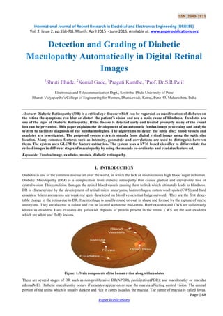
  • 1. ISSN 2349-7815 International Journal of Recent Research in Electrical and Electronics Engineering (IJRREEE) Vol. 2, Issue 2, pp: (68-71), Month: April 2015 - June 2015, Available at: www.paperpublications.org Page | 68 Paper Publications Detection and Grading of Diabetic Maculopathy Automatically in Digital Retinal Images 1 Shruti Bhude, 2 Komal Gade, 3 Pragati Kamthe, 4 Prof. Dr.S.R.Patil Electronics and Telecommunication Dept., Savitribai Phule University of Pune Bharati Vidyapeeths’s College of Engineering for Women, Dhankawadi, Katraj, Pune-43, Maharashtra, India Abstract: Diabetic Retinopathy (DR) is a critical eye disease which can be regarded as manifestation of diabetes on the retina the symptoms can blur or distort the patient’s vision and are a main cause of blindness. Exudates are one of the signs of Diabetic Retinopathy. If the disease is detected early and treated promptly many of the visual loss can be prevented. This paper explains the development of an automatic fundus image processing and analytic system to facilitate diagnosis of the ophthalmologists. The algorithms to detect the optic disc; blood vessels and exudates are investigated. The proposed system extracts macula from digital retinal image using the optic disc location. Many common features such as intensity, geometric and correlations are used to distinguish between them. The system uses GLCM for feature extraction. The system uses a SVM based classifier to differentiate the retinal images in different stages of maculopathy by using the macula co-ordinates and exudates feature set. Keywords: Fundus image, exudates, macula, diabetic retinopathy. I. INTRODUCTION Diabetes is one of the common disease all over the world, in which the lack of insulin causes high blood sugar in human. Diabetic Maculopathy (DM) is a complication from diabetic retinopathy that causes gradual and irreversible loss of central vision. This condition damages the retinal blood vessels causing them to leak which ultimately leads to blindness. DR is characterized by the development of retinal micro aneurysms, haemorrhages, cotton wool spots (CWS) and hard exudates. Micro aneurysms are weak red spots developed on blood vessels that bulge outward. They are the first detec- table change in the retina due to DR. Haemorrhage is usually round or oval in shape and formed by the rapture of micro aneurysms. They are also red in colour and can be located within the mid-retina. Hard exudates and CWS are collectively known as exudates. Hard exudates are yellowish deposits of protein present in the retina. CWS are the soft exudates which are white and fluffy lesions. Figure: 1. Main components of the human retina along with exudates There are several stages of DR such as non-proliferative DR(NPDR), proliferative(PDR), and maculopathy or macular edema(ME). Diabetic maculopathy occurs if exudates appear on or near the macula affecting central vision. The central portion of the retina which is usually darkest and rich in cones is called the macula. The centre of macula is called fovea.
  • 2. ISSN 2349-7815 International Journal of Recent Research in Electrical and Electronics Engineering (IJRREEE) Vol. 2, Issue 2, pp: (68-71), Month: April 2015 - June 2015, Available at: www.paperpublications.org Page | 69 Paper Publications The importance of detecting the macula is that it is used for early detection of various diseases. Figure 1 shows the main component of human retina and also the exudates present on the surface of the retina. II. PROPOSED METHODOLOGY Automated screening of human retina and d-etection of early signs can save patient’s vision so it is important to develop CAD systems for retinal diseases. This paper proposes a complete system for grading of maculopathy to save sudden vision loss. The proposed system consists of preprocessing, macula and exudates detection, feature extraction and finally grading of maculopathy using a SVM based classifier. The algorithm improves the quality of automated system by eliminating blood vessels and optic disc pixels to ensure the reduction in false positives in detailed feature set for accurate detection of exudates. Figure 2 shows the complete flow diagram of our system starting from input retinal image to its final grading. Figure: 2. Flow diagram of proposed system Pre-processing: The acquired retinal image may contains extra background pixels which are not required for further processing and add more time in over all processing. The purpose of pre-processing is to remove the background by differentiating between background and foreground pixels. Once the back-ground is detected, the smallest window containing foreground pixels
  • 3. ISSN 2349-7815 International Journal of Recent Research in Electrical and Electronics Engineering (IJRREEE) Vol. 2, Issue 2, pp: (68-71), Month: April 2015 - June 2015, Available at: www.paperpublications.org Page | 70 Paper Publications is retain by eliminating all other unnecessary pixels. Pre-processing consists RGB to Gray conversion, contrast enhancement, histogram equalisation. Exudate's Detection: Exudates are the bright lesions which appear on the surface of the retina if the leaking blood contains fats and proteins along with water.The presence of optic disc(OD) makes it difficult for an automated system to detect theexudates with high accuracy.The proposed system detects and removes OD pixels for accurate detection of exudates. The following steps are used for the exudates detection. 1. Take pre-processed image as an input and apply morphological closing to remove the effect of blood vessel and dark lesions. 2. Apply adaptive contrast enhancement technique to improve the contrast of exudates on retinal surface. 3. Optic disc detection AND Macula detection Macula detection is an important module for developing the computerized system for grading of diabetic maculopathy. It is the macular area of the eye that is affected in diabetic maculopathy upsetting the central vision of the eye and in severe cases leading to blindness.In the macula detection technique,the macula is first localized with the help of localized optic disk and enhanced blood vessels.Finally,the macula is detected by taking the distance from the centre of optic disk along with enhanced blood vessels image to locate the darkest pixel in this reason and making clusters of those pixels. The largest cluster formed is the macula.The outputs of the different modules that is pre-processing, exudates and macula detection is shown in GUI. Feature Extraction: The exudates region (i.e. the region of interest) detection phase extracts as many possible regions as it can for possible exudates. Feature extraction is the procedure of data reduction to find a subset of helpful variables based on the image.In this work, seven textural features based on the gray level co-occurence matrix (GLCM) are extracted from each image. The seven Haralick texture descriptors are extracted from each co-occurrence matrices which are computed in each of four angles. • Angular Second Moment (ASM) / Energy. • Contrast. • Inverse Difference Moment (IDM) / Homogenety. • Dissimilarity. • Entropy • Maximum Probability • Inverse SVM Classifier: Support vector machine are supervised learning model with associative learning algorithm that analyses data and recognize pattern, used for classification. This will classify images (0-25%) as healthy,(25-50%) as non-CSME and CSME in two parts (50-75%) and (75-100%) . In order to classify ROI as exudate and non-exudate region, we use a SVM classifier using Gabor filter. Gabor filter extract III. RESULT The evaluation of automated diagnostic system is very important and should be done carefully. Hence we have used image database from a reputed eye institute, through which evaluation and validation of the proposed system is been done successfully .This system is able to provide about 87.33 % accurate results when tested with 100 such input fundus images.
  • 4. ISSN 2349-7815 International Journal of Recent Research in Electrical and Electronics Engineering (IJRREEE) Vol. 2, Issue 2, pp: (68-71), Month: April 2015 - June 2015, Available at: www.paperpublications.org Page | 71 Paper Publications IV. CONCLUSION Diabetic maculopathy is an advance level of retinal abnormalities which may be present in diabetes sufferers.This may cause total blindness if not detected and treated in time.We presented a computerised diagnostic system for automated screening of diabetic maculopathy and providing a expert advice. The proposed system performed retinal image analysis for grading of maculopathy using SVM based classifier. Our proposed system consists of pre-processing, exudates region detection followed by macula detection. This system is a unique software solution where in a doctor does not need to be opthalmist,but still can give a proper advise with this support system. REFERENCES [1] Junichiro Hayashi, Takamitsu Kunieda, Joshua Cole, Ryusuke Soga, Yuji Hatanaka, Miao Lu, Takeshi Hara and Hiroshi Fujita: A development of computer-aided diagnosis systemusing fundus images. Proceeding of the 7th International Conference on Virtual Systems and Multimedia (VSMM 2001), pp. 429-438 (2001). [2] Iqbal, M.I (771207-8638) Aibinu, A.M (730109-P554) Gubbal, N.S (820727-P639) Khan, A(801029-P212) AUTOMATIC DIAGNOSIS OF DIABETIC MACULOPATHY USING FUNDUS IMAGES [3] P. Mohanaiah, P. Sathyanarayana, L. GuruKumar Image Texture Feature Extraction Using GLCM Approach International Journal of Scientific and Research Publications, Volume 3, Issue 5, May 2013 1 ISSN 2250-3153 [4] Angel Suero, Diego Marin, Manuel E. Gegundez-Arias, and Jose M. Bravo Locating the Optic Disc [5] in Retinal Images Using Morphological Techniques IWBBIO 2013. Proceedings Granada, 18- 20 pp no.593-600 March, 2013 [6] Y. Kanagasingama, A. Bhuiyan, M.D. Abràmoff, R.T. Smith , L. Goldschmidt , T. Y. Wong., “ Progress on retinal image analysis for age related macular Degeneration”.,ELSEVIER . Progress in Retinal and Eye Research 38,pp. 20- 42, 2014. [7] A.Sopharaka, B.Uyyanonvaraa, S.Barmanb, T.H. Williamson., “Automatic detection of diabetic retinopathy exudates from non-dilated retinal images using mathematical morphology methods”., ELSEVIER. Computerized Medical Imaging and Graphics 32, pp.720-727, 2008. [8] R.J. Qureshi , L.Kovacs , B.Harangi , B.Nagy , T.Peto , A.Hajdu “ Combining algorithms for automatic detection of optic disc and macula in fundus images ” , ELSEVIER. Computer Vision and Image Understanding 116. pp. 138– 145, 2012. [9] N. S. Datta, R. Banerjee, H. S. Dutta, S. Mukhopadhyay. , “Hardware based analysis on automated early detection of Diabetic-Retinopathy ” . ELSEVIER .Procedia Technology 4, pp. 256 – 260, 2012. [10] P.Chowriappa , S.Duaa, U. R. Acharya , M. Muthu Rama Krishnan. “Ensemble selection for feature-based classification of diabetic maculopathy images”., ELSEVIER Computers in Biology and Medicine 43., pp. 2156– 2162, 2013. [11] Neijer M, Ginneken BV, Russell SR, Suttorp- Schuleten MS, Abrmoff MD: Automated detection and differentiation of drusen, exudates and cotton-wool spots in digital color fundus phoyographs for diabetic retinopathy diagnosis. Invest Ophthalmology Vis Sci48(5): pp.2260-2267,2007. [12] Akram MU, Khan MU: Automated detection of dark and bright lesions in retinal images for early detection of diabetic retinopathy. J Medical Systems 36(5):pp.3151-3162, 2012. [13] Alireza O, Shadgar B, Markham R: Computational-intelligence based approach for detection of exudates in diabetic retinopathy images.IEEE Transactions on Information Technology in Biomedicine13 (4):pp.535-545, 2009. [14] Lim ST,Zaki WMBW, Hussain A, Lim SL, Kusalavan S: Automatic classification of diabetic macular edema in digital fundus images.2011 IEEE Colloquium on Humanities, Science and Engineering(CHUSER),pp.265-269,2011. [15] Tariq A,Akram MU: An automated system for colored retinal image background and noise Segmentation. IEEE Symposium on Industrial Electronics and Applications (ISIEA 2010), Pp.405-409, 2010.