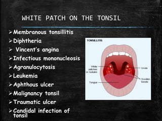
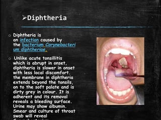
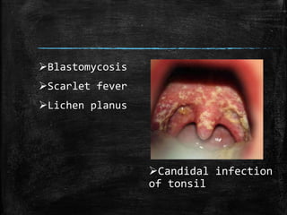
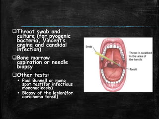
WHITE PATCH ON THE TONSIL
– DIFFERENTIAL DIAGNOSIS
ANATOMY OF THE PALATINE
TONSILS
Occupies the
tonsillar sinus or
fossa between the
palatoglossal and the
palatopharyngeal
arches.
Two surfaces:
medial and lateral
Two borders:
anterior and
posterior
Two poles: upper
and lower
Anatomy contd....
Anterior border: Palatoglossal arch
Posterior border: Palatolpharyngeal arch
Plica triangularis: Vestigial fold of mucous
membrane covering the anteroinferior part
of the tonsil.
Plica semilunaris: semilunar fold that may
cross the upper part of the tonsil.
Intratonsillar cleft: Largest crypt of the
tonsil.
Arterial Supply of Tonsil
Histology
Oral aspect is
covered by
stratified non
keratinized
epithelium which
dips into the
underlying tissue
to form crypts.
The lymphocytes
lie on the sides of
the crypt in the
form of nodules.
Development
Functions of the Tonsil
▪ Act as sentinels to
guard against foreign
intruders.
▪ Two mechanisms:
1. Providing local
immunity.
2. Providing a
surveillance
mechanism so that
the entire body is
prepared for
WHITE PATCH ON THE TONSIL
Membranous tonsillitis
Diphtheria
 Vincent’s angina
Infectious mononucleosis
Agranulocytosis
Leukemia
Aphthous ulcer
Malignancy tonsil
Traumatic ulcer
Candidal infection of
tonsil
Membranous tonsillitis
o Acute membranous
tonsillitis. It is a stage
ahead of acute
follicular tonsillitis when
exudation from the
crypts coalesces to
form a membrane on
the surface
of tonsil.
o It occurs due to
pyogenic organisms. An
exudative membrane
forms over the medial
surface of the tonsils,
along with the features
of acute tonsillitis, like
red and swollen tonsils
with marked hyperaemia
of the pillars, uvula and
soft palate.
Diphtheria
o Diphtheria is
an infection caused by
the bacterium Corynebacteri
um diphtheriae.
o Unlike acute tonsillitis
which is abrupt in onset,
diphtheria is slower in onset
with less local discomfort.
the membrane in diphtheria
extends beyond the tonsils,
on to the soft palate and is
dirty grey in colour. It is
adherent and its removal
reveals a bleeding surface.
Urine may show albumin.
Smear and culture of throat
swab will reveal
Vincent’s Angina
o Vincent's Angina is an acute
necrotizing infection of the
pharynx caused by a
combination of fusiform bacilli
(Fusiformis fusiformis - a Gram
-ve bacillus) and spirochetes
(Borrelia vincentii )
o It is insidious in onset with less
fever and less discomfort in
throat. Membrane, which
usually forms over one tonsil,
can be asily removed, revealing
an irregular ulcer on the tonsil.
Throat swab will show both the
organisms typical of the
disease, namely fusiform bacilli
and spirochaetes.
Infectious mononucleosis
o Infectious
mononucleosis (IM), also
known as glandular
fever, is an infection
commonly caused by
the Epstein–Barr
virus (EBV)
o This often attacks young
adults. Both tonsils are
very much enlarged,
congested and covered
with membrane. Local
discomfort is marked.
Lymph nodes are enlarged
in the posterior triangle
of the neck along with
splenomegaly. Blood
smear may show more
than 50% lymphocytes,
of which about 10% are
Agranulocytosis
o Agranulocytosis, also known as agranulosis
or granulopenia, is an acute condition
involving a severe and dangerous
leukopenia (lowered white blood cell
count), most commonly of neutrophils
causing a neutropenia in the circulating
blood. It is a severe lack of one major
class of infection-fighting white blood
cells.
o It presents with ulcerative necrotic
lesions not only on the tonsils but
elsewhere in the oropharynx. Patient is
severely ill.
Leukaemia
o Leukemia, s a group of cancers that usually begin
in the bone marrow and result in high numbers of
abnormal white blood cells
o Because leukemia prevents the immune system
from working normally, some patients experience
frequent infection, ranging from
infected tonsils, sores in the mouth,
or diarrhea to life-
threatening pneumonia or opportunistic infections.
o In children, 75% of the leukaemias are acute
lymphoblastic and 25% acute myelogenous or
chronic, while in adults, only 20 % of the
leukaemias are lymphocytic and the remaining 80%
non-lymphocytic. Peripheral blood shows
TLC>100,000 per cu. mm. It may be normal or
less than normal. Anaemia is always present and
may be progressive. Blast cells are seen on
examination of the bone marrow.
Aphthous ulcers
o Aphthous ulcers are
typically recurrent round
or oval sores or ulcers
inside the mouth on areas
where the skin is not
tightly bound to the
underlying bone, such as
on the inside of the lips
and cheeks or underneath
the tongue.
o Sometimes, it is solitary
and may involve the
tonsils and pillars. It may
be small or quite large
and may be alarming.
Malignancy tonsil
o The tonsil is the most common
site of squamous cell carcinoma
in the oropharynx.
o Main risk factors of developing
carcinoma tonsil include
tobacco smoking and regular
intake of high amount of
alcohol. It has also been
linked to a virus called Human
Papilloma Virus (HPV type
HPV16).[
o Persistent sore throat,
difficulty in swallowing, pain in
the ear or lump in the neck
are the presenting symptoms.
Palpation of tonsillar area is
done to determine the extent
Traumatic ulcer
o Traumatic injuries
involving the oral
cavity may typically
lead to the formation
of surface ulcerations.
The injuries may
result from events
such as accidentally
biting oneself while
talking, sleeping, or
secondary to
mastication. Other
forms of mechanical
trauma, as well
as chemical, electrical
, or thermal insults,
may also be involved.
o Membrane appears
Candidal infection
of tonsil
Blastomycosis
Scarlet fever
Lichen planus
Diagnosis of ulcero-membranous
lesion of throat requires:
History
Physical
examination
Total and
differential counts
(for
agranulocytosis,
leukemia and
infectious
mononucleosis)
Blood smear (for
atypical cells)
Throat swab and
culture (for pyogenic
bacteria, Vincent’s
angina and candidal
infection)
Bone marrow
aspiration or needle
biopsy
Other tests:
 Paul Bunnell or mono
spot test(for infectious
mononucleosis)
 Biopsy of the lesion(for
carcinoma tonsil).
White patch on the tonsil – differential diagnosis

White patch on the tonsil – differential diagnosis

  • 1.
    WHITE PATCH ONTHE TONSIL – DIFFERENTIAL DIAGNOSIS
  • 4.
    ANATOMY OF THEPALATINE TONSILS Occupies the tonsillar sinus or fossa between the palatoglossal and the palatopharyngeal arches. Two surfaces: medial and lateral Two borders: anterior and posterior Two poles: upper and lower
  • 5.
    Anatomy contd.... Anterior border:Palatoglossal arch Posterior border: Palatolpharyngeal arch Plica triangularis: Vestigial fold of mucous membrane covering the anteroinferior part of the tonsil. Plica semilunaris: semilunar fold that may cross the upper part of the tonsil. Intratonsillar cleft: Largest crypt of the tonsil.
  • 7.
  • 8.
    Histology Oral aspect is coveredby stratified non keratinized epithelium which dips into the underlying tissue to form crypts. The lymphocytes lie on the sides of the crypt in the form of nodules.
  • 9.
  • 10.
    Functions of theTonsil ▪ Act as sentinels to guard against foreign intruders. ▪ Two mechanisms: 1. Providing local immunity. 2. Providing a surveillance mechanism so that the entire body is prepared for
  • 12.
    WHITE PATCH ONTHE TONSIL Membranous tonsillitis Diphtheria  Vincent’s angina Infectious mononucleosis Agranulocytosis Leukemia Aphthous ulcer Malignancy tonsil Traumatic ulcer Candidal infection of tonsil
  • 13.
    Membranous tonsillitis o Acutemembranous tonsillitis. It is a stage ahead of acute follicular tonsillitis when exudation from the crypts coalesces to form a membrane on the surface of tonsil. o It occurs due to pyogenic organisms. An exudative membrane forms over the medial surface of the tonsils, along with the features of acute tonsillitis, like red and swollen tonsils with marked hyperaemia of the pillars, uvula and soft palate.
  • 14.
    Diphtheria o Diphtheria is aninfection caused by the bacterium Corynebacteri um diphtheriae. o Unlike acute tonsillitis which is abrupt in onset, diphtheria is slower in onset with less local discomfort. the membrane in diphtheria extends beyond the tonsils, on to the soft palate and is dirty grey in colour. It is adherent and its removal reveals a bleeding surface. Urine may show albumin. Smear and culture of throat swab will reveal
  • 15.
    Vincent’s Angina o Vincent'sAngina is an acute necrotizing infection of the pharynx caused by a combination of fusiform bacilli (Fusiformis fusiformis - a Gram -ve bacillus) and spirochetes (Borrelia vincentii ) o It is insidious in onset with less fever and less discomfort in throat. Membrane, which usually forms over one tonsil, can be asily removed, revealing an irregular ulcer on the tonsil. Throat swab will show both the organisms typical of the disease, namely fusiform bacilli and spirochaetes.
  • 16.
    Infectious mononucleosis o Infectious mononucleosis(IM), also known as glandular fever, is an infection commonly caused by the Epstein–Barr virus (EBV) o This often attacks young adults. Both tonsils are very much enlarged, congested and covered with membrane. Local discomfort is marked. Lymph nodes are enlarged in the posterior triangle of the neck along with splenomegaly. Blood smear may show more than 50% lymphocytes, of which about 10% are
  • 17.
    Agranulocytosis o Agranulocytosis, alsoknown as agranulosis or granulopenia, is an acute condition involving a severe and dangerous leukopenia (lowered white blood cell count), most commonly of neutrophils causing a neutropenia in the circulating blood. It is a severe lack of one major class of infection-fighting white blood cells. o It presents with ulcerative necrotic lesions not only on the tonsils but elsewhere in the oropharynx. Patient is severely ill.
  • 18.
    Leukaemia o Leukemia, sa group of cancers that usually begin in the bone marrow and result in high numbers of abnormal white blood cells o Because leukemia prevents the immune system from working normally, some patients experience frequent infection, ranging from infected tonsils, sores in the mouth, or diarrhea to life- threatening pneumonia or opportunistic infections. o In children, 75% of the leukaemias are acute lymphoblastic and 25% acute myelogenous or chronic, while in adults, only 20 % of the leukaemias are lymphocytic and the remaining 80% non-lymphocytic. Peripheral blood shows TLC>100,000 per cu. mm. It may be normal or less than normal. Anaemia is always present and may be progressive. Blast cells are seen on examination of the bone marrow.
  • 19.
    Aphthous ulcers o Aphthousulcers are typically recurrent round or oval sores or ulcers inside the mouth on areas where the skin is not tightly bound to the underlying bone, such as on the inside of the lips and cheeks or underneath the tongue. o Sometimes, it is solitary and may involve the tonsils and pillars. It may be small or quite large and may be alarming.
  • 20.
    Malignancy tonsil o Thetonsil is the most common site of squamous cell carcinoma in the oropharynx. o Main risk factors of developing carcinoma tonsil include tobacco smoking and regular intake of high amount of alcohol. It has also been linked to a virus called Human Papilloma Virus (HPV type HPV16).[ o Persistent sore throat, difficulty in swallowing, pain in the ear or lump in the neck are the presenting symptoms. Palpation of tonsillar area is done to determine the extent
  • 21.
    Traumatic ulcer o Traumaticinjuries involving the oral cavity may typically lead to the formation of surface ulcerations. The injuries may result from events such as accidentally biting oneself while talking, sleeping, or secondary to mastication. Other forms of mechanical trauma, as well as chemical, electrical , or thermal insults, may also be involved. o Membrane appears
  • 22.
  • 23.
    Diagnosis of ulcero-membranous lesionof throat requires: History Physical examination Total and differential counts (for agranulocytosis, leukemia and infectious mononucleosis) Blood smear (for atypical cells)
  • 24.
    Throat swab and culture(for pyogenic bacteria, Vincent’s angina and candidal infection) Bone marrow aspiration or needle biopsy Other tests:  Paul Bunnell or mono spot test(for infectious mononucleosis)  Biopsy of the lesion(for carcinoma tonsil).