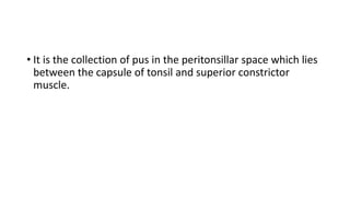
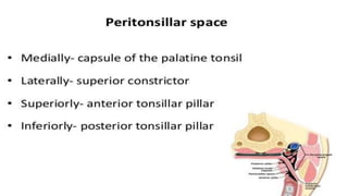
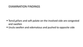
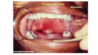
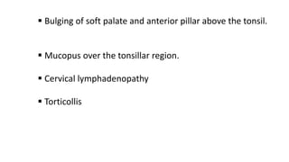
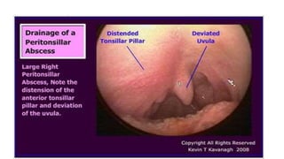
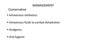
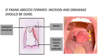
PERITONSILLAR ABSCESS
(QUINSY)
• It is the collection of pus in the peritonsillar space which lies
between the capsule of tonsil and superior constrictor
muscle.
• Peritonsillar abscess usually follows acute tonsillitis though it may
arise de novo without previous history of sore throats .
• First, one of the tonsillar crypts , usually the crypta magna get
infected
AETIOLOGY
• It forms an intratonsillar abscess which then bursts through the
tonsillar capsule to set up peritonsillitis and then an abscess.
• Strept. pyogenes, Staph- aureus or anaerobic organism
CLINICAL FEATURES
• Usually adults are infected.
• Usually unilateral
• GENERAL FEATURES :
- Fever
- Body aches
- Headache
- nausea
• LOCAL FEATURES
-throat pain
-odynophagia
-muffled and thick speech- hot potato voice
-foul breath
-ipsilateral earache
-trismus
EXAMINATION FINDINGS
 Tonsil,pillars and soft palate on the involved side are congested
and swollen
 Uvula swollen and edematous and pushed to opposite side
 Bulging of soft palate and anterior pillar above the tonsil.
 Mucopus over the tonsillar region.
 Cervical lymphadenopathy
 Torticollis
INVESTIGATIONS
Contrast enhanced CT or MRI- shows abscess and its
contents.
MANAGEMENT
Conservative
 Intravenous antibiotics.
 Intravenous fluids to combat dehydration
 Analgesics
 Oral hygiene
INCISION AND DRAINAGE
• Done at the point of maximum bulge above the upper pole of tonsil or just
lateral to the point of junction of anterior pillar with a line drawn through
the base of uvula .
• With the help of a guarded knife(quinsy forceps), a small stab incision is
made and then a sinus forceps inserted to open the abscess.
IF FRANK ABSCESS FORMED- INCISION AND DRAINAGE
SHOULD BE DONE.
• INTERVAL TONSILLECTOMY :
Tonsils are removed four to six weeks following an attack
of quinsy
COMPLICATIONS
• Parapaharyngeal abscess
• Odema of larynx
• Septicemia
• Pneumonitis or lung abscess
• Jugular vein thrombosis
• Spontaneous haemorrhage from carotid artery or jugular vein.
MEMBRANE OVER TONSIL
DIFFERENTIAL DIAGNOSIS
1. Membranous tonsillitis
2. Diphtheria
3. Vincents angina
4. Infectious mononucleosis
5. Agranulocytosis
6. Leukemia
7. Aphthous uclers
8. Malignancy
9. Traumatic ulcer
DIAGNOSIS OF MEMBRANE OVER TONSIL
REQUIRES:
1. History
2. Physical examination
3. Total and differential counts (for agranulocytosis, leukemia,
neutropenia, infectious mononucleosis)
4. Blood smear
5. Throat swab and culture (for pyogenic bacteria, Vincent angina,
diphtheria and Candida infection)
MEMBRANOUS TONSILLITIS
• Stage ahead of follicular tonsillitis
• Occurs due to pyogenic organisms
• Forms an exudative membrane over the tonsil along with features of
acute tonsillitis
DIPHTHERIA
• Causative organism: Corynebacterium diphtheria
• Children more commonly affected but no age group is immune
• Oropharynx is commonly involved, but larynx and nasal cavity may
also be affected
• Greyish white membrane forms over the tonsils and spreads to soft
palate and posterior pharyngeal wall
• Cervical lymph nodes, particularly jugulodigastric, become enlarged
and tender, presenting with “bull-neck” appearance.
• Patient is ill and toxaemic.
VINCENT’S ANGINA
• Acute necrotizing infection of pharynx caused by combination of
fusiform bacilli
• Membrane usually forms over one tonsil and can be removed easily
revealing irregular ulcer over the tonsil.
INFECTIOUS MONONUCLEOSIS
• Also known as glandular fever
• Causative organism: Epstein-Barr virus(EBV)
• Often affects young adults
• Both tonsils are very much enlarged, congested and covered with
membrane
• Lymph nodes are enlarged in the posterior triangle along with
splenomegaly
• Blood smear shows 50% lymphocytes of which about 10% are atypical
• Paul-Bunnel test will show high titre of heterophil antibody
AGRANULOCYTOSIS
• Presents with ulcerative necrotic lesion not only on the tonsil but
elsewhere in the oropharynx
• Patient is severely ill
• Total leucocytic count is decreased to <2000/cu mm
LEUKEMIA
• In children, 75% of leukemias are acute lymphoblastic and 25% are
acute myelogenous or chronic
• In adults 20% of acute leukemias are lymphoblastic and 80% non-
lymphoblastic
• Peripheral blood shows TLC >100,000/cu mm
APHTHOUS ULCER
• May involve any part of oral cavity or oropharynx
• Sometimes, it is solitary and may involve tonsil and pillars
MALIGNANCY OF TONSIL
• Tonsil is the most common site of squamous cell carcinoma in the
oropharynx
• Risk factor include tobacco smoking and regular intake of high
amount of alcohol
• Also has been linked to Human Papilloma Virus (HPV type 16)
• Persistent sore throat, pain in the ear or lump in the neck are
presenting symptoms
TRAUMATIC ULCER
• Any injury to oropharynx heals by formation of a membrane
• Trauma to tonsil area may occur accidently when hit with a
toothbrush or finger in throat
• Membrane appears within 24h
LUDWIG’S ANGINA
1.Rapidlyprogressingcellulitisof submandibular space (i.e. sublingual&
submaxillaryspace)
2.Mixedflora (poly-microbial)
3.Mayresultintolife-threateningairway obstruction
• Subdivisions of submandibular space
1. SUBLINGUALSPACE:above mylohyoid muscle
2. SUBMAXILLARYSPACE:below mylohyoid muscle
• CONTENTS : - Submandibular salivary gland,
• -Lymph nodes
ETIOLOGY
1.Dental infection: 80% cases
• Tooth (lower molars & premolars)
•Roots of premolars lie above mylohyoid sublingual space
infection
• Roots of molars lie below mylohyoid submaxillary space
infection
2.Injury to floor of mouth
3.Submandibular sialadenitis
CAUSATIVE AGENTS
• Mixed aerobic &anaerobic infection
1. Streptococcus pyogenes
2. Streptococcus viridans
3. Streptococcus pneumoniae
4. Staphylococcus
5. Fusobacterium
6. Bacteroides
7. Peptostreptococcus
CLINICAL FEATURES
- Toothache, fever, odynophagia, drooling of saliva
- Floor of mouth swelling + tongue elevation
- Submental swelling: Brawny induration
- Trismus
- Stridor: falling back of tongue causing upper airway obstruction
- Initially cellulitis (no frank pus) - pus formation (only at
late stage)
CLINICAL FEATURES
- Parapharyngeal abscess
- Retropharyngeal abscess
- Acute airway obstruction (within hours):
• due to falling back of tongue
- Aspiration pneumonia
- Septicemia
- Death
• MANAGEMENT:
1.I.V. antibiotics: Ceftriaxone +Metronidazole / Clindamycin
2.IV fluid for adequate hydration
3.Monitor vital signs regularly eg. assessment for disease
progression & airway compromise
4.Airway obstruction: Intubation / tracheostomy
5.Incision & drainage
Transverse incision from one angle of mandible to opposite angle
of mandible

PERITONSILLAR ABSCESS.pptx ug ppt slideshow

  • 1.
  • 2.
    • It isthe collection of pus in the peritonsillar space which lies between the capsule of tonsil and superior constrictor muscle.
  • 6.
    • Peritonsillar abscessusually follows acute tonsillitis though it may arise de novo without previous history of sore throats . • First, one of the tonsillar crypts , usually the crypta magna get infected AETIOLOGY
  • 7.
    • It formsan intratonsillar abscess which then bursts through the tonsillar capsule to set up peritonsillitis and then an abscess. • Strept. pyogenes, Staph- aureus or anaerobic organism
  • 8.
    CLINICAL FEATURES • Usuallyadults are infected. • Usually unilateral • GENERAL FEATURES : - Fever - Body aches - Headache - nausea
  • 9.
    • LOCAL FEATURES -throatpain -odynophagia -muffled and thick speech- hot potato voice -foul breath -ipsilateral earache -trismus
  • 10.
    EXAMINATION FINDINGS  Tonsil,pillarsand soft palate on the involved side are congested and swollen  Uvula swollen and edematous and pushed to opposite side
  • 12.
     Bulging ofsoft palate and anterior pillar above the tonsil.  Mucopus over the tonsillar region.  Cervical lymphadenopathy  Torticollis
  • 14.
    INVESTIGATIONS Contrast enhanced CTor MRI- shows abscess and its contents.
  • 15.
    MANAGEMENT Conservative  Intravenous antibiotics. Intravenous fluids to combat dehydration  Analgesics  Oral hygiene
  • 16.
    INCISION AND DRAINAGE •Done at the point of maximum bulge above the upper pole of tonsil or just lateral to the point of junction of anterior pillar with a line drawn through the base of uvula . • With the help of a guarded knife(quinsy forceps), a small stab incision is made and then a sinus forceps inserted to open the abscess.
  • 17.
    IF FRANK ABSCESSFORMED- INCISION AND DRAINAGE SHOULD BE DONE.
  • 18.
    • INTERVAL TONSILLECTOMY: Tonsils are removed four to six weeks following an attack of quinsy
  • 19.
    COMPLICATIONS • Parapaharyngeal abscess •Odema of larynx • Septicemia • Pneumonitis or lung abscess • Jugular vein thrombosis • Spontaneous haemorrhage from carotid artery or jugular vein.
  • 20.
  • 21.
    DIFFERENTIAL DIAGNOSIS 1. Membranoustonsillitis 2. Diphtheria 3. Vincents angina 4. Infectious mononucleosis 5. Agranulocytosis 6. Leukemia 7. Aphthous uclers 8. Malignancy 9. Traumatic ulcer
  • 22.
    DIAGNOSIS OF MEMBRANEOVER TONSIL REQUIRES: 1. History 2. Physical examination 3. Total and differential counts (for agranulocytosis, leukemia, neutropenia, infectious mononucleosis) 4. Blood smear 5. Throat swab and culture (for pyogenic bacteria, Vincent angina, diphtheria and Candida infection)
  • 23.
    MEMBRANOUS TONSILLITIS • Stageahead of follicular tonsillitis • Occurs due to pyogenic organisms • Forms an exudative membrane over the tonsil along with features of acute tonsillitis
  • 24.
    DIPHTHERIA • Causative organism:Corynebacterium diphtheria • Children more commonly affected but no age group is immune • Oropharynx is commonly involved, but larynx and nasal cavity may also be affected • Greyish white membrane forms over the tonsils and spreads to soft palate and posterior pharyngeal wall
  • 25.
    • Cervical lymphnodes, particularly jugulodigastric, become enlarged and tender, presenting with “bull-neck” appearance. • Patient is ill and toxaemic.
  • 26.
    VINCENT’S ANGINA • Acutenecrotizing infection of pharynx caused by combination of fusiform bacilli • Membrane usually forms over one tonsil and can be removed easily revealing irregular ulcer over the tonsil.
  • 27.
    INFECTIOUS MONONUCLEOSIS • Alsoknown as glandular fever • Causative organism: Epstein-Barr virus(EBV) • Often affects young adults • Both tonsils are very much enlarged, congested and covered with membrane • Lymph nodes are enlarged in the posterior triangle along with splenomegaly • Blood smear shows 50% lymphocytes of which about 10% are atypical • Paul-Bunnel test will show high titre of heterophil antibody
  • 29.
    AGRANULOCYTOSIS • Presents withulcerative necrotic lesion not only on the tonsil but elsewhere in the oropharynx • Patient is severely ill • Total leucocytic count is decreased to <2000/cu mm
  • 30.
    LEUKEMIA • In children,75% of leukemias are acute lymphoblastic and 25% are acute myelogenous or chronic • In adults 20% of acute leukemias are lymphoblastic and 80% non- lymphoblastic • Peripheral blood shows TLC >100,000/cu mm
  • 31.
    APHTHOUS ULCER • Mayinvolve any part of oral cavity or oropharynx • Sometimes, it is solitary and may involve tonsil and pillars
  • 32.
    MALIGNANCY OF TONSIL •Tonsil is the most common site of squamous cell carcinoma in the oropharynx • Risk factor include tobacco smoking and regular intake of high amount of alcohol • Also has been linked to Human Papilloma Virus (HPV type 16) • Persistent sore throat, pain in the ear or lump in the neck are presenting symptoms
  • 33.
    TRAUMATIC ULCER • Anyinjury to oropharynx heals by formation of a membrane • Trauma to tonsil area may occur accidently when hit with a toothbrush or finger in throat • Membrane appears within 24h
  • 34.
  • 35.
    1.Rapidlyprogressingcellulitisof submandibular space(i.e. sublingual& submaxillaryspace) 2.Mixedflora (poly-microbial) 3.Mayresultintolife-threateningairway obstruction
  • 36.
    • Subdivisions ofsubmandibular space 1. SUBLINGUALSPACE:above mylohyoid muscle 2. SUBMAXILLARYSPACE:below mylohyoid muscle • CONTENTS : - Submandibular salivary gland, • -Lymph nodes
  • 37.
    ETIOLOGY 1.Dental infection: 80%cases • Tooth (lower molars & premolars) •Roots of premolars lie above mylohyoid sublingual space infection • Roots of molars lie below mylohyoid submaxillary space infection 2.Injury to floor of mouth 3.Submandibular sialadenitis
  • 38.
    CAUSATIVE AGENTS • Mixedaerobic &anaerobic infection 1. Streptococcus pyogenes 2. Streptococcus viridans 3. Streptococcus pneumoniae 4. Staphylococcus 5. Fusobacterium 6. Bacteroides 7. Peptostreptococcus
  • 39.
    CLINICAL FEATURES - Toothache,fever, odynophagia, drooling of saliva - Floor of mouth swelling + tongue elevation - Submental swelling: Brawny induration - Trismus - Stridor: falling back of tongue causing upper airway obstruction - Initially cellulitis (no frank pus) - pus formation (only at late stage)
  • 40.
    CLINICAL FEATURES - Parapharyngealabscess - Retropharyngeal abscess - Acute airway obstruction (within hours): • due to falling back of tongue - Aspiration pneumonia - Septicemia - Death
  • 41.
    • MANAGEMENT: 1.I.V. antibiotics:Ceftriaxone +Metronidazole / Clindamycin 2.IV fluid for adequate hydration 3.Monitor vital signs regularly eg. assessment for disease progression & airway compromise 4.Airway obstruction: Intubation / tracheostomy 5.Incision & drainage Transverse incision from one angle of mandible to opposite angle of mandible