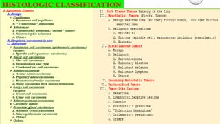
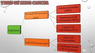
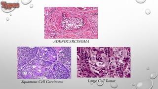
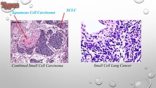
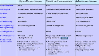
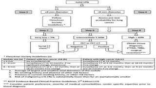
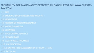
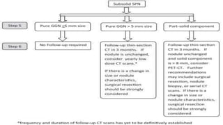
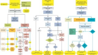
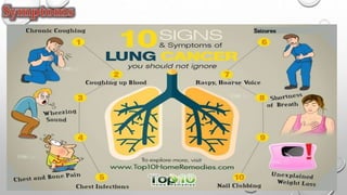
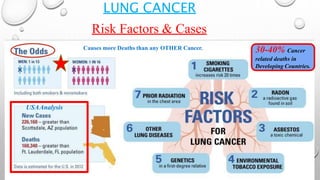
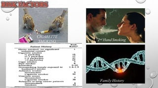
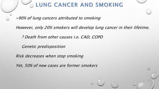
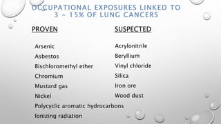
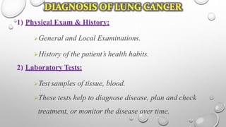
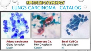
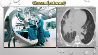
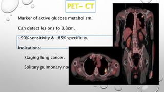
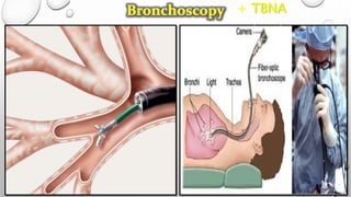
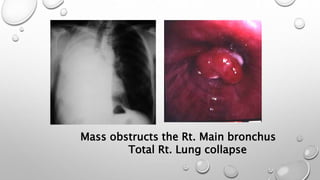
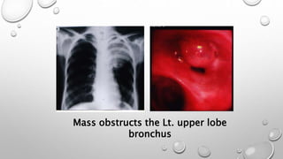
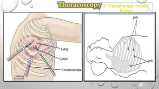
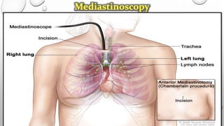
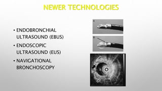
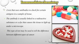
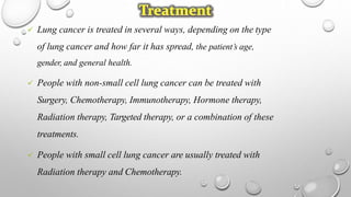
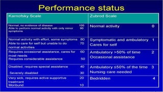
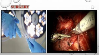
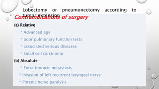
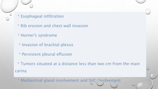
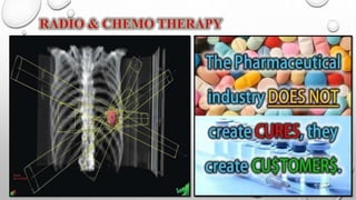
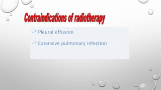
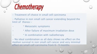
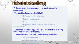
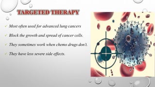
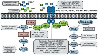
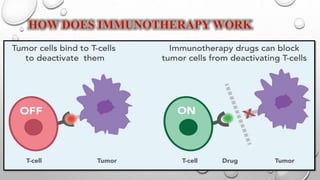
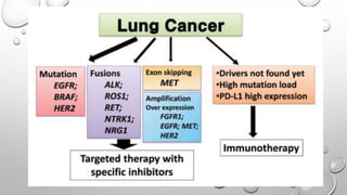
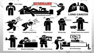
3. Abnormal growth of cells.
Tends to proliferate in an uncontrolled way.
Cancer is not one disease.
Involve in any tissue of the body.
Have many different forms in each body area.
Hippocrates discovered cancer.
4. THERE ARE MANY TYPES OF CANCER BUT SOME
MAJOR TYPES ARE…
5
7. Uncontrolled cell growth in tissues of lungs.
Also termed as Bronchogenic Carcinomas.
Begins in the lungs .
May spread to lymph nodes or other organs.
When cancer cells spread from one organ to another, called metastasis.
12. • 1- LOW DOSE SPIRAL CT CHEST: SERIAL CT FU ACCORDING TO SIZE OF NODULE
OR GGO.
• 2- DIGITAL CHEST RADIOGRAPHY: EMERGING TECHNOLOGICAL ADVANCEMENTS
IN DIGITAL CHEST RADIOGRAPHY, INCLUDING COMPUTER-AIDED DIAGNOSIS,
TEMPORAL SUBTRACTION, AND DUAL ENERGY SUBTRACTION METHODS, MAY
SIGNIFICANTLY IMPROVE THE ABILITY OF CHEST RADIOGRAPHY TO DETECT
SMALL LUNG NODULES
• 3- SPUTUM CYTOLOGY AND ADVANCED SPUTUM ANALYSIS: SENS 20-30%(C >
P)
BIOMARKER ANALYSIS BIOLOGICAL ROLE APPLICATION
HNRNP A2/B1 PROTEIN I IN SPUTUM MRNA PROCESSING EARLY DETECTION/TUM
Screenin
g
13. • 4- CONVENTIONAL, AF AND VIRTUAL BRONCHOSCOPY:
• CONVENTIONAL BRONCHOSCOPY IS A VALUABLE TECHNIQUE FOR LOCALIZING
PREINVASIVE LUNG CANCER WITHIN THE AIRWAYS. IN GENERAL, CONVENTIONAL
BRONCHOSCOPY CAN DETECT NODULAR OR POLYPOID LESIONS 2 MM IN SIZE AND FLAT
OR SUPERFICIALLY SPREADING LESIONS 2 CM IN DIAMETER . WITH REGARD TO
CARCINOMA IN SITU, 75% OF LESIONS ARE SUPERFICIAL OR FLAT AND 25% ARE NODULAR
OR POLYPOID.
• AF INVOLVES ILLUMINATING THE BRONCHIAL SURFACE WITH VIOLET OR BLUE LIGHT (400
TO 440 NM) IN ORDER TO DISTINGUISH NORMAL FROM ABNORMAL TISSUES. UPON SUCH
ILLUMINATION, DYSPLASTIC LESIONS AND CARCINOMA IN SITU WILL SHOW A DIMINUTION
IN THE INTENSITY OF AUTOFLUORESCENCE.
• VIRTUAL BRONCHOSCOPY (VB) IS A NOVEL NONINVASIVE METHOD FOR ASSESSING THE
AIRWAYS WHICH COMBINES HELICAL COMPUTED TOMOGRAPHY DATA AND VIRTUAL
REALITY COMPUTING IN ORDER TO CREATE THREE-DIMENSIONAL ENDOBRONCHIAL
SIMULATIONS, HAS RELATIVELY HIGH SENSITIVITY (90%) FOR LESIONS 5 MM IN DIAMETER,
BUT WAS LIMITED BY A POOR SPECIFICITY (CAN’T DIFFERENTIATE BENIGN FROM
16. PROBABILITY FOR MALIGNANCY DETECTED BY CALCULATOR ON: WWW.CHESTX-
RAY.COM
• 1. AGE
• 2. SMOKING (EVER VS NEVER AND PACK-Y)
• 3. HEMOPTYSIS
• 4. HISTORY OF PRIOR MALIGNANCY
• 5. NODULE DIAMETER
• 6. LOCATION
• 7. EDGE CHARACTERISTICS
• 8. GROWTH RATE
• 9. CAVITY WALL THICKNESS
• 10. CALCIFICATION
• 11. CONTRAST ENHANCEMENT ON CT SCAN . 15 HU
• 12. PET SCAN
17.
18.
19.
20.
21. COMPRESSION ON SVC: DILATED VEINS ON THE UPPER PART OF
THE CHEST & CONGESTED NON PULSATING NECK VEINS.
22. Anything that increases chances of getting a disease.
Having a risk factor doesn't mean that you will get
the disease; not having risk factors doesn't mean that
you will never get the disease.``
23. LUNG CANCER
30-40% Cancer
related deaths in
Developing Countries.
USAAnalysis
Risk Factors & Cases
Causes more Deaths than any OTHER Cancer.
25. LUNG CANCER AND SMOKING
~90% of lung cancers attributed to smoking
However, only 20% smokers will develop lung cancer in their lifetime.
? Death from other causes i.e. CAD, COPD
Genetic predisposition
Risk decreases when stop smoking
Yet, 50% of new cases are former smokers
26. OCCUPATIONAL EXPOSURES LINKED TO
3 - 15% OF LUNG CANCERS
PROVEN SUSPECTED
Acrylonitrile
Beryllium
Vinyl chloride
Silica
Iron ore
Wood dust
Arsenic
Asbestos
Bischloromethyl ether
Chromium
Mustard gas
Nickel
Polycyclic aromatic hydrocarbons
Ionizing radiation
27. ASBESTOSIS & LUNG CANCER
Prolonged heavy exposure has relative risk between 2 - 10 of causing
lung cancer.
Peak incidence 15 - 24 years after exposure.
Fiber type is important:
Crocidolite & amosite > chrysotile & anthophyllite.
Risk of smoking & asbestos exposure is multiplied.
Mortality ratio:
Nonsmoking asbestos worker: 5.17
Smoker: 10.85
Smoker & asbestos worker: 53.24
28.
29.
30. PATHOPHYSIOLOGY
DUE TO ETIOLOGICAL FACTORS
DAMAGE TO THE CELL
CARCINOGEN BIND TO DAMAGED CELL DNA
MALIGNANT TRANSFORMATON FROM NORMAL EPITHELLIUM
CELLULAR CHANGES
PASSED TO THE DAUGHTER CELL
EVENTUALLY MALIGNANT CELL
CARCINOMA
31.
32. Regional Lymph Node Classification System
Supraclavicular nodes
1. Low cervical, supraclavicular and sternal notch nodes
Superior mediastinal nodes
2. Upper Paratracheal: above the aortic arch, but below the
clavicles.
3A. Pre-vascular: nodes not adjacent to the trachea like the
nodes in station 2, but anterior to the vessels.
3P. Pre-vertebral: nodes not adjacent to the trachea, but
behind the esophagus, which is prevertebral (3P).
Inferior Mediastinal nodes
4. Lower Paratracheal (including Azygos Nodes): below
upper margin of aortic arch down to level of main bronchus.
Aortic nodes
5. Subaortic (A-P window): nodes lateral to ligamentum
arteriosum. These nodes are not located between the aorta
and the pulmonary trunk, but lateral to these vessels.
6. Para-aortic (ascending aorta or phrenic): nodes lying
anterior and lateral to the ascending aorta and the aortic
arch.
Subcarinal nodes
7. Subcarinal.
Inferior Mediastinal nodes
8. Paraesophageal (below carina).
9. Pulmonary Ligament: nodes lying within the pulmonary
33.
34. SCLC STAGING
Limited Stage (1/3)
confined to 1 hemithorax
disease fits within a tolerable radiation port
Extensive Stage (2/3)
doesn’t fit
Recommend also use TNM staging, as for NSCLC
35. 1) Physical Exam & History:
General and Local Examinations.
History of the patient’s health habits.
2) Laboratory Tests:
Test samples of tissue, blood.
These tests help to diagnose disease, plan and check
treatment, or monitor the disease over time.
45. A test that uses antibodies to check for certain
antigens in a sample of tissue.
The antibody is usually linked to a radioactive
substance or a dye that causes the tissue to light up
under a microscope.
This type of test may be used to tell the difference
between different types of cancer.
46. Lung cancer is treated in several ways, depending on the type
of lung cancer and how far it has spread, the patient’s age,
gender, and general health.
People with non-small cell lung cancer can be treated with
Surgery, Chemotherapy, Immunotherapy, Hormone therapy,
Radiation therapy, Targeted therapy, or a combination of these
treatments.
People with small cell lung cancer are usually treated with
Radiation therapy and Chemotherapy.
47.
48.
49. Contraindications of surgery
(a) Relative
* Advanced age
* poor pulmonary function tests
* associated serious diseases
* Small cell carcinoma
(b) Absolute
* Extra thoracic metastasis
* Invasion of left recurrent laryngeal nerve
* Phrenic nerve paralysis
Lobectomy or pneumonectomy according to
tumor extension
50. * Esophageal infiltration
* Rib erosion and chest wall invasion
* Horner's syndrome
* Invasion of brachial plexus
* Persistent pleural effusion
* Tumors situated at a distance less than two cm from the main
carina
* Mediastinal gland involvement and SVC involvement
51.
52. Indications
•* Operable tumours with containdication to surgery
• * Inoperable tumors within the limit of the thorax
• the tumouricidal dose is 6000 rads over a period of
six weeks directed twards:
• * Primary growth
• * Hilar LN
• * Mediastinum
• * Safety margin 2 cm arround the visualized
tumour extension
53. Indications
•* Pain due to rib erosion
•* Hemoptysis
•* SVC obstruction
•* Cough
•* Dyspnea due to bronchial obstruction
•* Pancost tumour
•* Bone metastasis
Dose: 3000 rads over a period of three weeks
56. • Treatment of choice in small cell carcinoma
• Palliative in non small cell cancer extending beyond the
limit of thorax:-
• * Metastatic symptoms
• * After failure of maximum irradiation dose
• * In combination with radiotherapy
• No ideal combination as all data shows no effect on the
median survival in non small cell cancer and very minimal
effect on small cell carcinoma from 6- 9 months
57. •* Combination chemotherapy 3-4 drugs is better than
monotherapy
•*The combined regimen should fulfill
• * Different modes of action
• * Lack of cross resistant
• * Divergent toxicity
• * Superiorly when all drugs administered
simultaneously
•* Combined chemotherapy + whole body irradiation including
cranial irradiation lead to best response
•* Chemotherapy is relatively successful in palliation of
• * Malignant effusion
• * Endocrinal manifestations
• * Prior to radiotherapy
58. Most often used for advanced lung cancers
Block the growth and spread of cancer cells.
They sometimes work when chemo drugs don’t.
They have less severe side effects.
59.
60. Recently emerged as a new treatment option for certain lung
cancers.
While any cancer treatment can cause side effects, immunotherapy is
generally well-tolerated; this is in part due to its mechanism of
action.
Immunotherapy uses our own immune system as a treatment
against cancer.
61.
62.
63. •Its role is mainly palliative for severe
dyspnea by recanalization of the
patency of the airway and control of
life threatening hemoptysis by sealing