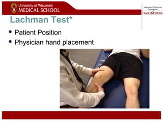
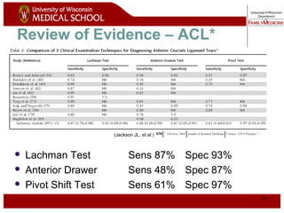
Musculoskeletal
Curriculum
History &
Physical Exam of
the Injured Knee
Copyright 2005
2
Authors
Kathleen Carr, MD
Madison Residency Program
Kathleen.Carr@fammed.wisc.edu
Dennis Breen, MD
Eau Claire Residency Program
Dennis.Breen@fammed.wisc.edu
Dan Smith, DO
3
Contributors
Marguerite Elliott, DO
Jeff Patterson, DO
Jerry Ryan, MD
4
Goal
 Learn a standardized, evidence-based history
and physical examination of patients with knee
injuries
WHICH WILL:
 Enable family medicine resident physicians to
accurately diagnose common knee problems
throughout the full age spectrum of patients
seen in family medicine
5
Competency-Based Objectives
 Patient care – focused history and exam
 Professionalism – respect, compassion
 Interpersonal and communication skills –
differential diagnosis
 Medical knowledge base – anatomy, injury
mechanisms
 Systems based practice – accuracy, time-efficiency
6
Assessing a knee injury
Components of the assessment include
 Focused history
 Attentive physical examination
 Thoughtfully ordered tests/studies
 for future discussion
Focused History
8
Focused History Questions
Onset of Pain
 Date of injury or when symptoms started
Location of pain*
 Anterior
 Medial
 Lateral
 Posterior
9
Focused History Questions2
 Mechanism of Injury -helps
predict injured structure
 Contact or noncontact injury?*
 If contact, what part of the knee was
contacted?
 Anterior blow?
 Valgus force?
 Varus force?
 Was foot of affected knee planted
on the ground?**
Valgus alignment =
distal segment
deviates lateral with
respect to proximal
segment.
Patellas Touch
http://moon.ouhsc.edu/dthompso/namics/varus.gif
10
Focused History Questions3
Injury-Associated Events*
 Pop heard or felt?
 Swelling after injury (immediate vs delayed)
 Catching / Locking
 Buckling / Instability (“giving way”)
11
Instability - Example
http://www.carletonsportsmed.com/Libraria_medicus/PF_patella_dislocat
ion.JPG
Patellar dislocation
12
Focused History Questions4
Degree of Immediate Dysfunction
|------------------------|
Unable to Antalgic Continued
Ambulate Gait* to Participate
13
Focused History Questions5
 Aggravating Factors
 Activities, changing positions, stairs, kneeling
 Relieving Factors/treatments tried
 Ice, medications, crutches
 History of previous knee injury or surgery
14
Historical Clues to Knee Injury
Diagnoses
Noncontact injury with “pop” ACL tear
Contact injury with “pop” MCL or LCL tear, meniscus
tear, fracture
Acute swelling ACL tear, PCL tear, fracture,
knee dislocation, patellar
dislocation
Lateral blow to the knee MCL tear
Medial blow to the knee LCL tear
Knee “gave out” or “buckled” ACL tear, patellar dislocation
Fall onto a flexed knee PCL tear
Physical Exam
16
Physical Exam - General
 Develop a standard routine*
 Alleviate the patient's fears
GENERAL STEPS
Inspection
Palpation
Range of motion
Strength testing
Special tests
17
Physical Exam - Exposure
 Adequate exposure - groin to
toes bilaterally
 Examine in supine position
 Compare knees
18
Observe – Static Alignment
 Patient stands facing examiner with
feet shoulder width apart
 Ankles, subtalar joints – pronation, supination
 Feet – pes planus, pes cavus
(http://www.steenwyk.com/pronsup.htm)
Pes planus Pes cavus
(http://www.arc.org.uk/about_arth/booklets/6012/images/6012_1.gif)
19
 Patient then
brings medial
aspects of knees
and ankles in
contact
 Knees – genu valgum
(I), genu varum (II)
Observe – Static Alignment
(http://www.orthoseek.com/articles/img/bowl1.gif)
Genu valgum Genu varum
20
Observe – Dynamic Alignment
Pronation/Supination may be
enhanced with ambulation
Antalgic gait indicates significant
problem (anti = against, algic = pain)
21
Inspect Knee
 Warmth
 Erythema
 Effusion*
Evidence of
local trauma
 Abrasions
 Contusions
 Lacerations
Patella position
Muscle atrophy
22
Inspect Knee-Related Muscles
 Quadriceps atrophy
 Long-standing problem
 Vastus medialis
atrophy
 After surgery
http://www.neuro.wustl.edu/neuromuscular/pics/people/patients/Hands/ibmquadatrsm.jpg
23
Normal Knee – Anterior, Extended
24
Surface Anatomy - Anterior, Extended*
Patella
Hollow
Indented
25
Normal Knee – Anterior, Flexed
26
Surface Anatomy - Anterior, Flexed
Head
Of
Fibula
Patella
Tibial
Tuberosity
27
Palpation – Anterior*
Patella:
Lateral and Medial Patellar Facets
Superior
And
Inferior
Patellar Facets
Patellar Tendon**
Lateral Fat Pad
Medial Fat
Pat
28
Surface Anatomy - Medial
Medial
Femoral
Condyle
Patella
Joint
Line
Medial
Tibial
Condyle
Tibial
Tuberosity
29
Palpation - Medial
Medial Collateral Ligament (MCL)*
Pes anserine
bursa**
Medial joint
line
30
Surface Anatomy – Lateral
Patella
Head
Of
Fibula
Tibial
Tuberosity
Quadriceps
31
Palpation – Lateral*
Lateral joint
line
Lateral Collateral
Ligament (LCL)**
32
Palpation - Posterior
 Popliteal fossa*
 Abnormal bulges
 Popliteal artery aneurysm
 Popliteal thrombophlebitis
 Baker’s cyst
33
Range Of Motion Testing
 Extension Flexion
0º 135º
 Describe loss of degrees of extension
 Example: “lacks 5 degrees of
extension”
 Locking* = patient unable to fully extend or flex
knee due to a mechanical blockage in the knee
(i.e., loose body, bucket-handle meniscus tear)
34
Strength Testing
 Test knee extensors (quadriceps) and knee
flexors (hamstrings)
 Can test both with patient in seated position,
knees bent over edge of table
 Ask patient to extend/straighten knee against your
resistance
 Then ask patient to flex/bend knee against your
resistance
 Compare to unaffected knee
35
Special Tests – Anterior Knee Pain
 Patellar apprehension test*
(http://www.sportsdoc.umn.edu/Clinical_Folder/Knee_Folder/Knee_Exam/lateral%20patellar
%20apprehension.htm)
 Patellofemoral grind test**
Starting
position
Push patella
laterally
36
Special Tests - Ligaments
 Assess stability
of 4 knee
ligaments via
applied
stresses*
Anterior Cruciate
Posterior
Cruciate
Lateral Collateral
Medial Collateral
37
Stress Testing of Ligaments
 Use a standard exam routine
 Direct, gentle pressure
 No sudden forces
 Abnormal test
1. Excessive motion = laxity
What is NORMAL motion?*
2. Soft/mushy end point**
38
Collateral Ligament Assessment
Patient and Examiner
Position*
39
Valgus Stress Test for MCL*
Note Direction Of Forces
40
Video of Valgus Stress Test
Click on
image for video
41
Varus Stress Test for LCL*
Note direction of forces
42
Video of Varus Stress Test
Click on
image for video
43
Lachman Test*
 Patient Position
 Physician hand placement
44
Lachman Test2
 View from lateral aspect*
Note direction of forces
45
Video of Lachman Test
Click on
image for video
46
Alternate Lachman Test
Click on
image for video
47
Anterior Drawer Test for ACL
 Physician Position & Movements*
 Patient Position
Note direction of forces
48
Posterior Drawer Testing- PCL*
Note direction of forces
49
Assess Meniscus – Knee Flexion
 Most sensitive test is full flexion*
 Examiner passively flexes the knee or has patient
perform a full two-legged squat to test for
meniscal injury
 Joint line tenderness**
 Flexion of the knee enhances palpation of the
anterior half of each meniscus
50
Tests that we do not
recommend routinely
 Pivot-Shift* - for ACL tear
 McMurray Test**- for meniscus tears
51
Review of Evidence – ACL*
 Lachman Test Sens 87% Spec 93%
 Anterior Drawer Sens 48% Spec 87%
 Pivot Shift Test Sens 61% Spec 97%
(Jackson JL, et al.)
52
Review of Evidence - Meniscus
 Joint Line Tenderness Sens 76% Spec 29%
 McMurray Test Sens 52% Spec 97%
(Jackson JL, et al.)
53
References
Calmbach WL, Hutchens M. Evaluation of Patients Presenting with Knee Pain: Part I.
History, Physical Examination, Radiographs, and Laboratory Tests. Am Fam
Physician 2003;68:907-12.
Ebell MH. A Tool for Evaluating Patients with Knee Injury. Family Practice Management.
March 2005:67-70.
Jackson JL, O’Malley PG, Kroenke K. Evaluation of Acute Knee Pain in Primary Care.
Ann Intern Med. 2003;139:575-588.
Malanga GA, Andrus S, Nadler SF, McLean J. Physical Examination of the Knee: A
Review of the Original Test Description and Scientific Validity of Common Orthopedic
Tests. Arch Phys Med Rehabil 2003;84:592-603.
Solomon DH, Simel DL, Bates DW, Katz JN. Does this patient have a torn meniscus or
ligament of the knee? Value of the Physical Examination. JAMA 2001;286:1610-
1620.
54
Video of Knee Exam
http://www.fammed.wisc.edu/our-department/media/musculoskeletal

Knee

  • 1.
    Musculoskeletal Curriculum History & Physical Examof the Injured Knee Copyright 2005
  • 2.
    2 Authors Kathleen Carr, MD MadisonResidency Program Kathleen.Carr@fammed.wisc.edu Dennis Breen, MD Eau Claire Residency Program Dennis.Breen@fammed.wisc.edu Dan Smith, DO
  • 3.
    3 Contributors Marguerite Elliott, DO JeffPatterson, DO Jerry Ryan, MD
  • 4.
    4 Goal  Learn astandardized, evidence-based history and physical examination of patients with knee injuries WHICH WILL:  Enable family medicine resident physicians to accurately diagnose common knee problems throughout the full age spectrum of patients seen in family medicine
  • 5.
    5 Competency-Based Objectives  Patientcare – focused history and exam  Professionalism – respect, compassion  Interpersonal and communication skills – differential diagnosis  Medical knowledge base – anatomy, injury mechanisms  Systems based practice – accuracy, time-efficiency
  • 6.
    6 Assessing a kneeinjury Components of the assessment include  Focused history  Attentive physical examination  Thoughtfully ordered tests/studies  for future discussion
  • 7.
  • 8.
    8 Focused History Questions Onsetof Pain  Date of injury or when symptoms started Location of pain*  Anterior  Medial  Lateral  Posterior
  • 9.
    9 Focused History Questions2 Mechanism of Injury -helps predict injured structure  Contact or noncontact injury?*  If contact, what part of the knee was contacted?  Anterior blow?  Valgus force?  Varus force?  Was foot of affected knee planted on the ground?** Valgus alignment = distal segment deviates lateral with respect to proximal segment. Patellas Touch http://moon.ouhsc.edu/dthompso/namics/varus.gif
  • 10.
    10 Focused History Questions3 Injury-AssociatedEvents*  Pop heard or felt?  Swelling after injury (immediate vs delayed)  Catching / Locking  Buckling / Instability (“giving way”)
  • 11.
  • 12.
    12 Focused History Questions4 Degreeof Immediate Dysfunction |------------------------| Unable to Antalgic Continued Ambulate Gait* to Participate
  • 13.
    13 Focused History Questions5 Aggravating Factors  Activities, changing positions, stairs, kneeling  Relieving Factors/treatments tried  Ice, medications, crutches  History of previous knee injury or surgery
  • 14.
    14 Historical Clues toKnee Injury Diagnoses Noncontact injury with “pop” ACL tear Contact injury with “pop” MCL or LCL tear, meniscus tear, fracture Acute swelling ACL tear, PCL tear, fracture, knee dislocation, patellar dislocation Lateral blow to the knee MCL tear Medial blow to the knee LCL tear Knee “gave out” or “buckled” ACL tear, patellar dislocation Fall onto a flexed knee PCL tear
  • 15.
  • 16.
    16 Physical Exam -General  Develop a standard routine*  Alleviate the patient's fears GENERAL STEPS Inspection Palpation Range of motion Strength testing Special tests
  • 17.
    17 Physical Exam -Exposure  Adequate exposure - groin to toes bilaterally  Examine in supine position  Compare knees
  • 18.
    18 Observe – StaticAlignment  Patient stands facing examiner with feet shoulder width apart  Ankles, subtalar joints – pronation, supination  Feet – pes planus, pes cavus (http://www.steenwyk.com/pronsup.htm) Pes planus Pes cavus (http://www.arc.org.uk/about_arth/booklets/6012/images/6012_1.gif)
  • 19.
    19  Patient then bringsmedial aspects of knees and ankles in contact  Knees – genu valgum (I), genu varum (II) Observe – Static Alignment (http://www.orthoseek.com/articles/img/bowl1.gif) Genu valgum Genu varum
  • 20.
    20 Observe – DynamicAlignment Pronation/Supination may be enhanced with ambulation Antalgic gait indicates significant problem (anti = against, algic = pain)
  • 21.
    21 Inspect Knee  Warmth Erythema  Effusion* Evidence of local trauma  Abrasions  Contusions  Lacerations Patella position Muscle atrophy
  • 22.
    22 Inspect Knee-Related Muscles Quadriceps atrophy  Long-standing problem  Vastus medialis atrophy  After surgery http://www.neuro.wustl.edu/neuromuscular/pics/people/patients/Hands/ibmquadatrsm.jpg
  • 23.
    23 Normal Knee –Anterior, Extended
  • 24.
    24 Surface Anatomy -Anterior, Extended* Patella Hollow Indented
  • 25.
    25 Normal Knee –Anterior, Flexed
  • 26.
    26 Surface Anatomy -Anterior, Flexed Head Of Fibula Patella Tibial Tuberosity
  • 27.
    27 Palpation – Anterior* Patella: Lateraland Medial Patellar Facets Superior And Inferior Patellar Facets Patellar Tendon** Lateral Fat Pad Medial Fat Pat
  • 28.
    28 Surface Anatomy -Medial Medial Femoral Condyle Patella Joint Line Medial Tibial Condyle Tibial Tuberosity
  • 29.
    29 Palpation - Medial MedialCollateral Ligament (MCL)* Pes anserine bursa** Medial joint line
  • 30.
    30 Surface Anatomy –Lateral Patella Head Of Fibula Tibial Tuberosity Quadriceps
  • 31.
    31 Palpation – Lateral* Lateraljoint line Lateral Collateral Ligament (LCL)**
  • 32.
    32 Palpation - Posterior Popliteal fossa*  Abnormal bulges  Popliteal artery aneurysm  Popliteal thrombophlebitis  Baker’s cyst
  • 33.
    33 Range Of MotionTesting  Extension Flexion 0º 135º  Describe loss of degrees of extension  Example: “lacks 5 degrees of extension”  Locking* = patient unable to fully extend or flex knee due to a mechanical blockage in the knee (i.e., loose body, bucket-handle meniscus tear)
  • 34.
    34 Strength Testing  Testknee extensors (quadriceps) and knee flexors (hamstrings)  Can test both with patient in seated position, knees bent over edge of table  Ask patient to extend/straighten knee against your resistance  Then ask patient to flex/bend knee against your resistance  Compare to unaffected knee
  • 35.
    35 Special Tests –Anterior Knee Pain  Patellar apprehension test* (http://www.sportsdoc.umn.edu/Clinical_Folder/Knee_Folder/Knee_Exam/lateral%20patellar %20apprehension.htm)  Patellofemoral grind test** Starting position Push patella laterally
  • 36.
    36 Special Tests -Ligaments  Assess stability of 4 knee ligaments via applied stresses* Anterior Cruciate Posterior Cruciate Lateral Collateral Medial Collateral
  • 37.
    37 Stress Testing ofLigaments  Use a standard exam routine  Direct, gentle pressure  No sudden forces  Abnormal test 1. Excessive motion = laxity What is NORMAL motion?* 2. Soft/mushy end point**
  • 38.
  • 39.
    39 Valgus Stress Testfor MCL* Note Direction Of Forces
  • 40.
    40 Video of ValgusStress Test Click on image for video
  • 41.
    41 Varus Stress Testfor LCL* Note direction of forces
  • 42.
    42 Video of VarusStress Test Click on image for video
  • 43.
    43 Lachman Test*  PatientPosition  Physician hand placement
  • 44.
    44 Lachman Test2  Viewfrom lateral aspect* Note direction of forces
  • 45.
    45 Video of LachmanTest Click on image for video
  • 46.
  • 47.
    47 Anterior Drawer Testfor ACL  Physician Position & Movements*  Patient Position Note direction of forces
  • 48.
    48 Posterior Drawer Testing-PCL* Note direction of forces
  • 49.
    49 Assess Meniscus –Knee Flexion  Most sensitive test is full flexion*  Examiner passively flexes the knee or has patient perform a full two-legged squat to test for meniscal injury  Joint line tenderness**  Flexion of the knee enhances palpation of the anterior half of each meniscus
  • 50.
    50 Tests that wedo not recommend routinely  Pivot-Shift* - for ACL tear  McMurray Test**- for meniscus tears
  • 51.
    51 Review of Evidence– ACL*  Lachman Test Sens 87% Spec 93%  Anterior Drawer Sens 48% Spec 87%  Pivot Shift Test Sens 61% Spec 97% (Jackson JL, et al.)
  • 52.
    52 Review of Evidence- Meniscus  Joint Line Tenderness Sens 76% Spec 29%  McMurray Test Sens 52% Spec 97% (Jackson JL, et al.)
  • 53.
    53 References Calmbach WL, HutchensM. Evaluation of Patients Presenting with Knee Pain: Part I. History, Physical Examination, Radiographs, and Laboratory Tests. Am Fam Physician 2003;68:907-12. Ebell MH. A Tool for Evaluating Patients with Knee Injury. Family Practice Management. March 2005:67-70. Jackson JL, O’Malley PG, Kroenke K. Evaluation of Acute Knee Pain in Primary Care. Ann Intern Med. 2003;139:575-588. Malanga GA, Andrus S, Nadler SF, McLean J. Physical Examination of the Knee: A Review of the Original Test Description and Scientific Validity of Common Orthopedic Tests. Arch Phys Med Rehabil 2003;84:592-603. Solomon DH, Simel DL, Bates DW, Katz JN. Does this patient have a torn meniscus or ligament of the knee? Value of the Physical Examination. JAMA 2001;286:1610- 1620.
  • 54.
    54 Video of KneeExam http://www.fammed.wisc.edu/our-department/media/musculoskeletal

Editor's Notes

  • #5 An important reason for achieving these skills is to provide patients with a correct and timely diagnosis, therefore giving them a greater chance of restoring normal pain-free use of their knee.
  • #6 PATIENT CARE: Perform a focused history and examination of a patient with a knee-related problem/complaint PROFESSIONALISM: Provide a respectful and compassionate evaluation of the patient with a knee complaint INTERPERSONAL & COMMUNICATION SKILLS: Present a provisional working diagnosis to the patient who presents with a knee problem MEDICAL KNOWLEDGE BASE: Understand anatomy and physiology of knee joint and the relationship to pathology of knee problems Understand mechanism of different types of injuries and use it to ascertain the proper diagnosis SYSTEMS-BASED PRACTICE: Appropriate utilization of imaging studies to augment the history and examination of a patient with a knee complaint Conduct an accurate evaluation in a time-efficient manner
  • #7 Notes on Ottawa Knee Rules if question arises from learners– 1. Age 55 or older 2. Point tenderness at patella (no bone tenderness of knee other than patella) 3. Tenderness at head of fibula. 4. Knee cannot be flexed to 90 degrees 5. Patient unable to bear weight for four steps immediately and in the emergency department or office. Tips for Accurate Usage: Tenderness of patella only counts if it is the only area of the bone tenderness in the knee Inability to bear weight means patient is unable to transfer weight twice onto each leg regardless of limping Sensitivity - 100% Negative predictive value 100% Specificity 49% Compared with examination, MRI more sensitive for ligamentous and meniscal damage but less specific. POTENTIAL BENEFITS Reduction in the proportion of patients referred for knee radiography. In a trial implementation study, there was a relative reduction of 26.4% in the proportion of patients referred for knee radiography in the intervention group (77.6% versus 57.1%; P < .001), but a relative reduction of only 1.3% in the control group (76.9% versus 75.9%; P=.60). These changes over time were significant when the intervention and control groups were compared (P<.001). Sensitivity and reliability of the rule for detecting knee fractures. In a prospective validation study, the rule was found to have a sensitivity of 1.0 (95% confidence interval for identifying 63 clinically important fractures). The same sensitivity results were found in a trial implementation study detecting 58 knee fractures. The k coefficient for interpretation of the rule in the prospective validation study was 0.77 (95% confident interval, 0.65 to 0.89) and in the trial implementation study was 0.91 (95% confidence interval, -.82-1.0). Reduction in waiting time for patients and health-care costs. In a trial implementation study, those discharged without radiography spent less time in the emergency department compared with nonfracture patients who underwent radiography during the after-intervention period, (85.7 minutes versus 118.8 minutes) and incurred lower estimated total medical charges for physician visits and radiography (US $80 versus US $183). Sources: Implementation of the Ottawa knee rule for the use of radiography in acute knee injuries. JAMA 1997 Dec 17;278(23):2075-9. Prospective validation of a decision rule for the use of radiography in acute knee injury. JAMA 1996 Feb 28;275(8):611-15.
  • #9 *Differential diagnosis by LOCATION: Anterior – Patellofemoral syndrome, bursitis, Osgood-Schlatter’s disease, patellar tendinitis, patellar fracture Medial – meniscus, MCL, DJD, pes anserine bursitis Lateral – Meniscus, LCL, DJD, iliotibial band friction syndrome, fibular head dysfunction Posterior – hamstring injury, tear of posterior horn of medial or lateral meniscus, Baker’s cyst, neurovascular injury (popliteal artery or nerve)
  • #10 *CONTACT INJURIES/DIRECT BLOWS: Commonly cause injury to: collateral ligaments, patellar dislocation, epiphyseal fractures in children with open growth plates Valgus forces are more common than varus-directed forces Blow to lateral aspect of knee resulting in stretch injury to soft tissues of medial knee (MCL more prone to injury than LCL) Pearl to help remember the difference between varus and valgus stress, Valgus has “L” as in lateral and patella. NONCONTACT INJURIES: Vulnerable structures: Cruciate ligaments (most common) Menisci Joint capsule **Think ACL INJURY any time you have a patient with a significant NON-CONTACT injury with foot planed on the ground (foot planted then knee twisted or body changed direction, felt a pop, immediate swelling, could not continue playing)
  • #11 POP FELT OR HEARD: Ligament or meniscus injury SWELLING AFTER INJURY=EFFUSION (intra-articular) Immediate vs delayed onset swelling/effusion Immediate refers to less than 6 hours after injury and correlates to: Cruciate ligament tear Articular fracture Knee dislocation Delayed swelling usually follows meniscal injuries 50% of patients with an acute ligament rupture will experience localized edema at injury site In instances where swelling is less than expected: Complete ligamentous or capsular disruption Fluid exudes through tear Localized swelling (rather than true joint swelling or effusion) can be caused by: Prepatellar bursitis Meniscal cystic changes Outgrowth of a Baker/popliteal cyst Dilation of an artery, such as a popliteal artery aneurysm Nontraumatic Effusion - septic arthritis, tumor, gout, degenerative arthritis, synovitis, symptomatic arthridities CATCHING / LOCKING Knee gets caught or stuck (“locked”) in a flexed position due to something blocking normal joint motion and person cannot voluntarily flex further. Often due to: Tear in meniscus Detached tissue lodging in knee joint Injury to cruciate ligament(s) Osteochondral fracture Pseudolocking is due to pain and muscle spasm secondary to increasing edema BUCKLING / INSTABILITY (“giving way”) Displacement of osseous components of the knee suggesting ligamentous laxity (tibia slides forward on femur when ACL deficient) or patellar instability (patella moves laterally when subluxed or dislocated) OR Quadriceps inhibition due to pain (such as during patellar subluxation or with meniscus tear) or weakness due to injury National Institute of Arthritis and Musculoskeletal and Skin DiseasesNational Institutes of Health, Article Created: 1999-05-06, Article Updated: 1999-05-07 Cf. arthrolith and arthrophyte.
  • #13 * Antalgic gait = A characteristic gait resulting from pain on weightbearing in which the stance phase of gait is shortened on the affected side.
  • #17 *Use of a standardized routine for the knee exam will help insure that a complete exam is done every time
  • #22 *“Pearl” - Persistent or recurrent effusion is NOT normal and most likely signals internal derangement such as: Cruciate Ligament Tear Meniscal Tear OCD - osteochondritis dessicans - localized osteocartilaginous separation at level of subchondral bone producing pain and swelling Chondral Defect Fracture
  • #25 Appears hollow on either side of patella There is a slight indentation above the patella A small amount of fluid will make these hollow-appearing areas disappear. Larger effusions are most conspicuous as a fullness proximal to the patella.
  • #28 *Assess for tenderness, edema, warmth **Palpate the insertion of the patellar tendon on tibial tubercle in adolescents (location of pain in Osgood-Schlatter syndrome in adolescents)
  • #30 *Assess for tenderness along entire course of ligament from origin on medial femoral condyle to insertion on proximal tibia. **Pes anserine bursa is about 3 finger widths inferior to the medial joint line and contains the insertion site for the sartorius, gracilis, and semitendinosis muscles
  • #32 * The LCL and joint line are more easily palpated with the knee in 90 degrees of flexion. ** LCL originates on lateral femoral epicondyle and inserts on fibular head
  • #33 *Popliteal artery is only palpable structure normally in this area
  • #34 *Locking vs Effusion Effusion can hinder extension and is often confused with locking
  • #36 *Patellar apprehension test: Apply firm, laterally-directed force toward medial aspect of patella Positive test is trepidation of the patient (pain or fear that patella will dislocate) Positive test implies a preceding episode of patellar instability (subluxation or dislocation) **Patellofemoral grind test Patient supine with knees extended Examiner’s thumb on superior patella Pt. contracts quadriceps muscle Examiner applies downward and inferior pressure Positive - pain with movement or unable to complete test Positive test suggests patellofemoral dysfunction (patellofemoral stress syndrome)
  • #37 * The stabilizing roles of each ligament include: The medial collateral ligament (MCL) prevents the knee from buckling inwards (valgus injury) The lateral collateral ligament (LCL) prevents the knee from buckling outwards (varus injury) The anterior cruciate ligament (ACL) prevents the tibia from sliding forward under the femur The posterior cruciate ligament (PCL) prevents the tibial from sliding backward under the femur
  • #38 *Normal Stability Medial and Lateral collateral ligaments Normal test is no motion with varus and/or valgus stress with knee in neutral and 30 degrees of flexion Anterior and Posterior Cruciate Ligements control anterior/posterior motion Lachman’s test assesses Anterior Cruciate Ligament: Normal test is <5mm of forward movement of tibia on femur with knee at 30 degrees of flexion Anterior and posterior drawer testing assesses ACL and PCL With knee in 90 degrees of flexion and foot stabilized, normal test will have <5mm of anterior motion (assessing ACL) or <5mm of posterior motion (assessing PCL) ** Normal end point of ligament that examiner feels with applied stress is FIRM. A soft or mushy end point implies ligament damage (stretching or complete tear).
  • #39 *Position patient supine on table with thigh resting on edge of exam table and foot supported by examiner Knee in 30 degrees of flexion – WHY? Increased laxity of medial side of knee in extension may indicate additional damage to posterior structures (posterior joint capsule & PCL)
  • #40 *VALGUS (MCL) stress Proximal hand on lateral aspect of knee holds and stabilizes thigh Distal hand directs ankle laterally Attempt to open knee joint on medial side Estimate the medial joint space and evaluate the stiffness of motion. Positive test = Significant gap in medial aspect of knee with valgus stress = MCL injury. Laxity is graded on a 1 to 4 scale: 1+, 5mm of medial joint space opening with a firm but abnormal endpoint; 2+, 10mm medial opening with a soft endpoint; 3+ (15mm) and 4+ (20mm) may be indicative of an associated cruciate ligament injury and must be carefully examined.
  • #42 *VARUS (LCL) Stress Supine position, with knee at 20 to 30 degrees of flexion and thigh supported. Stabilize medial aspect of knee and push ankle medially, trying to open knee joint on lateral side Disruption of LCL is indicated by difference in degree of lateral knee tautness with varus stress. Compare affected knee to uninjured side
  • #44 *Lachman Maneuver more sensitive and specific for ligamentous tears than drawer sign. Patient is supine Knee flexed to 20-30 degrees Hand placement: Grasp and stabilize patient’s thigh just proximal to patella With opposite hand, try to move proximal tibia forward on femur POSITIVE TEST = Excessive forward motion of tibia (>5mm) without firm endpoint indicates ACL damage Modification for patient with large thighs: Thigh placed over knee of examiner Push downward on femur with hand while other hand grasps proximal tibia, attempting to move it anteriorly
  • #45 *View from lateral aspect shows: Concave silhouette of knee, from tibial tubercle to superior aspect of patella, which obliterates with ACL damage/positive Lachman’s maneuver
  • #48 *Patient Position Supine Flex hip of affected knee to 45 degrees Bend knee to 90 degrees Patient's foot planted firmly on examination table Physician position: Sitting on dorsum of foot, place both hands behind knee Once hamstrings relaxed, try to displace proximal leg anteriorly Anterior drawer test is LESS SENSITIVE for ACL damage than Lachman’s Maneuver
  • #49 *Patient Position Supine Affected knee at 90 degrees of flexion Determine ‘neutral’ position by comparing resting position with unaffected knee Physician Position & Movements Patient's foot placed between examiner's legs while the palms of the hands are used to push the tibia posteriorly. Tester directs pressure backward upon proximal tibia, similar to Anterior Drawer Testing Interpretation of test: Posterior instability - PCL injury indicated by increased posterior tibial translation Confusion - trying to distinguish abnormal translation of tibia on femur - from excessive ACL or PCL laxity
  • #50 *Full flexion: Sensitivity 55-85%, Specificity 29-67% **Joint line tenderness This has a mean sensitivity of 76%, but mean specificity is 29%. (Jackson, Ann Int Med, 2003).
  • #51 * Pivot Shirt Test is seldom used, has questionable accuracy Substantiate capsular tears and injury to ACL Sensitivity = 35% awake, 85% under anesthesia; Specificity has not been reported in very many studies (Solomon, et al. JAMA 2001) **McMurray test is specific (97%) but has poor sensitivity (52%) for meniscal injury; it is difficult to perform accurately, and we advocate NOT performing this test routinely. Procedure: Positioning for medial meniscus Patient supine and knee in maximum flexion Examiner palpates posteromedial margin of affected knee joint with one hand and supports foot with opposite hand Examiner externally rotates lower leg as far as possible while cautiously extending the knee If medial meniscus tear, an audible, palpable, and painful clunk occurs as femur passes over damaged portion of meniscus Positioning for lateral meniscus Examiner places hand over posterolateral aspect of knee joint and internally rotates lower leg while fully extending the knee Clicks without pain or joint-line tenderness may represent a normal variant, especially during lateral meniscus testing
  • #52 *The reported specificities are from very small #s of studies, as most studies evaluated test results among patients known to have the injury.
  • #55 http://www.fammed.wisc.edu/our-department/media/musculoskeletal