Downloaded 233 times


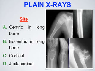
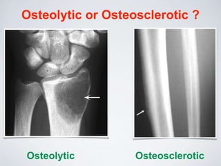
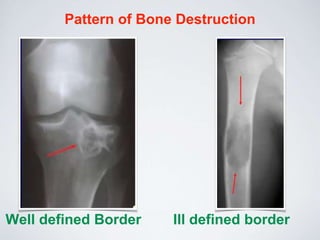
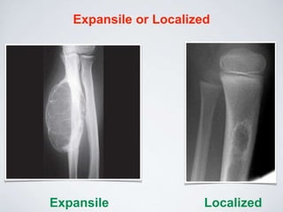

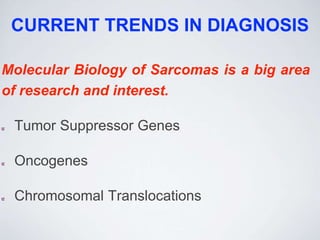
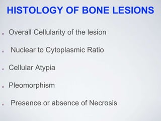
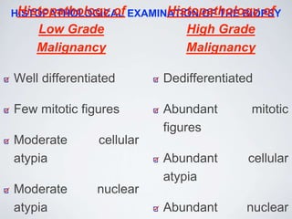
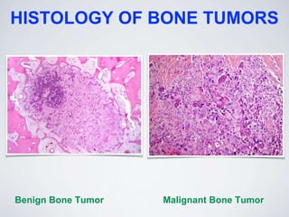

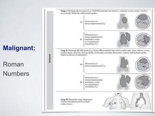
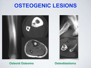
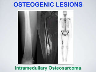
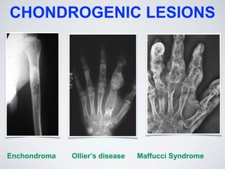
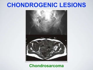
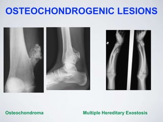
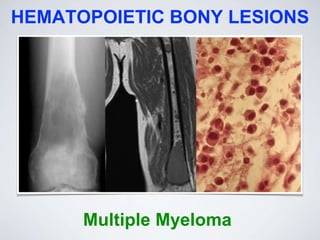
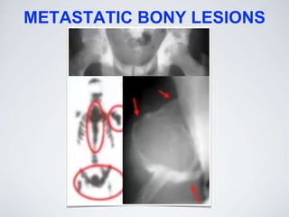


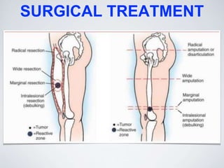
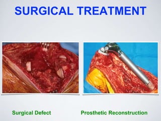
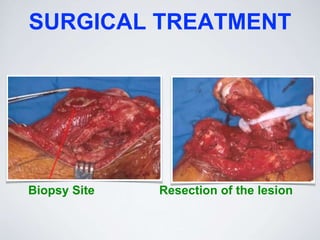
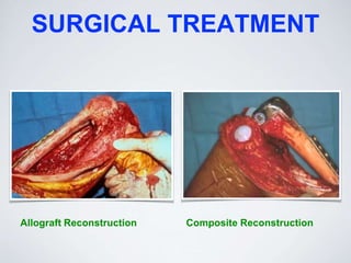


1. Benign bone tumors are more common than malignant bone tumors, though malignant tumors account for 5% of childhood cancers. The most common benign tumor is osteochondroma, while the most common primary malignant tumor is multiple myeloma and the most common bone tumor overall is metastatic carcinoma. 2. Pain and swelling are the most common presentations of bone tumors. Diagnostic evaluation begins with plain x-rays of the affected bone and surrounding area. Further imaging such as CT, bone scan and MRI are used if malignancy is suspected. 3. Biopsy is only performed after imaging to determine the appropriate surgical treatment, which is typically limb salvage rather than amputation. Adjuvant chemotherapy and radiotherapy are


![PERI-PROSTHETIC FRACTURE NAIL-PLATE CONSTRUCT [NPC].pptx](https://cdn.slidesharecdn.com/ss_thumbnails/drarunkumardrmohamedashrafperiprostheticfrasturenail-plateconstructnpc-260209164459-7e9d15a1-thumbnail.jpg?width=640&height=640&fit=bounds)


