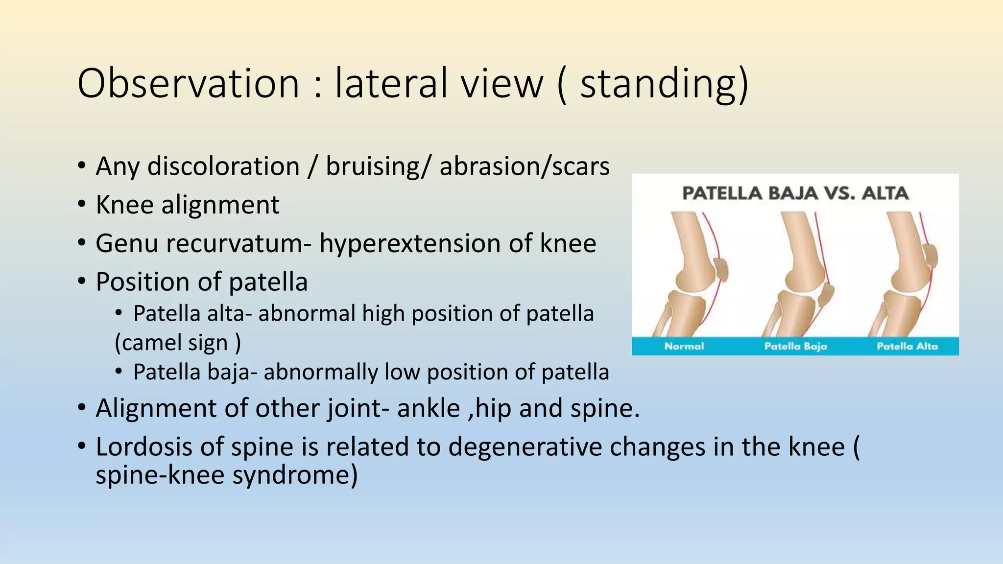
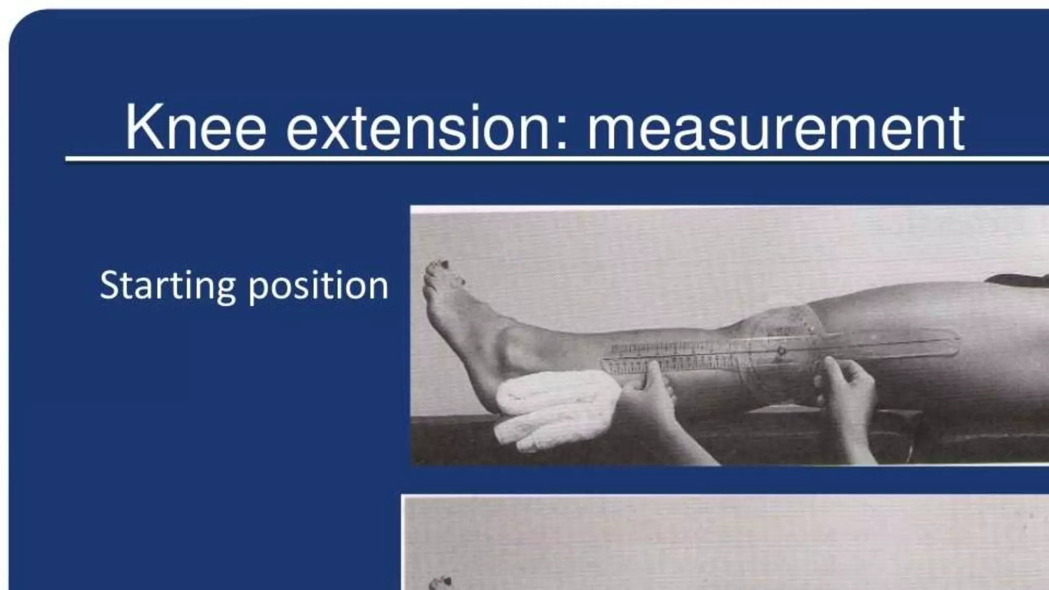
SlideShare a Scribd company logo
1 of 78
Pt. Assessment of Knee Joint
By :-
Dr. Taniya verma ( PT)
MPT-musculoskeletal
Knee joint
• Tibiofemoral joint
• Modified hinge joint having 3 degree of freedom
• Resting position- 25 degree flexion
• Closed pack position- full extension , lateral rotation of tibia
• Capsular pattern – flexion, extension
• Space between bones filled by menisci – medial (c shape) & lateral menisci (
O shape) ; thick along periphery and thinner along inner margin
• Patellofemoral joint
• Thickest layer of cartilage in patella; has 5 facets- superior, inferior,
lateral, medial and odd( frequently affected in patellofemoral
syndrome)
• malalignment of patellar movement can lead to patellofemoral
arthralgia
• Patella function as guide for quads tendon, decreases friction, control
capsular tension in knee, act as bony shield for cartilage of femoral
condyle
DEMOGRAPHIC DATA
• Age:
• Gender- conditions prone in
Female- OA ; ACL injuries
CHIEF COMPLAINTS
• IN PATIENT’S OWN WORD
• Pain
• Swelling : immediate, delayed
• Stiffness
• Deformity
• Locking
• Giving away
• Limp
• Loss of function
HISTORY OF PRESENT ILLNESS
• ONSET OF PAIN
• Date of injuy or when symptoms started
• Gradual/Sudden
• Progression- getting better/getting worse/no change
• LOCATION OF PAIN
• Anterior
• Lateral
• Medial
• posterior
Anterior knee pain
Lateral knee pain
• IT band friction syndrome
• Biceps femoris tendonitis
• osteoarthritis
• Poplitius tendonitis.
Medial knee pain
Posterior knee pain
Mechanism of present illness-Helps predict
injured structure
• Contact or non contact injury?
• If contact, what part of knee was contacted?
• Anterior blow/ varus force/valgus force
• Was foot of affected knee plated on the ground? (closed vs open chain
injury )
• Bony avulsions- slow developing forces
• Ligament tear- Rapidly developing forces
• MCL injury- Valgus force
• LCL injury- Varus force
• ACL INJURY
• Common in wt.bearing, slight flexion and rotation
• Anterior translator force on proximal tibia
• Hyperextension injury
• PCL INJURY
• Posterior translation of tibia
• Pretibial trauma( Dashboard trauma)
• MENISCAL INJURY
• Twisting force (injury with rotation of tibia) in weight bearing
• Any ‘clicking’ or ‘pop’ sound during injury?
• Indicates ligament tear in knee joint
• Any ‘give away’ or ‘catch’?
• Give away= instability
• Catch= (lock) meniscal injury
• Swelling immediate or delayed
• Synovial swelling= takes 8-24 hrs to develop
• Immediate (0–2 hours) (hemarthrosis)= ACL rupture,patellar dislocation
• Delayed (6-24 hrs)(effusion)= meniscus
• No swelling = MCL sprain
• Localized swelling- extracapsular injury
• Generalised swelling- Intracapsular injury
• History of type of shoes patient normally uses?- flat/high heels
• Functional ability of the patient?
• Any disability in running, climbing stairs etc
• SPORTS SPECIFIC HISTORY
• Total duration of play
• Total practice/ training session
• Exercise porotocol- warmup/cool down
• Competition.
• Past history
• Any previous injury indicates recurrent injuries around knee
• Was the pain at the same site?
• Similar nature?
• Treatment given previously
PAIN ASSESSMENT
• Site of pain – retropatellar?
• Type of pain
• Aching pain = degenerative pain
• Sharp catching pain= mechanical problem
• Arthritic pain= associated with stiffness and eases with movement
• Pain at rest= is not mechanical pain
• Pain during activity= sublaxation, patellar tracking disorder
• Pain after activity= synovitis, tendinitis
• Constant pain irrespective of activity= tumor?
• Pain around knee= contusion, tear of ligament, muscle
• Ask if patient has pain during any specific activity or position or movement.
BODY CHART
• Aggrevating/ relieving factors
• Severity of pain
• VAS/NPRS
OBSERVATIONS
• VIEW FOR OBSERVATIONS
• Standing
• Anterior, posterior and lateral view
• Sitting
• Anterior and lateral view
• Lying
• Supine, side-lying and prone
• Patient should be exposed to see knee joint and a joint proximal and
distal to it. ( hip and ankle)
• Key points
• Consent
• Adequate explanation about the necessity of exposure during patient
assessment and its benefits in the treatment
• Confined place and environment to maintain patient’s privacy
Observation : anterior view ( standing)
• Knee joint alignment
• Genu valgum /genu varum/windstep deformity
• Children at age of 18-19 months: genu varus which is normal
• Age 3-4 knee attains valgum
• Swelling
• Quadriceps wasting?- disuse atrophy/quadriceps inhibition
• Position of patella
• Squinting of patella- medially facing patella
1.VARUS / 2.VALGUS/ 3.WINDSWEPT DEFORMITY
Miserable malalignment syndrome. The patient often presents with medial
parapatellar pain with no localizing findings or images indicating pathology:
A, when viewed from the front, the squinting patella on the right is well
visualized, with both femurs having greater than 50° anteversion;
B, what the patient sees looking down her legs
Observation : lateral view ( standing)
• Any discoloration / bruising/ abrasion/scars
• Knee alignment
• Genu recurvatum- hyperextension of knee
• Position of patella
• Patella alta- abnormal high position of patella
(camel sign )
• Patella baja- abnormally low position of patella
• Alignment of other joint- ankle ,hip and spine.
• Lordosis of spine is related to degenerative changes in the knee (
spine-knee syndrome)
Camel sign- because of high patella ( one hump), the
infrapatellar fat pad ( second hump) or inflamed infrapatellar
bursae becomes more prominent
Observation : Posterior view ( standing)
• Any abnormal swelling?
• Baker’s cyst
• Gastrocnemius- semimembranous bursae
• Popletial artery aneurysm
• Lymphadenopathy
• Soft tissue tumor
• Wasting of hamstring and calf
Observation :sitting (lateral and anterior view)
• Observe for position of patella
• Patella should face forward and should rest on the distal end of femur.
• In patella alta= patella rest in anterior surface of femur
• Frogeyes patella or grasshopper patella- patella face upward and outward
away from each other
• Grasshopper eyes or frog eyes is seen in patients whose patellae are
proximally displaced (patella alta) and externally or laterally rotated
(lateral patellar tilt)
• Enlarged tibial tubercle= Osgood schlatter disease.
• Fig-pre patellar bursitis.
General observation
• Built
• Facial expressions
• Posture
• Guarding/splinting
• Extra devices
• Gait – antalgic/ hand to knee gait in quadriceps weakness
• located more distally than
prepatellar bursitis and is often
caused by frequent kneeling in an
upright position
Pain and tenderness on the
inside of your knee,
approximately 2 to 3 inches
below the joint, are
common symptoms of pes
anserine bursitis of the
knee.
Osgood-Schlatter
disease
• It is an inflammation of the area just below the knee where the tendon
from the kneecap (patellar tendon) attaches to the shinbone (tibia).
• In Osgood-Schlatter disease, the enlarged, inflamed tibial tubercle is nearly
always tender when pressure is applied.
PALPATION
• ( WITH VARIOUS ANGLES OF KNEE FLEXION )
• Medio-patellar plica- on medial border of patella
• Jumper’s knee- at the apex of patella ( patellar tendon)
• Patellar tendinitis: palpate distally
• Housemaid’s knee: prepatellar bursitis.
• Patellar retinaculum :can be palpated on either side of patella
• Quadriceps muscle: can be palpated for any strain,wasting or hard
muscle(myositis ossificans)
• MCL: along the medial aspect of knee.
• Pes Anserine bursitis: medial and slight distal to tibial tuberosity.
• Temperature: proximal to distal
• Tenderness:
• For soft tissue and bony tenderness.
• Knee flexed at 90’ – examined for tenderness along medial an lateral joint
line.
• Knee extended
• Retropatellar area- by gliding movement of patella between thumb and index finger on
medial and lateral facets, try to palpate undersurface of patella.
• Patellofemoral crepitions.
• Distal pulsations
• Poplitieal pulse
Resisted isometric contractions
• Extension of the knee
• Flexion of the knee
• Ankle dorsiflexion
• Ankle plantar flexion
Muscle length testing
• Rectus femoris ( ely test )
• Distal hamstring length test
Joint play movement
• Backward movement of tibia on femur. ( similar to posterior drawer
test)
• Forward movement of tibia on femur (similar to the anterior drawer
test)
• Medial and lateral translation of tibia on femur.
• Medial and lateral displacement of patella.
• Anteroposterior Movement of Fibula on Tibia
Special test
TEST FOR LIGAMENT STABILITY
• One plane medial and lateral instability –valgus and varus stress test
• One plane anterior and posterior instability- posterior sag sign, Lachman test, drawer
sign, active drawer test
• Anteromedial and anterolateral rotary instabilities - Lateral pivot shift maneuver,
Jerk test of hughston; cross over test of Arnold, slocum test
• Posteromedial and posterolateral rotary instabilities - Posteromedial pivot shift
Jakob test (reverse pivot shift maneuver), Hughston posterolateral and posteromedial
drawer sign .
• Tests for meniscus injury- McMurray test , Apley’s test.
• Plica test - Mediopatellar plica test
• Tests for patellofemoral dysfunction - Clarke’s sign ; McConnell test for
chondromalacia patella
• Tests for swelling -Brush, stroke, or bulge test ; Fluctuation test ; Patellar tap test
(ballotable patella)
• Test for patellar dislocation- fairbank apprehension test
Valgus and Varus stress test
Posterior sag sign
Lachman test
Drawer sign
Active drawer test
Anterolateral instability
• Lateral pivot shift test • Jerk test of hughston
Cross over test of Arnold slocum test
Jackob test (reverse pivot shift maneuver)
Postero-medial pivot shift test
Hughston’s posteromedial and posterolateral
drawer test
Meniscus injury
• McMurray test • Apley’s test
Mediopatellar plica test (Mital-hayden test)
CLARKE’S SIGN
• PURPOSE -To assess for patellofemoral dysfunction.
• PATIENT POSITION - supine with the knee extended.
• EXAMINER POSITION -The examiner stands adjacent to the patient’s knee.
• TEST PROCEDURE
• The unaffected leg is tested first. The thenar web space of one of the examiner’s hands
is placed slightly proximal to the upper pole or base of the patella. The examiner presses
down several times with increasing force until pain is produced on the proximal aspect of
the patella. The patient then is asked to contract the quadriceps muscles after the
examiner pushes down. The test is repeated with the painful knee, and the amounts of
pressure that cause pain are compared.
• INDICATIONS OF A POSITIVE TEST A positive test result is indicated if the test causes
retro patellar pain and the patient cannot hold a contraction. If the patient can complete
and maintain the contraction without pain, the test result is considered negative, as long
as the same test on the unaffected leg does not cause pain.
• CAUTIONS • Because a positive test result can be obtained on anyone if sufficient
pressure is applied to the patella, the amount of pressure applied must be carefully
controlled and compared with the opposite side. This is best done by repeating the
procedure several times, increasing the pressure each time and comparing the results
with those on the unaffected side.
• • To test different parts of the patella, the knee may be tested in 30°, 60°, and 90° of
flexion and in full extension.
MCCONNELL TEST FOR CHONDROMALACIA
PATELLA
• The unaffected leg is tested first. After the examiner has positioned the knee at 120°, 90°, 60°,
30°, and 0° (full extension), the patient performs isometric quadriceps contractions with the
femur laterally rotated, with each contraction held for 10 seconds. If pain is produced during any
of the contractions, the patient’s leg is passively returned to full extension. The leg then is fully
supported on the examiner’s knee, and the examiner pushes the patella medially. The medial
glide is maintained while the knee is returned to the painful angle, and the patient performs an
isometric contraction again, with the patella held medially. The two legs are compared.
• If the pain is decreased when the patella is held medially, the pain is patellofemoral in origin.
BRUSH, STROKE, OR BULGE TEST for effusion
• PURPOSE - To assess minimal effusion in the knee.
• PATIENT POSITION -The patient lies supine.
• EXAMINER POSITION - The examiner is positioned adjacent to the test knee.
• TEST PROCEDURE
• The examiner places one hand over the anteromedial tibia. Starting just below the joint line on
the medial side of the patella and using the palm and ngers, the examiner strokes proximally
toward the patient’s hip, as far as the suprapatellar pouch, two or three times. With the opposite
hand, the examiner strokes down the lateral side of the patella.
• INDICATIONS OF A POSITIVE TEST A positive test result is indicated when a wave of fluid passes
along the medial side of the joint and bulges just below the medial distal portion or border of the
patella during the down stroke. The wave of fluid may take up to 2 seconds to appear.
• CLINICAL NOTES • Also called the wipe test, this test assesses minimal effusion. • Normally, the
knee contains 1 to 7 ml of synovial fluid. This test can show as little as 4 to 8 ml of extra fluid
within the knee.
Fluctuation test
• PATIENT POSITION -supine.
• EXAMINER POSITION -adjacent to the test knee.
• TEST PROCEDURE - The examiner places the palm of one hand over the
suprapatellar pouch and the palm of the other hand anterior to the joint at
the joint line, with the thumb and index nger just beyond the margins of
the patella.Pressing down with one hand and then the other, the examiner
may feel the synovial fluid fluctuate under the hands and move from one
hand to the other.
• INDICATIONS OF A POSITIVE TEST- Increased fluctuation of fluid in the
knee compared to the contralateral knee indicates signicant effusion.
Patellar tap test
• PATIENT POSITION -The patient lies supine.
• EXAMINER POSITION -The examiner is positioned adjacent to the
test knee.
• TEST PROCEDURE - With the patient’s knee extended or flexed to
discomfort, the examiner applies a slight tap or pressure over the
patella.
• INDICATIONS OF A POSITIVE TEST- When the tap is applied, the
patella should be felt to fl oat; this is sometimes called the “dancing
patella” sign.
FAIRBANK’S APPREHENSION TEST
• PURPOSE -To assess for dislocation of the patella.
SUSPECTED INJURY Dislocation or subluxation of the patella
• PATIENT POSITION
-The patient lies supine with the quadriceps muscles relaxed and the knee
flexed to 30º over the examiner’s leg.
• EXAMINER POSITION - The examiner sits directly adjacent to the test knee.
• TEST PROCEDURE - The unaffected leg is tested first. The examiner’s
thumbs are placed on the medial border of the patella, and the ngers are
placed on the lateral aspect of the patient’s knee. The examiner carefully
and slowly pushes the patella laterally with the thumbs. The two legs are
compared.
• INDICATIONS OF A POSITIVE TEST If the patient feels that the patella is
going to dislocate, he or she will contract the quadriceps muscles to bring
the patella “back into line.” This action indicates a positive test result. The
patient also will have an apprehensive look.
Differential diagnosis by location
•ANTERIOR- patellofemoral syndrome, bursitis, Osgood
schalatter’s disease, patellar tendinitis, patellar fractur
•MEDIAL- meniscus, MCL, OA, pes anserine bursitis
•LATERAL- Meniscus, LCL, OA, iliotibal friction band
syndrome, Fibular head dysfunction
•POSTERIOR- Hamstring injury, Baker’s cyst,
Neurovascular injury ( popliteal artery or nerve. )
Differential diagnosis by location
anterior knee pain
Condition Typical finding
Tibial apophysitis Tendar and swollen tibial tuberosity
Resisted active extension and passive hyperflexion of
knee painful
Patellar tendonitis Tender patellar tendon; resisted knee extension
painful
Slipped capital femoral epiphysis Displacement of epiphysis of femoral head in
radiographs; passive hip IR painful
PFPS Slight effusion along with patellar crepitus on ROM;
reproduction of patient’s pain by applying direct
pressure at anterior aspect of patella
ACL injury Positive anterior drawers test; Lachman test
Medial knee pain
Condition Typical findings
Medial plica syndrome Tenderness near medial patellar retinaculum that
worsen with knee flexion
MCL tear Laxity; regional pain with valgus stress testing
Meniscal tear Tenderness over medial joint line; positive McMurry
test
OA Regional medial knee pain; tender joint line;
decreased ROM; degenerative changes in radiograph
Pes anserine bursitis Tenderness 2-4 cm below medial joint line; painful
resisted knee flexion in prone position
Medial collateral ligament sprain Point tenderness at medial joint line; positive valgus
stress test
Lateral knee pain
Condition typical findings
Iliotibial band tendonitis Tenderness at lateral epicondyle of femur approx. 3
cm proximal to the joint line
Positive noble test
Lateral collateral ligament sprain Point tenderness at lateral joint line; positive varus
stress testing
Posterior knee pain
Conditions Typical findings
Popliteal cyst Palpable fullness at medial aspect
of popletial area, at or near the
origin of medial head of
gastrocnemius
Hamstring tendonitis Localized tenderness over medial
hamstring tendon insertion
Comparison of clinical features of 3 common
cause of anterior knee pain
Signs Patellofemoral pain Patellar tendinopathy Fat pad impingement
Onset Running ( esp. downhill)
Steps/hills
Wt. bearing act. involving
knee flexion
Act. Involving
jumping/changing
directions
Often but not always,
sudden onset with
hyperextension injury
Pain Non specific/vague
Med./lat./infrapatellar
Increased by act. That
load PFJ
Around inferior pole of
patella
Inc. by jumping & early to
mid squat
Around inferior pole of
patella
Inc. by prolonged
standing, stairs, knee
extension
Inspection Generally normal
Or VMO wasting
Generally quadriceps
wasting
Puffiness may be
apparent around tendon
which may make pole of
patella appear to be
displaced posteriorly
Swelling May have small effusions, Rare, tendon may be May have a puffy
Signs Patellofemoral pain Patellar tendinopathy Fat pad impingement
Clicks Occasional No No
Crepitus Occasionally under patella No No
Giving way Occasional Occasionally due to
quadriceps inhibition
No
Knee ROM Usually normal
May be decreased
Usually normal,
No pain with overpressure
Active extension may be
painful in acute fat pad
impingement
Passive overpressure into ext.
is painful
PFJ movement May be restricted n any
direction ( commonly medial
glide due to tight lateral
structure)
May have normal PFJ
biomechanics
Normal or posteriorly
displaced inferior pole of
patella.
Functional testing Squats, stairs may aggravates
PFJ taping should decrease
pain
Decline squats aggravates
pain
PFJ taping has less effect
Aggravates by squats
PFJ taping should decrease
pain if inferior pole of patella
is tilted up.
References :-
• David J. Magee-orthopedic physical assessment 5th edition
• Peter brunker and karim khan- clinical sports medicine .3rd edition
• Muscle Function Testing : Vladimir Janda
• Measurement of Joint Motion A Guide to Goniometry : Cynthia C
norkin ( third edition )

More Related Content

What's hot

Coccydynia
Coccydynia Coccydynia
Coccydynia Ade Wijaya
 
Knee clinical examination for orthopaedic residents.dr mohamed ashraf TD medi...
Knee clinical examination for orthopaedic residents.dr mohamed ashraf TD medi...Knee clinical examination for orthopaedic residents.dr mohamed ashraf TD medi...
Knee clinical examination for orthopaedic residents.dr mohamed ashraf TD medi...drashraf369
 
Adhesive capsulitis case presentation physiotherapy
Adhesive capsulitis case presentation physiotherapyAdhesive capsulitis case presentation physiotherapy
Adhesive capsulitis case presentation physiotherapymanisha thakur
 
Meniscal injury
Meniscal injuryMeniscal injury
Meniscal injurymanoj das
 
Mulligan mobilization (MWM)
Mulligan mobilization (MWM)Mulligan mobilization (MWM)
Mulligan mobilization (MWM)Radhika Chintamani
 
Rotator cuff injuries
Rotator cuff injuriesRotator cuff injuries
Rotator cuff injuriespratigya deuja
 
An Introduction to the McKenzie Method
An Introduction to the McKenzie MethodAn Introduction to the McKenzie Method
An Introduction to the McKenzie MethodDeborah Currier
 
Posterior Cruciate Ligament Injury
Posterior Cruciate Ligament InjuryPosterior Cruciate Ligament Injury
Posterior Cruciate Ligament InjuryArslan Luqman
 
Radial nerve palsy
Radial nerve palsyRadial nerve palsy
Radial nerve palsymanoj das
 
Anterior cruciate ligament-Injury & management
Anterior cruciate ligament-Injury & managementAnterior cruciate ligament-Injury & management
Anterior cruciate ligament-Injury & managementAnand Rao
 
Clinical Examination of Hip
Clinical Examination of HipClinical Examination of Hip
Clinical Examination of HipVivek Mathew Philip
 
PRINCIPLES OF TENDON TRANSFERS
PRINCIPLES OF TENDON TRANSFERSPRINCIPLES OF TENDON TRANSFERS
PRINCIPLES OF TENDON TRANSFERSBenthungo Tungoe
 
The Cyriax Approach to Orthopaedic Manual Physical Therapy
The Cyriax Approach to Orthopaedic Manual Physical Therapy The Cyriax Approach to Orthopaedic Manual Physical Therapy
The Cyriax Approach to Orthopaedic Manual Physical Therapy Sreeraj S R
 
Clinical examination of spine
Clinical examination of spineClinical examination of spine
Clinical examination of spinePrasanthmuddada
 
Muscle Energy Technique (MET)
Muscle Energy Technique (MET)Muscle Energy Technique (MET)
Muscle Energy Technique (MET)Venus Pagare
 
Hand deformity in rheumatoid arthritis
Hand deformity in rheumatoid arthritisHand deformity in rheumatoid arthritis
Hand deformity in rheumatoid arthritissushilonlines
 

What's hot (20)

Coccydynia
Coccydynia Coccydynia
Coccydynia
 
Knee clinical examination for orthopaedic residents.dr mohamed ashraf TD medi...
Knee clinical examination for orthopaedic residents.dr mohamed ashraf TD medi...Knee clinical examination for orthopaedic residents.dr mohamed ashraf TD medi...
Knee clinical examination for orthopaedic residents.dr mohamed ashraf TD medi...
 
Adhesive capsulitis case presentation physiotherapy
Adhesive capsulitis case presentation physiotherapyAdhesive capsulitis case presentation physiotherapy
Adhesive capsulitis case presentation physiotherapy
 
Meniscal injury
Meniscal injuryMeniscal injury
Meniscal injury
 
Mulligan mobilization (MWM)
Mulligan mobilization (MWM)Mulligan mobilization (MWM)
Mulligan mobilization (MWM)
 
Pes planus
Pes planusPes planus
Pes planus
 
Rotator cuff injuries
Rotator cuff injuriesRotator cuff injuries
Rotator cuff injuries
 
An Introduction to the McKenzie Method
An Introduction to the McKenzie MethodAn Introduction to the McKenzie Method
An Introduction to the McKenzie Method
 
Examination of shoulder
Examination of shoulderExamination of shoulder
Examination of shoulder
 
Posterior Cruciate Ligament Injury
Posterior Cruciate Ligament InjuryPosterior Cruciate Ligament Injury
Posterior Cruciate Ligament Injury
 
Low back pain
Low back painLow back pain
Low back pain
 
Examination of hip joint
Examination of hip jointExamination of hip joint
Examination of hip joint
 
Radial nerve palsy
Radial nerve palsyRadial nerve palsy
Radial nerve palsy
 
Anterior cruciate ligament-Injury & management
Anterior cruciate ligament-Injury & managementAnterior cruciate ligament-Injury & management
Anterior cruciate ligament-Injury & management
 
Clinical Examination of Hip
Clinical Examination of HipClinical Examination of Hip
Clinical Examination of Hip
 
PRINCIPLES OF TENDON TRANSFERS
PRINCIPLES OF TENDON TRANSFERSPRINCIPLES OF TENDON TRANSFERS
PRINCIPLES OF TENDON TRANSFERS
 
The Cyriax Approach to Orthopaedic Manual Physical Therapy
The Cyriax Approach to Orthopaedic Manual Physical Therapy The Cyriax Approach to Orthopaedic Manual Physical Therapy
The Cyriax Approach to Orthopaedic Manual Physical Therapy
 
Clinical examination of spine
Clinical examination of spineClinical examination of spine
Clinical examination of spine
 
Muscle Energy Technique (MET)
Muscle Energy Technique (MET)Muscle Energy Technique (MET)
Muscle Energy Technique (MET)
 
Hand deformity in rheumatoid arthritis
Hand deformity in rheumatoid arthritisHand deformity in rheumatoid arthritis
Hand deformity in rheumatoid arthritis
 

Similar to Assessment of Knee Joint Pain

Examination of the Knee joint
Examination of the Knee jointExamination of the Knee joint
Examination of the Knee jointDr ABU SURAIH SAKHRI
 
mypptfoot-160706125859.pdf
mypptfoot-160706125859.pdfmypptfoot-160706125859.pdf
mypptfoot-160706125859.pdfa7med7amdy2
 
CLINICAL EXAMINATION OF HIP JOINT
CLINICAL EXAMINATION OF HIP JOINTCLINICAL EXAMINATION OF HIP JOINT
CLINICAL EXAMINATION OF HIP JOINTRITESHJAISWAL57
 
History and examintion of knee joint slideshare
History and examintion of knee joint slideshareHistory and examintion of knee joint slideshare
History and examintion of knee joint slideshareDr Ajay Shah IOM TUTH Nepal
 
APPROACH TO A PATIENT WITH CHRONIC KNEE PAIN.pdf
APPROACH TO A PATIENT WITH CHRONIC KNEE PAIN.pdfAPPROACH TO A PATIENT WITH CHRONIC KNEE PAIN.pdf
APPROACH TO A PATIENT WITH CHRONIC KNEE PAIN.pdfNingarajDyapur
 
Lower Patellofemoral Instability
Lower Patellofemoral InstabilityLower Patellofemoral Instability
Lower Patellofemoral InstabilityRizqi D Rosandi MD
 
PATELLOFEMORAL PAIN (Harleen kaur Nagi).pptx
PATELLOFEMORAL PAIN (Harleen kaur Nagi).pptxPATELLOFEMORAL PAIN (Harleen kaur Nagi).pptx
PATELLOFEMORAL PAIN (Harleen kaur Nagi).pptxHarleenNagi1
 
clinicalexaminationofshoulder.pptx
clinicalexaminationofshoulder.pptxclinicalexaminationofshoulder.pptx
clinicalexaminationofshoulder.pptxVaisHali822687
 
Common sports injuries.2560
Common sports injuries.2560Common sports injuries.2560
Common sports injuries.2560Ukris Ortho
 
Foot pain problems
Foot pain problems Foot pain problems
Foot pain problems mohamed aouini
 
4 a adult acquired flat foot - Derek Park
4 a adult acquired flat foot - Derek Park4 a adult acquired flat foot - Derek Park
4 a adult acquired flat foot - Derek ParkDerek Park
 
Postural deviation in different planes.pptx
Postural deviation in different planes.pptxPostural deviation in different planes.pptx
Postural deviation in different planes.pptxAvaniAkbari
 
Examination of knee joint
Examination of knee jointExamination of knee joint
Examination of knee jointFadzlina Zabri
 

Similar to Assessment of Knee Joint Pain (20)

Examination of the Knee joint
Examination of the Knee jointExamination of the Knee joint
Examination of the Knee joint
 
mypptfoot-160706125859.pdf
mypptfoot-160706125859.pdfmypptfoot-160706125859.pdf
mypptfoot-160706125859.pdf
 
CLINICAL EXAMINATION OF HIP JOINT
CLINICAL EXAMINATION OF HIP JOINTCLINICAL EXAMINATION OF HIP JOINT
CLINICAL EXAMINATION OF HIP JOINT
 
Foot Drop
Foot DropFoot Drop
Foot Drop
 
History and examintion of knee joint slideshare
History and examintion of knee joint slideshareHistory and examintion of knee joint slideshare
History and examintion of knee joint slideshare
 
APPROACH TO A PATIENT WITH CHRONIC KNEE PAIN.pdf
APPROACH TO A PATIENT WITH CHRONIC KNEE PAIN.pdfAPPROACH TO A PATIENT WITH CHRONIC KNEE PAIN.pdf
APPROACH TO A PATIENT WITH CHRONIC KNEE PAIN.pdf
 
Flat foot and Cavus foot
 Flat foot and Cavus foot Flat foot and Cavus foot
Flat foot and Cavus foot
 
Lower Patellofemoral Instability
Lower Patellofemoral InstabilityLower Patellofemoral Instability
Lower Patellofemoral Instability
 
PATELLOFEMORAL PAIN (Harleen kaur Nagi).pptx
PATELLOFEMORAL PAIN (Harleen kaur Nagi).pptxPATELLOFEMORAL PAIN (Harleen kaur Nagi).pptx
PATELLOFEMORAL PAIN (Harleen kaur Nagi).pptx
 
clinicalexaminationofshoulder.pptx
clinicalexaminationofshoulder.pptxclinicalexaminationofshoulder.pptx
clinicalexaminationofshoulder.pptx
 
Surgery 6th year, Tutorial (Dr. Ali A. Nabi)
Surgery 6th year, Tutorial (Dr. Ali A. Nabi)Surgery 6th year, Tutorial (Dr. Ali A. Nabi)
Surgery 6th year, Tutorial (Dr. Ali A. Nabi)
 
Common sports injuries.2560
Common sports injuries.2560Common sports injuries.2560
Common sports injuries.2560
 
Foot pain problems
Foot pain problems Foot pain problems
Foot pain problems
 
Dr tarek ankle pain1
Dr tarek ankle pain1Dr tarek ankle pain1
Dr tarek ankle pain1
 
Ctev
CtevCtev
Ctev
 
Gait Cycle.pptx
Gait Cycle.pptxGait Cycle.pptx
Gait Cycle.pptx
 
Gait Cycle.pptx
Gait Cycle.pptxGait Cycle.pptx
Gait Cycle.pptx
 
4 a adult acquired flat foot - Derek Park
4 a adult acquired flat foot - Derek Park4 a adult acquired flat foot - Derek Park
4 a adult acquired flat foot - Derek Park
 
Postural deviation in different planes.pptx
Postural deviation in different planes.pptxPostural deviation in different planes.pptx
Postural deviation in different planes.pptx
 
Examination of knee joint
Examination of knee jointExamination of knee joint
Examination of knee joint
 

Recently uploaded

Bangalore Call Girls Majestic 📞 9907093804 High Profile Service 100% Safe
Bangalore Call Girls Majestic 📞 9907093804 High Profile Service 100% SafeBangalore Call Girls Majestic 📞 9907093804 High Profile Service 100% Safe
Bangalore Call Girls Majestic 📞 9907093804 High Profile Service 100% Safenarwatsonia7
 
Call Girls In Andheri East Call 9920874524 Book Hot And Sexy Girls
Call Girls In Andheri East Call 9920874524 Book Hot And Sexy GirlsCall Girls In Andheri East Call 9920874524 Book Hot And Sexy Girls
Call Girls In Andheri East Call 9920874524 Book Hot And Sexy Girlsnehamumbai
 
Call Girls ITPL Just Call 7001305949 Top Class Call Girl Service Available
Call Girls ITPL Just Call 7001305949 Top Class Call Girl Service AvailableCall Girls ITPL Just Call 7001305949 Top Class Call Girl Service Available
Call Girls ITPL Just Call 7001305949 Top Class Call Girl Service Availablenarwatsonia7
 
Call Girl Coimbatore Prisha☎️ 8250192130 Independent Escort Service Coimbatore
Call Girl Coimbatore Prisha☎️  8250192130 Independent Escort Service CoimbatoreCall Girl Coimbatore Prisha☎️  8250192130 Independent Escort Service Coimbatore
Call Girl Coimbatore Prisha☎️ 8250192130 Independent Escort Service Coimbatorenarwatsonia7
 
Russian Call Girls Chickpet - 7001305949 Booking and charges genuine rate for...
Russian Call Girls Chickpet - 7001305949 Booking and charges genuine rate for...Russian Call Girls Chickpet - 7001305949 Booking and charges genuine rate for...
Russian Call Girls Chickpet - 7001305949 Booking and charges genuine rate for...narwatsonia7
 
Vip Call Girls Anna Salai Chennai 👉 8250192130 ❣️💯 Top Class Girls Available
Vip Call Girls Anna Salai Chennai 👉 8250192130 ❣️💯 Top Class Girls AvailableVip Call Girls Anna Salai Chennai 👉 8250192130 ❣️💯 Top Class Girls Available
Vip Call Girls Anna Salai Chennai 👉 8250192130 ❣️💯 Top Class Girls AvailableNehru place Escorts
 
Bangalore Call Girls Marathahalli 📞 9907093804 High Profile Service 100% Safe
Bangalore Call Girls Marathahalli 📞 9907093804 High Profile Service 100% SafeBangalore Call Girls Marathahalli 📞 9907093804 High Profile Service 100% Safe
Bangalore Call Girls Marathahalli 📞 9907093804 High Profile Service 100% Safenarwatsonia7
 
Call Girls Frazer Town Just Call 7001305949 Top Class Call Girl Service Avail...
Call Girls Frazer Town Just Call 7001305949 Top Class Call Girl Service Avail...Call Girls Frazer Town Just Call 7001305949 Top Class Call Girl Service Avail...
Call Girls Frazer Town Just Call 7001305949 Top Class Call Girl Service Avail...narwatsonia7
 
Call Girl Bangalore Nandini 7001305949 Independent Escort Service Bangalore
Call Girl Bangalore Nandini 7001305949 Independent Escort Service BangaloreCall Girl Bangalore Nandini 7001305949 Independent Escort Service Bangalore
Call Girl Bangalore Nandini 7001305949 Independent Escort Service Bangalorenarwatsonia7
 
Call Girls Thane Just Call 9910780858 Get High Class Call Girls Service
Call Girls Thane Just Call 9910780858 Get High Class Call Girls ServiceCall Girls Thane Just Call 9910780858 Get High Class Call Girls Service
Call Girls Thane Just Call 9910780858 Get High Class Call Girls Servicesonalikaur4
 
Call Girls Electronic City Just Call 7001305949 Top Class Call Girl Service A...
Call Girls Electronic City Just Call 7001305949 Top Class Call Girl Service A...Call Girls Electronic City Just Call 7001305949 Top Class Call Girl Service A...
Call Girls Electronic City Just Call 7001305949 Top Class Call Girl Service A...narwatsonia7
 
Ahmedabad Call Girls CG Road 🔝9907093804 Short 1500 💋 Night 6000
Ahmedabad Call Girls CG Road 🔝9907093804  Short 1500  💋 Night 6000Ahmedabad Call Girls CG Road 🔝9907093804  Short 1500  💋 Night 6000
Ahmedabad Call Girls CG Road 🔝9907093804 Short 1500 💋 Night 6000aliya bhat
 
Call Girls Jp Nagar Just Call 7001305949 Top Class Call Girl Service Available
Call Girls Jp Nagar Just Call 7001305949 Top Class Call Girl Service AvailableCall Girls Jp Nagar Just Call 7001305949 Top Class Call Girl Service Available
Call Girls Jp Nagar Just Call 7001305949 Top Class Call Girl Service Availablenarwatsonia7
 
Call Girls Hosur Just Call 7001305949 Top Class Call Girl Service Available
Call Girls Hosur Just Call 7001305949 Top Class Call Girl Service AvailableCall Girls Hosur Just Call 7001305949 Top Class Call Girl Service Available
Call Girls Hosur Just Call 7001305949 Top Class Call Girl Service Availablenarwatsonia7
 
Mumbai Call Girls Service 9910780858 Real Russian Girls Looking Models
Mumbai Call Girls Service 9910780858 Real Russian Girls Looking ModelsMumbai Call Girls Service 9910780858 Real Russian Girls Looking Models
Mumbai Call Girls Service 9910780858 Real Russian Girls Looking Modelssonalikaur4
 
Call Girls Service In Shyam Nagar Whatsapp 8445551418 Independent Escort Service
Call Girls Service In Shyam Nagar Whatsapp 8445551418 Independent Escort ServiceCall Girls Service In Shyam Nagar Whatsapp 8445551418 Independent Escort Service
Call Girls Service In Shyam Nagar Whatsapp 8445551418 Independent Escort Serviceparulsinha
 
Call Girl Surat Madhuri 7001305949 Independent Escort Service Surat
Call Girl Surat Madhuri 7001305949 Independent Escort Service SuratCall Girl Surat Madhuri 7001305949 Independent Escort Service Surat
Call Girl Surat Madhuri 7001305949 Independent Escort Service Suratnarwatsonia7
 
Artifacts in Nuclear Medicine with Identifying and resolving artifacts.
Artifacts in Nuclear Medicine with Identifying and resolving artifacts.Artifacts in Nuclear Medicine with Identifying and resolving artifacts.
Artifacts in Nuclear Medicine with Identifying and resolving artifacts.MiadAlsulami
 
Call Girls Hebbal Just Call 7001305949 Top Class Call Girl Service Available
Call Girls Hebbal Just Call 7001305949 Top Class Call Girl Service AvailableCall Girls Hebbal Just Call 7001305949 Top Class Call Girl Service Available
Call Girls Hebbal Just Call 7001305949 Top Class Call Girl Service Availablenarwatsonia7
 

Recently uploaded (20)

Bangalore Call Girls Majestic 📞 9907093804 High Profile Service 100% Safe
Bangalore Call Girls Majestic 📞 9907093804 High Profile Service 100% SafeBangalore Call Girls Majestic 📞 9907093804 High Profile Service 100% Safe
Bangalore Call Girls Majestic 📞 9907093804 High Profile Service 100% Safe
 
Call Girls In Andheri East Call 9920874524 Book Hot And Sexy Girls
Call Girls In Andheri East Call 9920874524 Book Hot And Sexy GirlsCall Girls In Andheri East Call 9920874524 Book Hot And Sexy Girls
Call Girls In Andheri East Call 9920874524 Book Hot And Sexy Girls
 
Call Girls ITPL Just Call 7001305949 Top Class Call Girl Service Available
Call Girls ITPL Just Call 7001305949 Top Class Call Girl Service AvailableCall Girls ITPL Just Call 7001305949 Top Class Call Girl Service Available
Call Girls ITPL Just Call 7001305949 Top Class Call Girl Service Available
 
Call Girl Coimbatore Prisha☎️ 8250192130 Independent Escort Service Coimbatore
Call Girl Coimbatore Prisha☎️  8250192130 Independent Escort Service CoimbatoreCall Girl Coimbatore Prisha☎️  8250192130 Independent Escort Service Coimbatore
Call Girl Coimbatore Prisha☎️ 8250192130 Independent Escort Service Coimbatore
 
Russian Call Girls in Delhi Tanvi ➡️ 9711199012 💋📞 Independent Escort Service...
Russian Call Girls in Delhi Tanvi ➡️ 9711199012 💋📞 Independent Escort Service...Russian Call Girls in Delhi Tanvi ➡️ 9711199012 💋📞 Independent Escort Service...
Russian Call Girls in Delhi Tanvi ➡️ 9711199012 💋📞 Independent Escort Service...
 
Russian Call Girls Chickpet - 7001305949 Booking and charges genuine rate for...
Russian Call Girls Chickpet - 7001305949 Booking and charges genuine rate for...Russian Call Girls Chickpet - 7001305949 Booking and charges genuine rate for...
Russian Call Girls Chickpet - 7001305949 Booking and charges genuine rate for...
 
Vip Call Girls Anna Salai Chennai 👉 8250192130 ❣️💯 Top Class Girls Available
Vip Call Girls Anna Salai Chennai 👉 8250192130 ❣️💯 Top Class Girls AvailableVip Call Girls Anna Salai Chennai 👉 8250192130 ❣️💯 Top Class Girls Available
Vip Call Girls Anna Salai Chennai 👉 8250192130 ❣️💯 Top Class Girls Available
 
Bangalore Call Girls Marathahalli 📞 9907093804 High Profile Service 100% Safe
Bangalore Call Girls Marathahalli 📞 9907093804 High Profile Service 100% SafeBangalore Call Girls Marathahalli 📞 9907093804 High Profile Service 100% Safe
Bangalore Call Girls Marathahalli 📞 9907093804 High Profile Service 100% Safe
 
Call Girls Frazer Town Just Call 7001305949 Top Class Call Girl Service Avail...
Call Girls Frazer Town Just Call 7001305949 Top Class Call Girl Service Avail...Call Girls Frazer Town Just Call 7001305949 Top Class Call Girl Service Avail...
Call Girls Frazer Town Just Call 7001305949 Top Class Call Girl Service Avail...
 
Call Girl Bangalore Nandini 7001305949 Independent Escort Service Bangalore
Call Girl Bangalore Nandini 7001305949 Independent Escort Service BangaloreCall Girl Bangalore Nandini 7001305949 Independent Escort Service Bangalore
Call Girl Bangalore Nandini 7001305949 Independent Escort Service Bangalore
 
Call Girls Thane Just Call 9910780858 Get High Class Call Girls Service
Call Girls Thane Just Call 9910780858 Get High Class Call Girls ServiceCall Girls Thane Just Call 9910780858 Get High Class Call Girls Service
Call Girls Thane Just Call 9910780858 Get High Class Call Girls Service
 
Call Girls Electronic City Just Call 7001305949 Top Class Call Girl Service A...
Call Girls Electronic City Just Call 7001305949 Top Class Call Girl Service A...Call Girls Electronic City Just Call 7001305949 Top Class Call Girl Service A...
Call Girls Electronic City Just Call 7001305949 Top Class Call Girl Service A...
 
Ahmedabad Call Girls CG Road 🔝9907093804 Short 1500 💋 Night 6000
Ahmedabad Call Girls CG Road 🔝9907093804  Short 1500  💋 Night 6000Ahmedabad Call Girls CG Road 🔝9907093804  Short 1500  💋 Night 6000
Ahmedabad Call Girls CG Road 🔝9907093804 Short 1500 💋 Night 6000
 
Call Girls Jp Nagar Just Call 7001305949 Top Class Call Girl Service Available
Call Girls Jp Nagar Just Call 7001305949 Top Class Call Girl Service AvailableCall Girls Jp Nagar Just Call 7001305949 Top Class Call Girl Service Available
Call Girls Jp Nagar Just Call 7001305949 Top Class Call Girl Service Available
 
Call Girls Hosur Just Call 7001305949 Top Class Call Girl Service Available
Call Girls Hosur Just Call 7001305949 Top Class Call Girl Service AvailableCall Girls Hosur Just Call 7001305949 Top Class Call Girl Service Available
Call Girls Hosur Just Call 7001305949 Top Class Call Girl Service Available
 
Mumbai Call Girls Service 9910780858 Real Russian Girls Looking Models
Mumbai Call Girls Service 9910780858 Real Russian Girls Looking ModelsMumbai Call Girls Service 9910780858 Real Russian Girls Looking Models
Mumbai Call Girls Service 9910780858 Real Russian Girls Looking Models
 
Call Girls Service In Shyam Nagar Whatsapp 8445551418 Independent Escort Service
Call Girls Service In Shyam Nagar Whatsapp 8445551418 Independent Escort ServiceCall Girls Service In Shyam Nagar Whatsapp 8445551418 Independent Escort Service
Call Girls Service In Shyam Nagar Whatsapp 8445551418 Independent Escort Service
 
Call Girl Surat Madhuri 7001305949 Independent Escort Service Surat
Call Girl Surat Madhuri 7001305949 Independent Escort Service SuratCall Girl Surat Madhuri 7001305949 Independent Escort Service Surat
Call Girl Surat Madhuri 7001305949 Independent Escort Service Surat
 
Artifacts in Nuclear Medicine with Identifying and resolving artifacts.
Artifacts in Nuclear Medicine with Identifying and resolving artifacts.Artifacts in Nuclear Medicine with Identifying and resolving artifacts.
Artifacts in Nuclear Medicine with Identifying and resolving artifacts.
 
Call Girls Hebbal Just Call 7001305949 Top Class Call Girl Service Available
Call Girls Hebbal Just Call 7001305949 Top Class Call Girl Service AvailableCall Girls Hebbal Just Call 7001305949 Top Class Call Girl Service Available
Call Girls Hebbal Just Call 7001305949 Top Class Call Girl Service Available
 

Assessment of Knee Joint Pain

  • 1. Pt. Assessment of Knee Joint By :- Dr. Taniya verma ( PT) MPT-musculoskeletal
  • 2. Knee joint • Tibiofemoral joint • Modified hinge joint having 3 degree of freedom • Resting position- 25 degree flexion • Closed pack position- full extension , lateral rotation of tibia • Capsular pattern – flexion, extension • Space between bones filled by menisci – medial (c shape) & lateral menisci ( O shape) ; thick along periphery and thinner along inner margin
  • 3. • Patellofemoral joint • Thickest layer of cartilage in patella; has 5 facets- superior, inferior, lateral, medial and odd( frequently affected in patellofemoral syndrome) • malalignment of patellar movement can lead to patellofemoral arthralgia • Patella function as guide for quads tendon, decreases friction, control capsular tension in knee, act as bony shield for cartilage of femoral condyle
  • 4.
  • 5. DEMOGRAPHIC DATA • Age: • Gender- conditions prone in Female- OA ; ACL injuries
  • 6. CHIEF COMPLAINTS • IN PATIENT’S OWN WORD • Pain • Swelling : immediate, delayed • Stiffness • Deformity • Locking • Giving away • Limp • Loss of function
  • 7. HISTORY OF PRESENT ILLNESS • ONSET OF PAIN • Date of injuy or when symptoms started • Gradual/Sudden • Progression- getting better/getting worse/no change • LOCATION OF PAIN • Anterior • Lateral • Medial • posterior
  • 9. Lateral knee pain • IT band friction syndrome • Biceps femoris tendonitis • osteoarthritis • Poplitius tendonitis.
  • 12.
  • 13. Mechanism of present illness-Helps predict injured structure • Contact or non contact injury? • If contact, what part of knee was contacted? • Anterior blow/ varus force/valgus force • Was foot of affected knee plated on the ground? (closed vs open chain injury ) • Bony avulsions- slow developing forces • Ligament tear- Rapidly developing forces • MCL injury- Valgus force • LCL injury- Varus force
  • 14.
  • 15. • ACL INJURY • Common in wt.bearing, slight flexion and rotation • Anterior translator force on proximal tibia • Hyperextension injury
  • 16. • PCL INJURY • Posterior translation of tibia • Pretibial trauma( Dashboard trauma)
  • 17. • MENISCAL INJURY • Twisting force (injury with rotation of tibia) in weight bearing
  • 18. • Any ‘clicking’ or ‘pop’ sound during injury? • Indicates ligament tear in knee joint • Any ‘give away’ or ‘catch’? • Give away= instability • Catch= (lock) meniscal injury • Swelling immediate or delayed • Synovial swelling= takes 8-24 hrs to develop • Immediate (0–2 hours) (hemarthrosis)= ACL rupture,patellar dislocation • Delayed (6-24 hrs)(effusion)= meniscus • No swelling = MCL sprain • Localized swelling- extracapsular injury • Generalised swelling- Intracapsular injury
  • 19. • History of type of shoes patient normally uses?- flat/high heels • Functional ability of the patient? • Any disability in running, climbing stairs etc • SPORTS SPECIFIC HISTORY • Total duration of play • Total practice/ training session • Exercise porotocol- warmup/cool down • Competition. • Past history • Any previous injury indicates recurrent injuries around knee • Was the pain at the same site? • Similar nature? • Treatment given previously
  • 20. PAIN ASSESSMENT • Site of pain – retropatellar? • Type of pain • Aching pain = degenerative pain • Sharp catching pain= mechanical problem • Arthritic pain= associated with stiffness and eases with movement • Pain at rest= is not mechanical pain • Pain during activity= sublaxation, patellar tracking disorder • Pain after activity= synovitis, tendinitis • Constant pain irrespective of activity= tumor? • Pain around knee= contusion, tear of ligament, muscle • Ask if patient has pain during any specific activity or position or movement.
  • 22. • Aggrevating/ relieving factors • Severity of pain • VAS/NPRS
  • 23. OBSERVATIONS • VIEW FOR OBSERVATIONS • Standing • Anterior, posterior and lateral view • Sitting • Anterior and lateral view • Lying • Supine, side-lying and prone
  • 24. • Patient should be exposed to see knee joint and a joint proximal and distal to it. ( hip and ankle) • Key points • Consent • Adequate explanation about the necessity of exposure during patient assessment and its benefits in the treatment • Confined place and environment to maintain patient’s privacy
  • 25. Observation : anterior view ( standing) • Knee joint alignment • Genu valgum /genu varum/windstep deformity • Children at age of 18-19 months: genu varus which is normal • Age 3-4 knee attains valgum • Swelling • Quadriceps wasting?- disuse atrophy/quadriceps inhibition • Position of patella • Squinting of patella- medially facing patella
  • 26. 1.VARUS / 2.VALGUS/ 3.WINDSWEPT DEFORMITY
  • 27. Miserable malalignment syndrome. The patient often presents with medial parapatellar pain with no localizing findings or images indicating pathology: A, when viewed from the front, the squinting patella on the right is well visualized, with both femurs having greater than 50° anteversion; B, what the patient sees looking down her legs
  • 28. Observation : lateral view ( standing) • Any discoloration / bruising/ abrasion/scars • Knee alignment • Genu recurvatum- hyperextension of knee • Position of patella • Patella alta- abnormal high position of patella (camel sign ) • Patella baja- abnormally low position of patella • Alignment of other joint- ankle ,hip and spine. • Lordosis of spine is related to degenerative changes in the knee ( spine-knee syndrome)
  • 29. Camel sign- because of high patella ( one hump), the infrapatellar fat pad ( second hump) or inflamed infrapatellar bursae becomes more prominent
  • 30. Observation : Posterior view ( standing) • Any abnormal swelling? • Baker’s cyst • Gastrocnemius- semimembranous bursae • Popletial artery aneurysm • Lymphadenopathy • Soft tissue tumor • Wasting of hamstring and calf
  • 31. Observation :sitting (lateral and anterior view) • Observe for position of patella • Patella should face forward and should rest on the distal end of femur. • In patella alta= patella rest in anterior surface of femur • Frogeyes patella or grasshopper patella- patella face upward and outward away from each other • Grasshopper eyes or frog eyes is seen in patients whose patellae are proximally displaced (patella alta) and externally or laterally rotated (lateral patellar tilt) • Enlarged tibial tubercle= Osgood schlatter disease.
  • 33. General observation • Built • Facial expressions • Posture • Guarding/splinting • Extra devices • Gait – antalgic/ hand to knee gait in quadriceps weakness
  • 34.
  • 35. • located more distally than prepatellar bursitis and is often caused by frequent kneeling in an upright position
  • 36. Pain and tenderness on the inside of your knee, approximately 2 to 3 inches below the joint, are common symptoms of pes anserine bursitis of the knee.
  • 37.
  • 38.
  • 39. Osgood-Schlatter disease • It is an inflammation of the area just below the knee where the tendon from the kneecap (patellar tendon) attaches to the shinbone (tibia). • In Osgood-Schlatter disease, the enlarged, inflamed tibial tubercle is nearly always tender when pressure is applied.
  • 40. PALPATION • ( WITH VARIOUS ANGLES OF KNEE FLEXION ) • Medio-patellar plica- on medial border of patella • Jumper’s knee- at the apex of patella ( patellar tendon) • Patellar tendinitis: palpate distally • Housemaid’s knee: prepatellar bursitis. • Patellar retinaculum :can be palpated on either side of patella • Quadriceps muscle: can be palpated for any strain,wasting or hard muscle(myositis ossificans) • MCL: along the medial aspect of knee. • Pes Anserine bursitis: medial and slight distal to tibial tuberosity.
  • 41. • Temperature: proximal to distal • Tenderness: • For soft tissue and bony tenderness. • Knee flexed at 90’ – examined for tenderness along medial an lateral joint line. • Knee extended • Retropatellar area- by gliding movement of patella between thumb and index finger on medial and lateral facets, try to palpate undersurface of patella. • Patellofemoral crepitions. • Distal pulsations • Poplitieal pulse
  • 42. Resisted isometric contractions • Extension of the knee • Flexion of the knee • Ankle dorsiflexion • Ankle plantar flexion
  • 43.
  • 44.
  • 45.
  • 46. Muscle length testing • Rectus femoris ( ely test ) • Distal hamstring length test
  • 47. Joint play movement • Backward movement of tibia on femur. ( similar to posterior drawer test) • Forward movement of tibia on femur (similar to the anterior drawer test) • Medial and lateral translation of tibia on femur. • Medial and lateral displacement of patella. • Anteroposterior Movement of Fibula on Tibia
  • 48.
  • 49. Special test TEST FOR LIGAMENT STABILITY • One plane medial and lateral instability –valgus and varus stress test • One plane anterior and posterior instability- posterior sag sign, Lachman test, drawer sign, active drawer test • Anteromedial and anterolateral rotary instabilities - Lateral pivot shift maneuver, Jerk test of hughston; cross over test of Arnold, slocum test • Posteromedial and posterolateral rotary instabilities - Posteromedial pivot shift Jakob test (reverse pivot shift maneuver), Hughston posterolateral and posteromedial drawer sign . • Tests for meniscus injury- McMurray test , Apley’s test. • Plica test - Mediopatellar plica test • Tests for patellofemoral dysfunction - Clarke’s sign ; McConnell test for chondromalacia patella • Tests for swelling -Brush, stroke, or bulge test ; Fluctuation test ; Patellar tap test (ballotable patella) • Test for patellar dislocation- fairbank apprehension test
  • 50. Valgus and Varus stress test
  • 55. Anterolateral instability • Lateral pivot shift test • Jerk test of hughston
  • 56. Cross over test of Arnold slocum test
  • 57. Jackob test (reverse pivot shift maneuver)
  • 59. Hughston’s posteromedial and posterolateral drawer test
  • 60. Meniscus injury • McMurray test • Apley’s test
  • 61. Mediopatellar plica test (Mital-hayden test)
  • 62. CLARKE’S SIGN • PURPOSE -To assess for patellofemoral dysfunction. • PATIENT POSITION - supine with the knee extended. • EXAMINER POSITION -The examiner stands adjacent to the patient’s knee. • TEST PROCEDURE • The unaffected leg is tested rst. The thenar web space of one of the examiner’s hands is placed slightly proximal to the upper pole or base of the patella. The examiner presses down several times with increasing force until pain is produced on the proximal aspect of the patella. The patient then is asked to contract the quadriceps muscles after the examiner pushes down. The test is repeated with the painful knee, and the amounts of pressure that cause pain are compared. • INDICATIONS OF A POSITIVE TEST A positive test result is indicated if the test causes retro patellar pain and the patient cannot hold a contraction. If the patient can complete and maintain the contraction without pain, the test result is considered negative, as long as the same test on the unaffected leg does not cause pain. • CAUTIONS • Because a positive test result can be obtained on anyone if sufcient pressure is applied to the patella, the amount of pressure applied must be carefully controlled and compared with the opposite side. This is best done by repeating the procedure several times, increasing the pressure each time and comparing the results with those on the unaffected side. • • To test different parts of the patella, the knee may be tested in 30°, 60°, and 90° of flexion and in full extension.
  • 63. MCCONNELL TEST FOR CHONDROMALACIA PATELLA • The unaffected leg is tested rst. After the examiner has positioned the knee at 120°, 90°, 60°, 30°, and 0° (full extension), the patient performs isometric quadriceps contractions with the femur laterally rotated, with each contraction held for 10 seconds. If pain is produced during any of the contractions, the patient’s leg is passively returned to full extension. The leg then is fully supported on the examiner’s knee, and the examiner pushes the patella medially. The medial glide is maintained while the knee is returned to the painful angle, and the patient performs an isometric contraction again, with the patella held medially. The two legs are compared. • If the pain is decreased when the patella is held medially, the pain is patellofemoral in origin.
  • 64. BRUSH, STROKE, OR BULGE TEST for effusion • PURPOSE - To assess minimal effusion in the knee. • PATIENT POSITION -The patient lies supine. • EXAMINER POSITION - The examiner is positioned adjacent to the test knee. • TEST PROCEDURE • The examiner places one hand over the anteromedial tibia. Starting just below the joint line on the medial side of the patella and using the palm and ngers, the examiner strokes proximally toward the patient’s hip, as far as the suprapatellar pouch, two or three times. With the opposite hand, the examiner strokes down the lateral side of the patella. • INDICATIONS OF A POSITIVE TEST A positive test result is indicated when a wave of fluid passes along the medial side of the joint and bulges just below the medial distal portion or border of the patella during the down stroke. The wave of fluid may take up to 2 seconds to appear. • CLINICAL NOTES • Also called the wipe test, this test assesses minimal effusion. • Normally, the knee contains 1 to 7 ml of synovial fluid. This test can show as little as 4 to 8 ml of extra fluid within the knee.
  • 65. Fluctuation test • PATIENT POSITION -supine. • EXAMINER POSITION -adjacent to the test knee. • TEST PROCEDURE - The examiner places the palm of one hand over the suprapatellar pouch and the palm of the other hand anterior to the joint at the joint line, with the thumb and index nger just beyond the margins of the patella.Pressing down with one hand and then the other, the examiner may feel the synovial fluid fluctuate under the hands and move from one hand to the other. • INDICATIONS OF A POSITIVE TEST- Increased fluctuation of fluid in the knee compared to the contralateral knee indicates signicant effusion.
  • 66. Patellar tap test • PATIENT POSITION -The patient lies supine. • EXAMINER POSITION -The examiner is positioned adjacent to the test knee. • TEST PROCEDURE - With the patient’s knee extended or flexed to discomfort, the examiner applies a slight tap or pressure over the patella. • INDICATIONS OF A POSITIVE TEST- When the tap is applied, the patella should be felt to fl oat; this is sometimes called the “dancing patella” sign.
  • 67. FAIRBANK’S APPREHENSION TEST • PURPOSE -To assess for dislocation of the patella. SUSPECTED INJURY Dislocation or subluxation of the patella • PATIENT POSITION -The patient lies supine with the quadriceps muscles relaxed and the knee flexed to 30Âş over the examiner’s leg. • EXAMINER POSITION - The examiner sits directly adjacent to the test knee. • TEST PROCEDURE - The unaffected leg is tested rst. The examiner’s thumbs are placed on the medial border of the patella, and the ngers are placed on the lateral aspect of the patient’s knee. The examiner carefully and slowly pushes the patella laterally with the thumbs. The two legs are compared. • INDICATIONS OF A POSITIVE TEST If the patient feels that the patella is going to dislocate, he or she will contract the quadriceps muscles to bring the patella “back into line.” This action indicates a positive test result. The patient also will have an apprehensive look.
  • 68. Differential diagnosis by location •ANTERIOR- patellofemoral syndrome, bursitis, Osgood schalatter’s disease, patellar tendinitis, patellar fractur •MEDIAL- meniscus, MCL, OA, pes anserine bursitis •LATERAL- Meniscus, LCL, OA, iliotibal friction band syndrome, Fibular head dysfunction •POSTERIOR- Hamstring injury, Baker’s cyst, Neurovascular injury ( popliteal artery or nerve. )
  • 69.
  • 70.
  • 71.
  • 72. Differential diagnosis by location anterior knee pain Condition Typical finding Tibial apophysitis Tendar and swollen tibial tuberosity Resisted active extension and passive hyperflexion of knee painful Patellar tendonitis Tender patellar tendon; resisted knee extension painful Slipped capital femoral epiphysis Displacement of epiphysis of femoral head in radiographs; passive hip IR painful PFPS Slight effusion along with patellar crepitus on ROM; reproduction of patient’s pain by applying direct pressure at anterior aspect of patella ACL injury Positive anterior drawers test; Lachman test
  • 73. Medial knee pain Condition Typical findings Medial plica syndrome Tenderness near medial patellar retinaculum that worsen with knee flexion MCL tear Laxity; regional pain with valgus stress testing Meniscal tear Tenderness over medial joint line; positive McMurry test OA Regional medial knee pain; tender joint line; decreased ROM; degenerative changes in radiograph Pes anserine bursitis Tenderness 2-4 cm below medial joint line; painful resisted knee flexion in prone position Medial collateral ligament sprain Point tenderness at medial joint line; positive valgus stress test
  • 74. Lateral knee pain Condition typical findings Iliotibial band tendonitis Tenderness at lateral epicondyle of femur approx. 3 cm proximal to the joint line Positive noble test Lateral collateral ligament sprain Point tenderness at lateral joint line; positive varus stress testing
  • 75. Posterior knee pain Conditions Typical findings Popliteal cyst Palpable fullness at medial aspect of popletial area, at or near the origin of medial head of gastrocnemius Hamstring tendonitis Localized tenderness over medial hamstring tendon insertion
  • 76. Comparison of clinical features of 3 common cause of anterior knee pain Signs Patellofemoral pain Patellar tendinopathy Fat pad impingement Onset Running ( esp. downhill) Steps/hills Wt. bearing act. involving knee flexion Act. Involving jumping/changing directions Often but not always, sudden onset with hyperextension injury Pain Non specific/vague Med./lat./infrapatellar Increased by act. That load PFJ Around inferior pole of patella Inc. by jumping & early to mid squat Around inferior pole of patella Inc. by prolonged standing, stairs, knee extension Inspection Generally normal Or VMO wasting Generally quadriceps wasting Puffiness may be apparent around tendon which may make pole of patella appear to be displaced posteriorly Swelling May have small effusions, Rare, tendon may be May have a puffy
  • 77. Signs Patellofemoral pain Patellar tendinopathy Fat pad impingement Clicks Occasional No No Crepitus Occasionally under patella No No Giving way Occasional Occasionally due to quadriceps inhibition No Knee ROM Usually normal May be decreased Usually normal, No pain with overpressure Active extension may be painful in acute fat pad impingement Passive overpressure into ext. is painful PFJ movement May be restricted n any direction ( commonly medial glide due to tight lateral structure) May have normal PFJ biomechanics Normal or posteriorly displaced inferior pole of patella. Functional testing Squats, stairs may aggravates PFJ taping should decrease pain Decline squats aggravates pain PFJ taping has less effect Aggravates by squats PFJ taping should decrease pain if inferior pole of patella is tilted up.
  • 78. References :- • David J. Magee-orthopedic physical assessment 5th edition • Peter brunker and karim khan- clinical sports medicine .3rd edition • Muscle Function Testing : Vladimir Janda • Measurement of Joint Motion A Guide to Goniometry : Cynthia C norkin ( third edition )